A 27-year-old woman presented with a malodorous discharge in the vagina which started a week earlier. On examination, Whiff's test is positive and the gram stain shows the presence of clue cells. This infection is commonly treated with which of the following?
A female presents with postcoital bleeding. Which of the following is the most appropriate investigation?
Which of the following is a tumor marker associated with ovarian solid-cystic masses?
A 45-year-old woman presents with a history of cervical erosion and spotting for the past 2 months. What is the next best step?
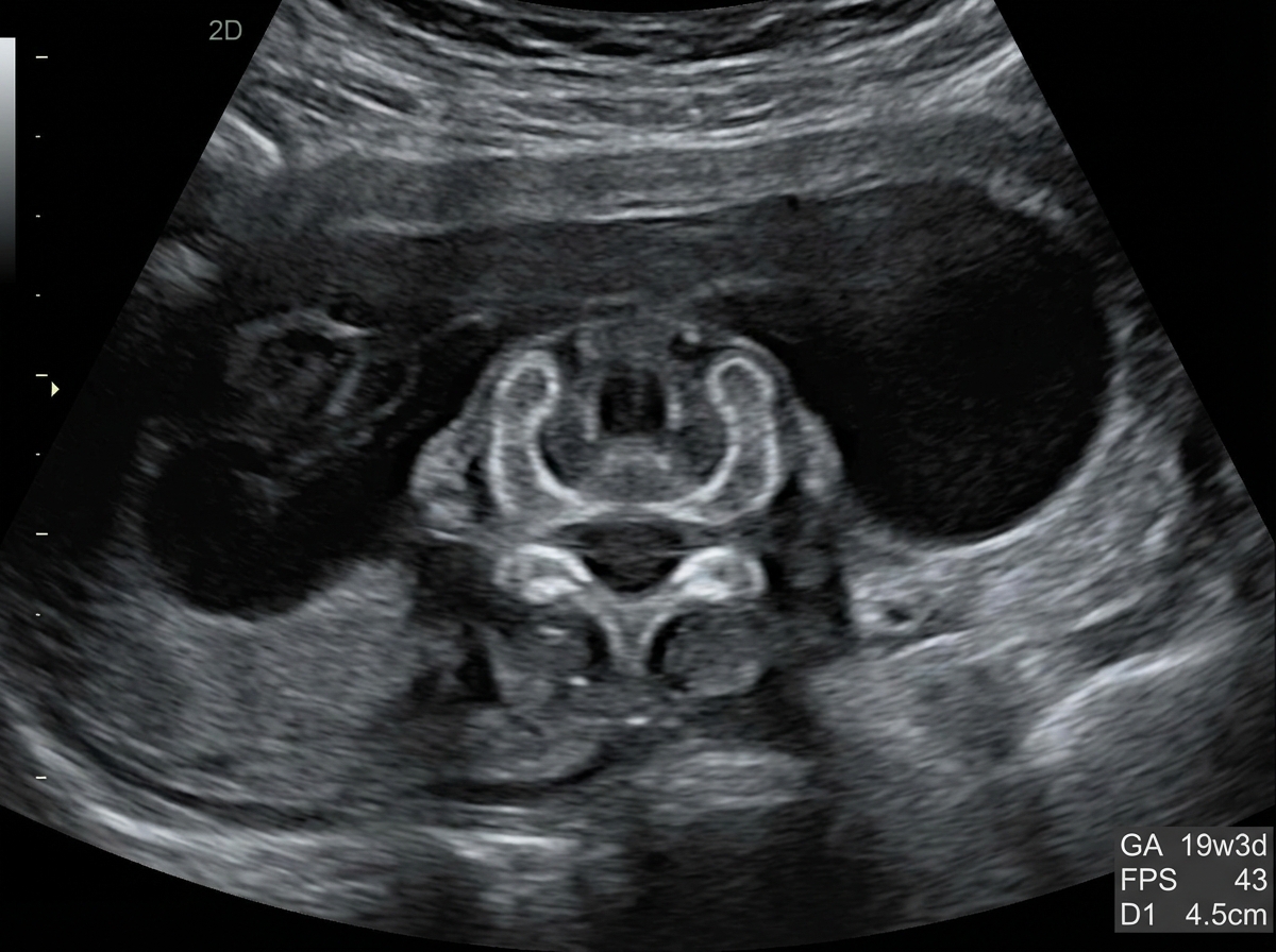
A 28-year-old married woman is anxious about conception and presents with complaints of profuse vaginal discharge. She has no history of itching. It has been 12 days since her last menstrual period (LMP). What is the most likely cause of her symptoms?
A 25-year-old woman presents with intense vaginal pruritus and pain for the past week. She says the pain is worse when she urinates. Her last menstrual period was 4 weeks ago. She is sexually active, has a single partner, and uses condoms infrequently. She denies any recent history of fevers, chills, abdominal or flank pain, or menstrual irregularities. Her past medical history is significant for systemic lupus erythematosus (SLE), diagnosed 5 years ago and managed medically. Her current medications include prednisone and oral contraceptives. The patient is afebrile and her vital signs are within normal limits. Physical examination is significant for a small amount of discharge from the vagina, along with severe inflammation and scarring. The discharge is thick, white, and has the consistency of cottage cheese. The vaginal pH is 4.1. The microscopic examination of potassium hydroxide (KOH) mount of the vaginal discharge reveals pseudohyphae. A urine pregnancy test is negative. Which of the following would be the most appropriate treatment for this patient’s condition?
A 35-year-old G0P1 female presents to her OB/GYN after 17 weeks gestation. A quad screen is performed revealing the following results: elevated inhibin and beta HCG, decreased aFP and estriol. An ultrasound was performed demonstrating increased nuchal translucency. When the fetus is born, what may be some common characteristics of the newborn if amniocentesis confirms the quad test results?
A 31-year-old G3P0020 presents to her physician for a prenatal visit at 12 weeks gestation. She does not smoke cigarettes and stopped drinking alcohol once she was diagnosed with pregnancy at 10 weeks gestation. An ultrasound examination showed the following: Ultrasound finding Measured Normal value (age-specified) Heart rate 148/min 137–150/min Crown-rump length 44 mm 45–52 mm Nasal bone visualized visualized Nuchal translucency 3.3 mm < 2.5 mm Which of the following statements regarding the presented patient is correct?
A 45-year-old primigravida woman at 13-weeks' gestation is scheduled for a prenatal evaluation. This is her first appointment, though she has known she is pregnant for several weeks. A quad screening is performed with the mother's blood and reveals the following: AFP (alpha-fetoprotein) Decreased hCG (human chorionic gonadotropin) Elevated Estriol Decreased Inhibin Elevated Ultrasound evaluation of the fetus reveals increased nuchal translucency. Which mechanism of the following mechanisms is most likely to have caused the fetus’s condition?
A 23-year-old woman comes to the physician for a routine health maintenance examination. She feels well. Menses have occurred at regular 30-day intervals and last for 5 days with normal flow. She has a history of gonorrhea that was treated at 20 years of age. She has smoked one pack of cigarettes daily for 3 years. She drinks one glass of wine daily. Her only medication is an oral contraceptive. Vital signs are within normal limits. Physical examination including pelvic examination shows no abnormalities. A Pap smear shows high-grade squamous epithelial lesion. Which of the following is the most appropriate next step in management?
Explanation: ***Metronidazole*** - This patient's symptoms (malodorous vaginal discharge, positive **Whiff test**, and presence of **clue cells** on Gram stain) are classic findings for **bacterial vaginosis (BV)**. - **Metronidazole** is the first-line and most effective antibiotic for treating bacterial vaginosis, as it targets the anaerobic bacteria overgrowing in the vagina. *Azithromycin* - **Azithromycin** is primarily used to treat infections like **chlamydia**, gonorrhea, or some respiratory tract infections. - It is **not effective** against the anaerobic bacteria responsible for bacterial vaginosis. *Nystatin pessary* - **Nystatin** is an **antifungal medication** specifically used to treat **vulvovaginal candidiasis (yeast infection)**. - The patient's presentation (malodorous discharge, positive Whiff test, clue cells) does not align with a yeast infection, making nystatin ineffective. *Tetracycline* - **Tetracycline** is a broad-spectrum antibiotic, but it is **not the preferred treatment** for bacterial vaginosis. - Its use is often associated with a higher risk of side effects and is typically reserved for other bacterial infections like **chlamydia**, acne, or Rocky Mountain spotted fever.
Explanation: **Pap smear, HPV DNA testing** - **Postcoital bleeding** is a classic symptom of **cervical cancer**, which can be identified by a **Pap smear** to detect abnormal cervical cells. - **HPV DNA testing** is essential as persistent infection with high-risk human papillomavirus (HPV) genotypes is the primary cause of cervical cancer. *Pap smear, HCV DNA, electrophoresis* - While a **Pap smear** is appropriate for cervical cytology, **HCV DNA testing** is for Hepatitis C virus infection and is not routinely indicated for postcoital bleeding. - **Electrophoresis** is used to analyze proteins (e.g., hemoglobinopathies) and has no direct role in evaluating postcoital bleeding or cervical pathology. *Liquid-based cytology, cervical biopsy* - **Liquid-based cytology** is a method of preparing a Pap smear, but it's not a standalone investigation. - A **cervical biopsy** is a more invasive procedure done *after* initial screening (like Pap smear with HPV testing) suggests abnormalities, not as a primary first-line investigation for postcoital bleeding unless there are visible lesions. *Cervical biopsy, HBV DNA* - A **cervical biopsy** is typically performed following an abnormal **Pap smear** or colposcopy findings, not as the initial diagnostic step for postcoital bleeding. - **HBV DNA testing** is for Hepatitis B virus infection and is irrelevant to the workup of postcoital bleeding.
Explanation: ***AFP (Alpha-fetoprotein)*** - **AFP** is a reliable tumor marker for differentiating **germ cell tumors**, especially **yolk sac tumors**, which often present as an ovarian solid-cystic mass. - Elevated **AFP** levels help in diagnosis, monitoring treatment response, and detecting recurrence of these specific ovarian malignancies. *CEA (Carcinoembryonic antigen)* - **CEA** is primarily associated with **gastrointestinal cancers**, such as colorectal cancer, and is less specific for ovarian masses. - While it can be elevated in some mucinous ovarian carcinomas, it's not the most specific marker for a general solid-cystic ovarian mass. *HCG (Human chorionic gonadotropin)* - **HCG** is a key marker for **gestational trophoblastic disease** and some **germ cell tumors**, such as choriocarcinoma. - It is not typically elevated in most common solid-cystic ovarian masses, especially those of epithelial origin. *HER2/neu (Human Epidermal growth factor Receptor 2)* - **HER2/neu** is primarily associated with **breast cancer** and some gastric cancers, playing a role in targeted therapy. - It is not a standard tumor marker for the general evaluation or diagnosis of ovarian solid-cystic masses.
Explanation: ***LBC + HPV*** - Cervical erosion and spotting are concerning for **cervical intraepithelial neoplasia (CIN)** or **cervical cancer**, making **Liquid-Based Cytology (LBC)** the appropriate screening method. - **Human Papillomavirus (HPV) testing** is crucial as persistent high-risk HPV infection is the primary cause of cervical cancer and helps in risk stratification and management. *Pap smear + HSV* - A **routine Pap smear** (conventional cytology) is less sensitive than LBC for detecting abnormal cervical cells and is generally being phased out by LBC. - **Herpes Simplex Virus (HSV)** causes genital herpes and is not directly associated with cervical cancer, thus testing for it in this context is not the most appropriate immediate next step. *Pap smear + HBV* - As mentioned, a **routine Pap smear** is not the preferred method for cervical cancer screening compared to LBC. - **Hepatitis B Virus (HBV)** causes liver disease and is entirely unrelated to cervical pathology; therefore, testing for it would be irrelevant to the patient's symptoms. *LBC + HSV* - While **LBC** is the correct advanced cytology method, adding **HSV testing** is not indicated as HSV does not cause cervical cancer or intraepithelial lesions that present with cervical erosion and spotting. - Focus should be on identifying potential malignancy or pre-malignant changes with HPV co-testing, not sexually transmitted infections unrelated to cancer risk.
Explanation: ***Physiological*** - **Physiological vaginal discharge** at mid-cycle (12 days post-LMP) is common and normal, often becoming profuse, clear, and elastic, indicating **ovulation**. - The absence of **itching** or other bothersome symptoms supports a non-pathological cause, especially given her anxiety about conception. *Candida* - **Candidal infections** typically present with a **thick, white, curdy discharge** and are characteristically associated with intense **itching**, which is absent in this case. - The discharge is usually not described as profuse or clear. *Trichomonas* - **Trichomoniasis** is associated with a **frothy, greenish-yellow discharge** and often causes **vaginal itching**, **burning**, and a **foul odor**, none of which are described. - While discharge can be profuse, the clinical picture does not align with Trichomonas. *Bacterial vaginosis* - **Bacterial vaginosis** typically presents with a **thin, grayish-white discharge** and a characteristic **"fishy" odor**, especially after intercourse. - **Itching** is less common than with Candida, but the discharge characteristics and the timing relative to ovulation do not fit this diagnosis.
Explanation: ***Oral fluconazole for the patient alone*** - The presence of **intense vaginal pruritus and pain**, **thick, white, cottage cheese-like discharge**, a **vaginal pH of 4.1**, and **pseudohyphae on KOH mount** are classic findings for **vulvovaginal candidiasis (VVC)**, also known as a yeast infection. - Oral fluconazole is an effective systemic antifungal treatment for VVC. Since VVC is generally not sexually transmitted, treating the partner is typically not necessary. *A single dose of azithromycin* - **Azithromycin** is an antibiotic primarily used to treat bacterial infections like **Chlamydia trachomatis**, which would present with different symptoms such as mucopurulent discharge and dysuria, but not typically the profound pruritus or characteristic discharge described. - It would be ineffective against a fungal infection and could disrupt the normal vaginal flora, potentially worsening VVC. *Oral metronidazole for the patient and her sexual partner* - **Metronidazole** is an antibiotic used to treat **bacterial vaginosis** (typically pH > 4.5, thin grey discharge, positive whiff test) or **trichomoniasis** (foamy yellow-green discharge, high pH). - Treating the partner for VVC is generally unnecessary, and metronidazole is not effective against fungal infections. *Oral fluconazole for the patient and her sexual partner* - While oral fluconazole is appropriate for the patient, **VVC is not considered a sexually transmitted infection**, so routine treatment of the sexual partner is not recommended unless the partner is symptomatic with balanitis. - There is no indication that the partner has symptoms of a fungal infection, making partner treatment unnecessary. *Topical antifungal cream for the patient alone* - Topical antifungal creams (e.g., miconazole, clotrimazole) are effective for **uncomplicated VVC**. - However, given the **"intense vaginal pruritus and pain," "severe inflammation and scarring,"** and the patient's history of **SLE managed with prednisone** (which can predispose to more severe or recurrent infections), a systemic treatment like oral fluconazole may be more appropriate for a potentially complicated or severe case, offering faster relief and better penetration.
Explanation: ***Epicanthal folds, macroglossia, flat profile, depressed nasal bridge, and simian palmar crease*** - The quad screen results (**elevated inhibin and beta HCG, decreased aFP and estriol**) and **increased nuchal translucency** are classical findings suggestive of **Down syndrome (Trisomy 21)**. - The listed characteristics are all common clinical features observed in newborns with **Down syndrome**, including distinctive facial features and a single palmar crease. *Microphthalmia, microcephaly, cleft lip/palate, holoprosencephaly, and polydactyly* - These features are indicative of **Patau syndrome (Trisomy 13)**. The quad screen findings for Patau syndrome would typically show **decreased beta-hCG** and **decreased PAPP-A**, which do not match the patient's results. - While nuchal translucency can be increased in Trisomy 13, the specific quad screen pattern points away from this diagnosis. *Epicanthal folds, high-pitched crying/mewing, and microcephaly* - The **high-pitched crying (cat-like cry)** is a hallmark feature of **Cri-du-chat syndrome (5p deletion)**, which is associated with microcephaly. - Quad screen abnormalities are not reliably associated with Cri-du-chat syndrome, and it's a chromosomal deletion, not a trisomy, which is implied by the quad screen pattern. *Elfin facies, low nasal bridge, and extreme friendliness with strangers* - These are characteristic features of **Williams syndrome (deletion on chromosome 7 impacting elastin gene)**. - Williams syndrome is a microdeletion syndrome not typically detected by quad screening, and the presented quad screen results are not consistent with this condition. *Rocker-bottom feet, micrognathia, clenched hands with overlapping fingers, and prominent occiput* - These features are classic physical findings associated with **Edwards syndrome (Trisomy 18)**. - The quad screen for Edwards syndrome would show **decreased beta-hCG**, **decreased AFP**, and **decreased unconjugated estriol**, which is different from the patient's elevated inhibin and beta HCG.
Explanation: ***To increase the diagnostic accuracy of this result, the levels of free beta-hCG and pregnancy-associated plasma protein A (PAPP-A) should be determined.*** - An elevated **nuchal translucency** at 12 weeks gestation is a soft marker for aneuploidy, particularly Down syndrome, making further biochemical screening appropriate. - Combining **free beta-hCG** and **PAPP-A** with nuchal translucency (first-trimester screening) significantly improves the detection rate for chromosomal abnormalities like trisomy 21. *To increase the diagnostic accuracy of this result, the levels of serum alpha-fetoprotein, hCG, and unconjugated estriol should be determined.* - **Alpha-fetoprotein (AFP)**, **hCG**, and **unconjugated estriol** are components of the second-trimester triple or quad screen, typically performed between 15-20 weeks, not 12 weeks. - Performing these markers at 12 weeks would yield less accurate results for genetic screening and is not the standard approach for first-trimester risk assessment. *At this gestational age, nuchal translucency has low diagnostic value.* - **Nuchal translucency (NT)** is highly valuable and diagnostically significant *between 11 weeks 2 days and 13 weeks 6 days gestation*, or when the crown-rump length (CRL) is 45-84 mm. - The measurement presented (3.3 mm) is **elevated (>2.5 mm)** for this gestational age, indicating increased risk and requiring further evaluation. *Pathology other than Down syndrome should be suspected because of the presence of a nasal bone.* - While the **presence of a nasal bone** can be a reassuring sign, it does not rule out Down syndrome, especially with a significantly elevated nuchal translucency (3.3 mm). - The combination of a thickened NT and a present nasal bone still warrants further investigation, as **nasal bone hypoplasia/absence** is a stronger, but not exclusive, marker for Down syndrome. *The observed ultrasound image is caused by the problems with the embryonic kidneys.* - **Nuchal translucency** is a collection of fluid under the skin in the fetal neck, and while multiple abnormalities can cause it, kidney problems are not a direct or primary cause. - Abnormally increased **NT** is associated with chromosomal abnormalities (e.g., trisomy 21, 18, 13), cardiac defects, and other genetic syndromes, not typically isolated embryonic kidney pathology at this stage.
Explanation: ***Nondisjunction*** - The quad screen results (**decreased AFP**, **elevated hCG**, **decreased estriol**, **elevated inhibin**) combined with **increased nuchal translucency** are highly suggestive of **Trisomy 21 (Down syndrome)**. - **Nondisjunction** is the most common mechanism leading to trisomies, such as Trisomy 21, where homologous chromosomes fail to separate during meiosis I or sister chromatids fail to separate during meiosis II. *Nucleotide excision repair defect* - This mechanism is associated with conditions like **xeroderma pigmentosum**, characterized by extreme sensitivity to UV light and increased risk of skin cancer. - It does not explain the quad screen findings or increased nuchal translucency indicative of a chromosomal aneuploidy. *Mismatch repair* - Defects in **mismatch repair** are linked to conditions like **Lynch syndrome**, which predisposes individuals to various cancers, particularly colorectal cancer. - This mechanism is not related to the genesis of chromosomal abnormalities like Trisomy 21. *Mosaicism* - **Mosaicism** occurs when an individual has at least two genetically distinct cell lines originating from a single zygote, meaning some cells have Trisomy 21 and others do not. - While it can result in Down syndrome, **nondisjunction** is the primary event that leads to the initial trisomy in the majority of cases, and mosaicism is considered a less common cause or a secondary event. *Robertsonian translocation* - A **Robertsonian translocation** involves the fusion of two acrocentric chromosomes, which can lead to Trisomy 21 if an individual inherits an extra copy of chromosome 21 in addition to the translocated chromosome. - While it accounts for a minority of Down syndrome cases (about 4%), **nondisjunction** is the most frequent cause, responsible for approximately 95% of cases.
Explanation: ***Colposcopy*** - A **colposcopy** is the appropriate next step for a **high-grade squamous intraepithelial lesion (HSIL)** on a Pap smear, as it allows for direct visualization of the cervix and targeted biopsies. - This procedure helps confirm the diagnosis and determine the extent and severity of the lesion. *Endometrial sampling* - **Endometrial sampling** is used to investigate abnormal uterine bleeding or evaluate for endometrial hyperplasia/carcinoma, not cervical abnormalities. - The patient has regular menses and no other symptoms suggestive of endometrial pathology. *Repeat cytology in 6 months* - **Repeat cytology in 6 months** is usually reserved for low-grade squamous intraepithelial lesions (LSIL) or atypical squamous cells of undetermined significance (ASC-US), especially in younger patients. - An HSIL finding warrants immediate colposcopy due to the higher risk of progression to cancer. *Loop electrosurgical excision* - **Loop electrosurgical excision procedure (LEEP)** is a treatment modality used after a significant lesion (e.g., CIN 2/3) has been identified and localized by colposcopy and biopsy. - It is not the initial diagnostic step following an abnormal Pap smear showing HSIL. *Cervical biopsy* - A **cervical biopsy** is typically performed as part of a colposcopy, where abnormal areas are directly visualized and sampled. - Performing a random cervical biopsy without colposcopic guidance might miss the high-grade lesion or lead to inadequate sampling.
Explanation: ***Single-dose PO metronidazole*** - The symptoms of **increased vaginal discharge**, **dysuria**, and **cervical petechiae** (strawberry cervix) in a sexually active woman, along with motile, flagellated organisms on wet mount, are classic for **Trichomonas vaginalis** infection. - The recommended treatment for trichomoniasis is a single 2-gram oral dose of **metronidazole**, or 500 mg orally twice daily for 7 days. *Vaginal metronidazole* - While metronidazole is the correct drug, the **vaginal formulation** is less effective than oral metronidazole for treating trichomoniasis. - **Oral metronidazole** achieves higher systemic concentrations and treats potential involvement of the **urethra** and **paraurethral glands**, which vaginal formulations may not adequately address. *PO fluconazole* - **Fluconazole** is an antifungal medication primarily used to treat **yeast infections** (candidiasis), not parasitic infections like trichomoniasis. - The clinical presentation and wet mount findings (motile flagellated organisms) are not consistent with a yeast infection. *Vaginal clindamycin* - **Clindamycin** is an antibiotic used to treat **bacterial vaginosis** (BV). - While BV can cause vaginal discharge, the presence of **motile flagellated organisms** and **cervical petechiae** are specific to trichomoniasis, and not typically seen in BV. *IM benzathine penicillin* - **Benzathine penicillin** is the standard treatment for **syphilis**. - The patient's symptoms and wet mount findings are not indicative of syphilis, which presents with chancres, rashes, or neurological symptoms depending on the stage.
Explanation: ***Combination oral contraceptives*** - This patient presents with symptoms highly suggestive of **polycystic ovary syndrome (PCOS)**, including irregular menses, hirsutism, acne, obesity, and elevated blood glucose. **Combination oral contraceptives (COCs)** are the first-line treatment for PCOS to regulate menstrual cycles and manage hyperandrogenism. - COCs suppress **gonadotropin-releasing hormone (GnRH)**, reducing ovarian androgen production, and increase **sex hormone-binding globulin (SHBG)**, which lowers free testosterone levels. *Insulin* - While the patient has **elevated blood glucose (190 mg/dL)**, this is likely secondary to **insulin resistance** associated with PCOS. Insulin therapy is typically reserved for more severe hyperglycemia or diabetes unresponsive to oral agents. - Treating insulin resistance with **metformin** or lifestyle changes is usually preferred before initiating insulin in PCOS patients. *Leuprolide* - **Leuprolide** is a **GnRH agonist** that initially stimulates and then down-regulates pituitary gonadotropin release, leading to a hypogonadal state. It is primarily used for conditions like endometriosis or uterine fibroids. - While it can suppress androgen production, it is not the first-line treatment for **PCOS** due to potential side effects and the immediate goal of cycle regulation and symptom management with COCs. *Danazol* - **Danazol** is a synthetic androgen that suppresses gonadotropin release, leading to an anovulatory state and decreased estrogen production. It is used in conditions like **endometriosis** and **fibrocystic breast disease**. - It has significant **androgenic side effects**, which would exacerbate the patient's existing hirsutism and acne, making it an inappropriate choice for PCOS. *Metformin* - **Metformin** is an **insulin sensitizer** that is often used in PCOS patients, especially those with insulin resistance and an elevated BMI, to improve glucose metabolism and ovulation. - While metformin could be a valuable adjunct, the primary and most appropriate initial pharmacotherapy for managing irregular menses, hirsutism, and acne in PCOS is **combination oral contraceptives**.
Explanation: ***Mammography*** - The patient is 47 years old, and a **baseline mammogram** is recommended for women aged 40-49, with annual screening starting at 50, although some guidelines recommend annual screening from 40. - Given her age and absence of recent screening, ordering a mammogram is the **most appropriate next step** for breast cancer screening. *Colonoscopy* - **Colorectal cancer screening** with colonoscopy typically begins at age **45 years for average-risk individuals** (per latest guidelines), or earlier if there are specific risk factors such as a family history of colorectal cancer. - While important, this patient's primary presentation is for a **Pap smear**, indicating a focus on gynecological and associated screenings. Without further symptoms pertaining to the colon, a mammogram is a more immediate priority as a general health screening, considering the timing of her visit. *Colposcopy* - A colposcopy is performed to **further evaluate abnormal Pap smear results**, such as atypical squamous cells of undetermined significance (ASCUS) or higher. - The question states the visit is for a **routine Pap smear**, implying the results are not yet known or are expected to be normal, making colposcopy premature. *Mammography in 3 years* - Waiting three years for a mammogram would **delay routine screening** beyond recommended guidelines for a 47-year-old woman. - Current guidelines suggest either **annual or biennial screening** for women in their 40s. *Breast self-examination* - While breast self-examination (BSE) can promote **breast awareness**, it is **not considered a primary screening tool** for breast cancer due to a lack of evidence show that it reduces mortality. - **Clinical breast exams** (CBE) performed by a healthcare provider, along with mammography, are the recommended screening methods.
Explanation: ***Begin 3-year interval cervical cancer screening via Pap smear at age 21*** - Current guidelines from organizations like the **USPSTF** and **ACOG** recommend starting cervical cancer screening at age 21, regardless of the age of sexual debut. - For women aged 21-29, the standard recommendation is to undergo **cytology (Pap smear) alone every 3 years**. *Begin 2-year interval cervical cancer screening via Pap smear at 19 years of age* - This recommendation is too early and the interval is not consistent with current guidelines for routine screening based on age. - Starting screening at 19 years old is not supported by major medical organizations, which uniformly advise starting at age 21. *Offer to administer the HPV vaccine so that Pap smears can be avoided* - While the **HPV vaccine is highly recommended** for this age group to prevent cervical cancer, it does not eliminate the need for cervical cancer screening. - Vaccinated individuals can still be at risk for HPV types not covered by the vaccine or for infections acquired prior to vaccination, thus requiring screening. *Begin 2-year interval cervical cancer screening via Pap smear today* - This is incorrect as screening is not recommended until age 21, regardless of sexual activity. - Initiating screening at 16 years old is associated with **overdiagnosis** and overtreatment of benign HPV infections that would likely clear spontaneously, without clinical benefit. *Begin 5-year interval cervical cancer screening via Pap smear at age 21* - A 5-year interval for screening with Pap smear alone is not standard for women aged 21-29; this interval is typically reserved for **co-testing (HPV and cytology) in women aged 30-65**. - For a 21-year-old, the recommended interval for cytology alone is 3 years.
Explanation: ***Metronidazole*** - The image shows **trichomonads** (flagellated protozoa), and the question describes a **yellow, odorous vaginal discharge** with itching and soreness, consistent with **trichomoniasis**. - **Metronidazole** is the drug of choice for treating trichomoniasis. *Fluconazole* - **Fluconazole** is an antifungal medication primarily used to treat **candidiasis** (yeast infections), which typically presents with a thick, white, "cottage cheese" discharge, not a yellow, odorous discharge. - The lab results confirm a protozoal infection, not fungal. *Nystatin* - **Nystatin** is also an antifungal medication, usually available as a topical or oral rinse, used for **mucocutaneous candidiasis**. - It would not be effective against a **protozoal infection** like trichomoniasis. *Acyclovir* - **Acyclovir** is an antiviral medication used to treat **herpes simplex virus (HSV)** infections, such as genital herpes. - Genital herpes presents with painful vesicles and ulcers, which are not described in this patient's symptoms. *Ampicillin* - **Ampicillin** is an antibiotic used to treat bacterial infections. - It would not be effective against **protozoal infections** like trichomoniasis, and broad-spectrum antibiotics are not indicated here.
Explanation: ***Spina bifida cystica*** - Markedly elevated **maternal serum α-fetoprotein (MSAFP)**, with otherwise normal quadruple screen results, is highly indicative of an **open neural tube defect**, such as spina bifida cystica. - Spina bifida cystica involves an **incomplete closure of the spinal column**, allowing fetal cerebrospinal fluid and tissue to leak into the amniotic fluid, thereby elevating MSAFP. *Holoprosencephaly* - This condition involves incomplete division of the **forebrain** and is typically associated with **normal or low MSAFP**, not elevated levels. - It often presents with severe facial anomalies and is sometimes linked to trisomy 13. *Trisomy 18* - **Trisomy 18 (Edwards syndrome)** typically presents with a characteristic quadruple screen pattern of **low MSAFP**, low β-hCG, and low estriol, with normal inhibin A. - The presented elevated MSAFP and otherwise normal values rule out trisomy 18. *Fetal alcohol syndrome* - **Fetal alcohol syndrome** is a developmental disorder caused by prenatal alcohol exposure, leading to microcephaly, facial dysmorphia, and neurodevelopmental issues. - It does not directly cause an **elevated MSAFP**; MSAFP is used to screen for neural tube defects and certain chromosomal abnormalities. *Trisomy 21* - **Trisomy 21 (Down syndrome)** is characterized by a quadruple screen showing **elevated β-hCG** and **inhibin A**, with **low MSAFP** and estriol. - The elevated MSAFP and normal β-hCG and inhibin A in this case make trisomy 21 an unlikely diagnosis.
Explanation: ***Human papillomavirus infection*** - The patient's symptoms, including **post-coital bleeding**, **foul-smelling discharge**, and the presence of a **deformed cervix with a 4-cm exophytic, necrotizing mass**, are highly indicative of **cervical cancer**. - **High-risk human papillomavirus (HPV) infection** is established as the primary cause of cervical cancer, accounting for over 99% of cases. The patient's lack of recent gynecologic evaluation and long-term IUD use (which does not protect against STIs) are also relevant. *Exposure to radioactive iodine* - **Radioactive iodine ablation** for Graves' disease primarily affects the thyroid gland and is not a known cause of cervical cancer or vaginal masses. - While radiation exposure can increase cancer risk, the type of radiation used for thyroid ablation specifically targets thyroid tissue with minimal systemic impact on other organs like the cervix. *IUD complication* - Complications from a **levonorgestrel-releasing IUD** typically involve altered bleeding patterns (lighter periods or amenorrhea), pain, or infection, but rarely cause a large, necrotizing exophytic cervical mass. - While long-term IUD use without gynecologic follow-up can occasionally mask symptoms or lead to unnoticed infections, it does not directly cause cervical cancer or a mass of this description. *Exposure to heavy metals* - Occupational exposure to **heavy metals** (e.g., in a machinery operator) has not been consistently linked to an increased risk of cervical cancer. - While certain environmental toxins can be carcinogenic, heavy metals are not recognized as a primary risk factor for the specific presentation of cervical cancer. *Hyperestrogenemia* - **Hyperestrogenemia** is primarily associated with an increased risk of **endometrial cancer** and, to a lesser extent, breast cancer. - It does not significantly increase the risk of cervical cancer, which is almost exclusively linked to HPV infection.
Explanation: ***Gastroschisis*** - The elevated maternal serum **α-fetoprotein (MSAFP)** and the ultrasound finding of a **paraumbilical abdominal wall defect** with **bowel herniation** freely suspended in amniotic fluid are classic signs of gastroschisis. - Gastroschisis is a congenital malformation frequently associated with **isolated defects**, and typically *not* with chromosomal abnormalities or other syndromes, differentiating it from omphalocele. *Chromosomal trisomy* - While elevated MSAFP can be seen in some chromosomal abnormalities, the specific ultrasound findings of a **freely floating bowel** and a **defect lateral to the umbilical cord** are inconsistent with the typical presentation of chromosomal trisomies, which are more often associated with omphalocele. - **Omphalocele**, not gastroschisis, is frequently associated with chromosomal abnormalities such as **trisomy 13, 18, and 21**, as well as other genetic syndromes. *Spina bifida* - Spina bifida is a **neural tube defect** characterized by a defect in the closure of the spinal column, often with a meningeal or myelomeningeal sac. - Although it also causes **elevated MSAFP**, the ultrasound finding would show a **spinal defect**, not an abdominal wall defect with herniated bowel. *Bladder exstrophy* - Bladder exstrophy involves the **eversion of the bladder** through an anterior abdominal wall defect, exposing the bladder mucosa. - While it is an anterior abdominal wall defect, the ultrasound would show a **missing bladder**, not herniated bowel, and MSAFP elevation is not a consistent finding. *Beckwith-Wiedemann syndrome* - This syndrome is associated with **omphalocele**, macroglossia, and organomegaly, but not typically gastroschisis. - Although omphalocele can cause elevated MSAFP, the specific defect described (**right of the umbilical cord, free-floating bowel**) is characteristic of gastroschisis, which is usually *not* associated with Beckwith-Wiedemann syndrome.
Explanation: ***Breast ultrasound*** - In women under 30 with a palpable breast mass, **ultrasound** is the primary imaging modality due to higher breast density, which limits the effectiveness of mammography. - Ultrasound can differentiate between **solid and cystic masses** and guide further diagnostic procedures if needed. *Reassurance* - While many breast masses are benign, a **newly discovered palpable mass** in a young woman always warrants further investigation to rule out malignancy, especially given the "hard" description. - Simply providing reassurance without imaging could delay diagnosis of a potentially serious condition. *Mammogram* - **Mammography** is less sensitive in younger women due to the **dense glandular tissue** of their breasts, which can obscure masses. - It involves radiation exposure, which should be minimized in younger patients unless specifically indicated. *Incision and drainage* - **Incision and drainage** is a procedure typically reserved for **infected cysts or abscesses**, which usually present with signs of inflammation such as pain, redness, and warmth. - The patient's mass is described as **nontender** and lacks other signs of infection. *Mastectomy* - **Mastectomy** is a surgical procedure for **breast cancer** and is a definitive treatment, not a diagnostic step. - It would be highly inappropriate to consider mastectomy without a definitive diagnosis and staging of malignancy.
Explanation: ***Colposcopy*** - The presence of **atypical squamous cells of undetermined significance (ASC-US)** with a **positive high-risk HPV test** warrants further investigation to rule out cervical dysplasia. - **Colposcopy** allows for direct visual examination of the cervix, identification of abnormal areas, and targeted biopsies, which is essential given the positive high-risk HPV. *Endometrial biopsy* - This procedure is indicated for evaluating the **endometrium**, typically in cases of abnormal uterine bleeding or suspicion of endometrial hyperplasia or cancer. - It is not relevant for addressing **cervical cytological abnormalities** like ASC-US or positive high-risk HPV. *Loop electrosurgical excision* - This is a **therapeutic procedure** used to remove abnormal cervical tissue, usually after a diagnosis of **high-grade squamous intraepithelial lesion (HSIL)** or cervical cancer is confirmed by biopsy. - It is not the initial diagnostic step for ASC-US with positive HPV. *Follow-up after 3 years and repeat cytology by Pap smear* - This extended follow-up period is typically recommended for patients with **normal co-testing (negative cytology and negative HPV)**, not for those with ASC-US and positive high-risk HPV. - Waiting three years would delay the diagnosis of potential cervical precancerous lesions. *Follow-up after 1 year and repeat cytology by Pap smear and HPV testing* - This approach, known as **"reflex testing"** or **"co-testing"**, is generally used for women with negative cytology but positive HPV, or for certain ASC-US cases with negative HPV. - However, for ASC-US with a **positive high-risk HPV result**, current guidelines recommend immediate colposcopy, especially in women over 30.
Explanation: ***Test for HPV*** - For women aged 21-29 with **atypical squamous cells of undetermined significance (ASC-US)** Pap smear results, **reflex HPV testing** is the preferred next step in management. - If the HPV test is positive, **colposcopy** is indicated; if negative, the patient can return to routine screening in 3 years. *Resume routine screening schedule* - This is incorrect because an ASC-US result indicates a potential abnormality that requires further investigation, not immediate return to routine screening. - **HPV co-testing** or a repeat Pap smear is needed to rule out underlying cervical dysplasia. *Repeat Pap smear in 3 years* - This is incorrect because for an ASC-US result, a 3-year interval is only appropriate if the **HPV test is negative**; otherwise, colposcopy or earlier re-evaluation is needed. - Waiting 3 years risks progression of potential high-grade lesions if HPV is positive. *Perform colposcopy* - This is incorrect as **colposcopy** is generally reserved for patients with a positive **HPV test** following an ASC-US result or for more severe Pap smear abnormalities (e.g., HSIL, ASC-H). - Performing colposcopy without prior HPV testing for ASC-US may lead to unnecessary procedures and costs, as many HPV-negative ASC-US cases resolve spontaneously. *Obtain a vaginal smear* - A vaginal smear is generally used to evaluate cells from the vaginal wall, often for infections like **bacterial vaginosis** or **candidiasis**, or for a history of hysterectomy. - It is not the appropriate next step for an ASC-US Pap smear, which specifically concerns cervical cell abnormalities and requires HPV evaluation for risk stratification.
Explanation: ***lecithin:sphingomyelin < 1.5*** - A lecithin:sphingomyelin (L:S) ratio less than 2:1 (or 1.5 in some clinical contexts) indicates **fetal lung immaturity** and a **high risk for respiratory distress syndrome (RDS)**. - The **lecithin level increases** significantly in the amniotic fluid during the third trimester as fetal lungs mature, while **sphingomyelin levels remain relatively constant**. *lecithin:phosphatidylserine < 1.5* - While **phosphatidylserine** is a component of surfactant, the **Lecithin:Sphingomyelin (L:S) ratio** is the established and most commonly used marker for fetal lung maturity. - There is **no widely recognized or clinically validated threshold** for a lecithin:phosphatidylserine ratio in predicting respiratory distress syndrome. *lecithin:sphingomyelin > 1.5* - An L:S ratio **greater than 2:1 (or 1.5, in some labs)** generally indicates **fetal lung maturity** and a low risk for respiratory distress syndrome. - Therefore, this ratio would suggest a **lower likelihood of pulmonary distress**, which contradicts the aim of identifying risk. *lecithin:phosphatidylserine > 3.0* - As with an L:S ratio, a higher ratio would generally indicate **lung maturity**, not increased risk for pulmonary distress. - There is **no clinical standard for lecithin:phosphatidylserine ratio** to assess lung maturity for preventing RDS. *lecithin:sphingomyelin > 3.0* - An L:S ratio of **greater than 2:1 (or 3.0 in certain clinical scenarios)** is a strong indicator of **fetal lung maturity**, meaning the risk of respiratory distress syndrome is low. - The question asks for a ratio that would be **predictive of pulmonary distress**, whereas this ratio indicates the opposite.
Explanation: ***Triple test*** - This 21-year-old patient with **pre-existing diabetes** and an **HbA1c of 8.3%** has a significantly increased risk of fetal neural tube defects and other chromosomal abnormalities. The triple test, performed between **15 and 20 weeks**, can screen for these risks by measuring **alpha-fetoprotein (AFP)**, **human chorionic gonadotropin (hCG)**, and **unconjugated estriol (uE3)**. - Given the patient's **poor glycemic control** (HbA1c 8.3% indicates consistently high blood glucose levels), the triple test offers a non-invasive screening method to assess these elevated risks. *Chorionic villus sampling* - While CVS can detect chromosomal abnormalities and some genetic disorders, it is an **invasive procedure** associated with a risk of miscarriage and is typically performed earlier in pregnancy (10-13 weeks). - It is usually reserved for cases with **higher risk factors** identified through non-invasive screening or a history of genetic disorders, which are not explicitly stated as the primary concern here compared to the hyperglycemia-related defects. *Serum creatinine* - **Serum creatinine** is used to assess kidney function and is essential in diabetic patients to monitor for nephropathy, but it is **not a screening test for fetal abnormalities**. - While important for the mother's health management, it does not directly address the immediate concern of fetal risk due to uncontrolled diabetes during pregnancy. *C-peptide assessment* - **C-peptide** is a marker of endogenous insulin production and is useful in classifying diabetes type or assessing residual beta-cell function; however, this patient is a known **type 1 diabetic** taking insulin. - While it has diagnostic utility for the mother's condition, it does not provide information about fetal well-being or the risk of congenital anomalies. *Oral glucose tolerance test* - An **oral glucose tolerance test (OGTT)** is used to diagnose **gestational diabetes mellitus** (GDM) in women without pre-existing diabetes, or to confirm it in those with borderline values. - This patient already has a confirmed diagnosis of **type 1 diabetes**; therefore, an OGTT is not indicated for her as she is already being treated for diabetes.
Explanation: **Repeat Pap smear in 12 months** * For women aged 21-24 years with a **low-grade squamous intraepithelial lesion (LSIL)**, the recommended management is a repeat Pap test in **12 months**, as many of these lesions spontaneously regress. * Given her age and that she is **asymptomatic**, watchful waiting with repeat cytology is a safe and appropriate approach. *Colposcopy with endocervical and endometrial sampling* * **Colposcopy** is generally reserved for persistent LSIL, **high-grade squamous intraepithelial lesions (HSIL)**, or certain HPV co-testing results in older women. * **Endometrial sampling** is not indicated in this case as there are no symptoms suggestive of endometrial pathology, nor does LSIL on a Pap smear necessitate it. *Colposcopy with endocervical sampling* * As above, **colposcopy** is not the initial step for LSIL in this age group, unless persistence is noted. * **Endocervical sampling** (ECC) is performed during colposcopy to evaluate the endocervical canal, but the initial management for LSIL in young women is observation. *Loop electrosurgical excision procedure* * **Loop electrosurgical excision procedure (LEEP)** is an **ablative or excisional procedure** used to remove cervical lesions, primarily for persistent HSIL or CIN2/3. * It is an **overtreatment** for an initial diagnosis of LSIL in a 24-year-old woman, especially given the high rate of spontaneous regression. *Repeat Pap smear in 3 years* * While some guidelines allow for less frequent screening in women with normal Pap results and negative HPV co-testing, a **3-year interval** is too long for follow-up of an LSIL result. * The risk of progression, although low, requires closer monitoring within the **12-month timeframe** to identify persistent lesions.
Explanation: ***Anencephaly*** - **Elevated maternal serum α-fetoprotein (MSAFP)** and **amniotic fluid α-fetoprotein (AFAFP)**, along with elevated **acetylcholinesterase (AChE)** in amniotic fluid, are classic markers for **open neural tube defects**. Anencephaly, characterized by the **absence of a major portion of the brain and skull**, is an open neural tube defect. - The **increased amniotic fluid volume (polyhydramnios)** is due to the fetus's inability to swallow amniotic fluid, a common finding in anencephaly. *Holoprosencephaly* - This condition involves incomplete separation of the **prosencephalon (forebrain)**, leading to **severe facial abnormalities** and brain malformations. - While it is a severe brain malformation, it is typically a **closed neural tube defect** or a developmental anomaly not involving an open lesion, and therefore, it is usually not associated with elevated MSAFP, AFAFP, or AChE. *Spina bifida occulta* - This is the **mildest form of spina bifida**, involving a small gap in the vertebrae without protrusion of the spinal cord or meninges. - It is a **closed neural tube defect** and is typically asymptomatic, often not associated with elevated MSAFP or AFAFP levels. *Myelomeningocele* - While a **myelomeningocele** is an **open neural tube defect** that would cause elevated MSAFP, AFAFP, and AChE, it is characterized by the protrusion of the spinal cord and meninges through a vertebral defect. - The primary characteristic of anencephaly (absence of a major portion of the brain/skull) better fits the severe degree of neural tube defect suggested by the findings, particularly the polyhydramnios due to absent swallowing reflex. *Lissencephaly* - This is a brain malformation characterized by a **lack of gyri and sulci**, resulting in a smooth brain surface. - It is a brain development defect, not an **open neural tube defect**, and as such, it is not associated with elevated MSAFP, AFAFP, or AChE.
Explanation: ***CA-125*** - The patient's symptoms of **fatigue, constipation, decreased appetite/thirst, abdominal distension, and weight loss** in an older female are highly concerning for **ovarian cancer**, for which **CA-125** is a tumor marker. - Ovarian cancer can cause **paraneoplastic syndromes** or **mass effect** leading to these diffuse, non-specific symptoms, and unexplained falls/light-headedness. *Seasonal viral infection* - While viral infections can cause fatigue and decreased appetite, they typically present with more acute symptoms like **fever, cough, or myalgias**, which are absent here. - Viral infections are unlikely to cause **significant weight loss** (10 lbs) and **abdominal distension** without other clear infectious signs. *Cardiac arrhythmia* - An arrhythmia could explain light-headedness and falls, but the patient's other symptoms like **constipation, abdominal distension, and significant weight loss** are not typical. - The "odd sensation in her chest" is vague and could be benign, and her pulse of 92/min is not diagnostic of a specific arrhythmia. *Vagal response* - A vagal response can cause light-headedness and falls, especially during straining (e.g., using the bathroom), but it is usually a **transient event**. - It does not explain the **chronic symptoms** of fatigue, constipation, abdominal distension, weight loss, and recurrent episodes suggesting an underlying systemic issue. *Dehydration* - Dehydration can cause light-headedness and falls, especially in older adults with decreased thirst, but it is unlikely to be the primary cause of such **significant weight loss** (10 pounds) and **abdominal distension**. - The patient's blood pressure is stable (122/88 mmHg), which is not typical for significant dehydration causing frequent falls.
Explanation: ***White blood cells alone*** - The patient's symptoms (mucoid secretion, cervical motion tenderness, clear urethral discharge, history of unprotected intercourse with multiple partners) are highly suggestive of **Chlamydia trachomatis infection**. - **Chlamydia** is an obligate intracellular bacterium that does not show up on a routine Gram stain of urethral or cervical discharge, so only **white blood cells** would be seen, indicating inflammation. *White blood cells + gram-negative rod* - The presence of **gram-negative rods** in the urinary tract would suggest a common bacterial urinary tract infection (UTI) caused by organisms like *E. coli* or *Klebsiella*. - This typically presents with **dysuria** and more purulent discharge, which is not consistent with the patient's presentation of mucoid discharge and absence of dysuria. *White blood cells + gram-negative diplococci* - **Gram-negative diplococci** are characteristic of **Neisseria gonorrhoeae** infection. While gonorrhea can cause cervicitis and urethritis, the discharge is usually **purulent** and opaque, not clear and mucoid as described. - While coinfection is common, the most fitting single description for the *reported* discharge and lack of specific gram-stainable organisms points away from gonorrhea as the sole microscopic finding. *White blood cells + gram-negative coccobacilli* - **Gram-negative coccobacilli** could suggest infections from organisms like *Gardnerella vaginalis* (bacterial vaginosis) or *Haemophilus ducreyi* (chancroid). - These conditions typically present with different clinical pictures, such as a **fishy odor** for bacterial vaginosis or **painful genital ulcers** for chancroid, none of which are described here. *White blood cells + motile flagellates* - The presence of **motile flagellates** in a vaginal or urethral swab is characteristic of **Trichomonas vaginalis** infection. - While *Trichomonas* can cause urethritis and cervicitis, the discharge is typically **frothy**, yellow-green, and malodorous, which differs from the clear, mucoid secretion described in this case.
Explanation: ***Physiologic leukorrhea*** - The presentation of **thin, white, odorless vaginal discharge** in an adolescent with normal vital signs and no other symptoms is highly suggestive of **physiologic leukorrhea**. - **Physiologic leukorrhea** is a normal discharge that varies in consistency, color, and amount throughout the menstrual cycle due to fluctuating hormone levels. The absence of abnormalities on wet mount further supports this diagnosis. *Vaginal foreign body* - A **vaginal foreign body** would typically present with a **foul-smelling, often blood-tinged discharge**, which is not described here. - While possible, the characteristics of the discharge and lack of additional symptoms make this diagnosis less likely. *Bacterial vaginosis* - **Bacterial vaginosis** is characterized by a **thin, gray-white discharge with a foul, fishy odor**, particularly after intercourse. - Diagnosis typically involves a positive whiff test and the presence of **clue cells** on wet mount, neither of which were observed. *Vaginal candidiasis* - **Vaginal candidiasis** (yeast infection) presents with a **thick, white, curd-like discharge**, often accompanied by **pruritus, erythema**, and dysuria. - The patient's discharge is described as thin and white, not curd-like, and there are no other associated symptoms. *Trichomoniasis* - **Trichomoniasis** typically causes a **yellow-green, frothy, malodorous discharge**, often associated with vaginal itching, burning, and dyspareunia. - Microscopy would reveal motile **trichomonads**, which were not seen on the wet mount.
Explanation: ***Herpes simplex virus type 2*** - The presence of **red, vesicular ulcers on the labia** that are painful, along with **tender inguinal lymphadenopathy**, fevers, headaches, and lethargy, are classic signs of a primary HSV-2 infection. - HSV-2 is the most common cause of **genital herpes**, leading to painful vesicular or ulcerative lesions in the genital area. *Chlamydia trachomatis* - This pathogen typically causes **urethritis**, **cervicitis**, or **pelvic inflammatory disease (PID)**, often with minimal or no symptoms, or with discharge or dysuria. - It does not typically present with **painful vesicular or ulcerative lesions** and systemic symptoms like fever and headache are less common. *Klebsiella granulomatis* - This bacterium causes **granuloma inguinale (donovanosis)**, characterized by progressive, painless, beefy red **ulcers** with rolled borders, not vesicles. - The lesions are typically **not painful** and do not usually present with systemic symptoms like fever and headache in the initial stages. *Treponema pallidum* - The primary stage of syphilis, caused by *Treponema pallidum*, presents as a **painless chancre**, which is a firm, indurated ulcer. - The lesions in this patient are described as **painful and vesicular**, which is inconsistent with a syphilitic chancre. *Herpes simplex virus type 1* - While HSV-1 can cause genital herpes, it is more commonly associated with **oral herpes (cold sores)**. - Genital HSV-1 infections are typically **less severe** and **recur less frequently** than HSV-2 infections.
Explanation: ***Duct ectasia*** - The patient's presentation with **gray-green nipple discharge**, an **inverted nipple**, and **tubular calcifications on mammogram** are classic signs of duct ectasia in a postmenopausal woman. - The presence of **dilated subareolar ducts** in the contralateral breast further supports this diagnosis, as it is a benign condition characterized by widening of the breast ducts. *Periareolar fistula* - This condition is typically associated with recurrent **subareolar abscesses** and chronic drainage, often from a nipple piercing or previous infection, which are not described here. - Periareolar fistulas rarely present solely with gray-green discharge and tubular calcifications without a clear history of infection or abscess. *Intraductal papilloma* - Intraductal papillomas usually present with **serous or bloody nipple discharge**, rather than the gray-green discharge seen in this patient. - While they can cause nipple discharge, they are not typically associated with **inverted nipples** or **tubular calcifications** on mammogram. *Periductal mastitis* - Periductal mastitis is an inflammatory condition that can cause nipple inversion and discharge, but the discharge is usually **purulent or inflammatory**, and it is often accompanied by signs of infection like pain, redness, and swelling, which are absent in this case. - It is more commonly seen in **smokers**, whereas this patient is a non-smoker. *Phyllodes tumor* - Phyllodes tumors usually present as a **rapidly growing palpable breast mass**, which may be benign or malignant, but they are not typically associated with nipple discharge or tubular calcifications. - The description of **gray-green discharge** and **tubular calcifications** does not align with the typical presentation of a phyllodes tumor.
Explanation: ***T. pallidum hemagglutination assay*** - A positive **Venereal Disease Research Laboratory (VDRL)** test indicates potential exposure to syphilis, but it is a **non-treponemal test** and can yield false positives. - A **treponemal test** such as the *T. pallidum* hemagglutination assay (TPHA) or fluorescent treponemal antibody-absorbed (FTA-ABS) is required to confirm the diagnosis of syphilis. *Full serum panel for HBV* - The **hepatitis B surface antigen (HBsAg)** is negative, and **anti-HBsAg (HBsAb)** is positive, indicating either prior vaccination or resolved infection with immunity. - This patient does not have active hepatitis B infection, so a full serum panel for HBV is not necessary. *PCR for HBV DNA* - Similar to the above, the serological markers indicate immunity to HBV, not active infection. - **PCR for HBV DNA** would only be indicated if there were signs of active infection or **occult HBV**. *HBV vaccination* - The patient already has **protective antibodies (anti-HBsAg)** against Hepatitis B, indicating immunity. - Vaccination would be redundant as she is already immune. *Prescription of benzylpenicillin* - While **benzylpenicillin** is the treatment for syphilis, a definitive diagnosis has not yet been made. - Confirmation with a **treponemal test** is crucial before initiating treatment to avoid unnecessary antibiotic exposure.
Explanation: ***Condylomata acuminata*** - The image likely depicts a **koilocyte**, a key indicator of **Human Papillomavirus (HPV) infection**, which causes condylomata acuminata. - Koilocytes are characterized by **perinuclear cytoplasmic vacuolization** and nuclear atypia, directly linked to HPV. *Bacterial vaginosis* - Characterized by a **shift in vaginal flora**, presenting with "clue cells" (vaginal epithelial cells covered in bacteria) and discharge, not koilocytes. - While common, bacterial vaginosis does not cause the **cytopathic changes** seen with HPV infection. *Trichomoniasis* - Caused by the **protozoan parasite** *Trichomonas vaginalis*, leading to a frothy, green-yellow discharge and cervical inflammation (strawberry cervix). - Diagnosis involves identifying the **motile trichomonads** on wet mount, not koilocytes on a Pap smear. *Genital herpes* - Caused by **herpes simplex virus (HSV)**, resulting in painful vesicular lesions that ulcerate. - Cytologic findings include **multinucleated giant cells** with nuclear molding and intranuclear inclusions, distinctly different from koilocytes. *Syphilitic chancre* - A primary lesion of syphilis caused by **_Treponema pallidum_**, presenting as a painless ulcer. - Diagnosis is made by **darkfield microscopy** or serologic tests; cytology is not used to identify syphilitic chancres.
Explanation: ***Twice-yearly clinical breast exams, annual mammography, annual breast MRI, and breast self-exams*** - For patients with **BRCA1 or BRCA2 mutations**, an intensive breast cancer screening protocol is recommended due to their highly increased lifetime risk of breast cancer. - This typically includes **semiannual clinical breast exams**, **annual mammography**, and **annual breast MRI**, often starting at a young age. *Order magnetic resonance imaging of the breast* - While MRI is a crucial part of screening for high-risk individuals, it is **not sufficient as a standalone screening modality**. - A comprehensive approach combining multiple screening methods is needed to maximize detection rates. *Annual ultrasound, annual mammography, and monthly self-breast exams* - **Breast ultrasound** is generally used as an adjunct to mammography when specific abnormalities are found or in women with dense breasts, not as a routine primary screening tool for BRCA carriers. - While **mammography** and **self-breast exams** are included, this option lacks the crucial **annual MRI** and **twice-yearly clinical breast exams** recommended for BRCA carriers. *Annual clinical breast exams, annual mammography, and monthly self-breast exams* - This protocol is **less intensive** than what is recommended for women with BRCA mutations. - It omits the essential **annual breast MRI** and the **twice-yearly clinical breast exams** that are critical for early detection in this high-risk population. *Refer to radiation therapy* - **Radiation therapy** is a treatment modality for existing cancer, not a screening approach for cancer prevention or early detection. - Referring for radiation therapy would be appropriate only after a diagnosis of breast cancer, not as a primary screening strategy.
Explanation: ***Pelvic ultrasound*** - A **non-mobile right adnexal mass** in a postmenopausal woman, along with vague symptoms like **abdominal distension** and **weight gain**, raises suspicion for **ovarian cancer**. - A **pelvic ultrasound** is the initial, non-invasive, and cost-effective imaging modality to characterize adnexal masses, assessing size, morphology, and vascularity. *Abdominal MRI* - While **MRI** offers excellent soft tissue contrast, it is typically used as a **secondary imaging modality** for further characterization of adnexal masses when ultrasound findings are inconclusive or for surgical planning, not as the initial step. - Its higher cost and longer scan time make it less suitable for initial screening compared to ultrasound. *PET-CT* - **PET-CT** is primarily used for **staging malignancies** and detecting metastatic disease, or in cases of unknown primary, and is not the initial diagnostic test for an adnexal mass. - It involves radiation exposure and is generally reserved for situations where malignancy is already highly suspected or confirmed. *Exploratory laparotomy and debulking* - **Exploratory laparotomy** and **debulking** are surgical procedures performed for the definitive diagnosis, staging, and treatment of ovarian cancer, but only *after* a thorough initial workup has been completed. - It is an invasive procedure and should not be the first step in the investigation of an adnexal mass. *CA-125 level* - Measuring **CA-125** levels is useful as a **tumor marker** in the workup of suspected ovarian cancer, particularly in symptomatic postmenopausal women, and for monitoring treatment response. - However, it has **low specificity** (can be elevated in benign conditions) and should be ordered in conjunction with imaging, not as the sole initial diagnostic step.
Explanation: ***45 X0*** - The presence of **cystic hygroma** and **fetal hydrops** strongly suggests **Turner syndrome (45, X0)**, as these are classic sonographic findings. - The history of **recurrent early pregnancy losses** is also consistent with chromosomal aneuploidies, with 45, X0 being a common cause of such losses. *Monosomy 18* - **Monosomy 18** is a very rare and usually lethal chromosomal abnormality, typically resulting in **early miscarriage**. - Its clinical presentation, if live-born, is distinct and does not primarily feature **cystic hygroma** or **hydrops** as the main diagnostic clues. *Trisomy 13* - **Trisomy 13 (Patau syndrome)** is associated with severe malformations, including **cleft lip/palate**, **polydactyly**, and **holoprosencephaly**. - While it can cause fetal hydrops and other structural anomalies, **cystic hygroma** is not its most characteristic or common sonographic marker in the way it is for Turner syndrome. *Trisomy 21* - **Trisomy 21 (Down syndrome)** is characterized by **nuchal translucency** and **cardiac defects**, but **cystic hygroma** and **hydrops** are less common and less severe than in Turner syndrome. - The constellation of findings in this case points more strongly to Turner syndrome. *Monosomy 13* - **Monosomy 13** is an extremely rare and usually **lethal** chromosomal anomaly, often leading to early spontaneous abortion. - It would typically result in more severe generalized developmental defects rather than the specific combination of **cystic hygroma** and **hydrops** seen here.
Explanation: ***Azithromycin*** - A **yellow cervical discharge** and **negative *Neisseria* species NAAT** strongly suggest a *Chlamydia trachomatis* infection, for which azithromycin is a first-line treatment. - Given the patient's symptoms of **vaginal discharge and itchiness**, along with the high prevalence of chlamydia, empirical treatment is appropriate while awaiting further test results. *Ceftriaxone* - This antibiotic is primarily used to treat **gonorrhea**, which has been ruled out by the negative *Neisseria* NAAT. - While sometimes given in combination therapy, it is not the best single agent in this scenario. *Azithromycin and ceftriaxone* - This combination is typically used for **empirical treatment of cervicitis** when both gonorrhea and chlamydia are suspected. - However, since **gonorrhea has been excluded** by NAAT, adding ceftriaxone is unnecessary at this point. *Fluconazole* - Fluconazole is an **antifungal medication** used to treat **yeast infections** (*Candida* species). - While the patient reports itchiness, a **yellow discharge** is more characteristic of a bacterial infection like chlamydia, rather than the typically white, cottage-cheese-like discharge of a yeast infection. *Cervical cultures* - While collecting cervical cultures for other pathogens (e.g., **Trichomonas**) might be considered, the immediate priority in a symptomatic patient with suspected chlamydia is to **initiate treatment to prevent complications** and reduce transmission. - **NAATs are highly sensitive and specific** for chlamydia and gonorrhea, making them preferred over traditional cultures for these infections.
Explanation: ***Age of sexual debut*** - An early **age of sexual debut** (before 17 years old) is a significant risk factor for **HPV infection** and subsequently, cervical dysplasia. This patient's sexual debut at 16 years old falls within this high-risk category. - Early sexual activity, especially with multiple partners, increases the likelihood of exposure to **human papillomavirus (HPV)**, the primary cause of cervical cancer and its precursor lesions. *Ovarian surgery* - **Ovarian surgery**, such as a right ovary resection for a follicular cyst, is not a known risk factor for **cervical intraepithelial neoplasia (CIN)** or **cervical cancer**. - This aspect of her medical history is unrelated to the development of cervical dysplasia. *History of cocaine abuse* - While **cocaine abuse** can be associated with other health complications and risky behaviors, it is not a direct or independent **risk factor** for **cervical intraepithelial neoplasia (CIN)**. - There is no established physiological link between cocaine use and the development of cervical dysplasia. *Patient age* - Although the incidence of HPV infection peaks in younger women, the risk of developing **high-grade cervical intraepithelial neoplasia (HGSIL)** and invasive cancer increases with age, particularly after 30 due to persistent HPV infection. - However, at 34, her age is not as strong a contributing risk factor as an early **age of sexual debut** for the initial development of the underlying condition. *Age at first pregnancy* - **Early age at first pregnancy** (before 20) can increase the risk of cervical cancer in some studies, possibly due to hormonal changes in the cervix making it more vulnerable to **HPV infection**. - This patient's first pregnancy at 26 is not considered an early age at first pregnancy and therefore is not a significant risk factor in this context.
Explanation: ***Trisomy 21*** - The quadruple test results of **low alpha-fetoprotein**, **low unconjugated estriol**, **high beta-hCG**, and **high inhibin A** are highly characteristic of **Trisomy 21 (Down syndrome)**. - These specific maternal serum analyte patterns are used in **second-trimester screening** to assess the risk of chromosomal abnormalities. *Trisomy 18* - Trisomy 18 (Edwards syndrome) would typically show **low alpha-fetoprotein**, **low unconjugated estriol**, and **low beta-hCG**, often with **normal or low inhibin A**. - The elevated beta-hCG and inhibin A in the patient's results make Trisomy 18 less likely. *Neural tube defect* - Neural tube defects (NTDs) are associated with **elevated alpha-fetoprotein** levels in maternal serum. - The patient's results show **low alpha-fetoprotein**, which argues against an NTD. *Congenital toxoplasmosis* - Congenital toxoplasmosis is an infection and does not typically present with a specific pattern of abnormal maternal serum markers like those seen in the quadruple test. - Diagnosis would involve specific **serological testing for Toxoplasma antibodies** or ultrasound findings. *Trophoblastic disease* - Trophoblastic disease, such as a **hydatidiform mole**, is associated with **extremely high levels of beta-hCG**, often much higher than what would be seen in Trisomy 21. - It would also typically be detected earlier in pregnancy and present with distinct ultrasound findings, which were normal in this case.
Explanation: **Endometrial biopsy** - **Abnormal uterine bleeding (AUB)** in an **obese patient over 45 (or 35 with risk factors)** warrants evaluation for **endometrial hyperplasia or carcinoma**. - The patient's age (39), obesity (BMI 34), and change in bleeding pattern make endometrial biopsy the most appropriate initial diagnostic step to rule out malignancy. *Abdominal ultrasonography* - While ultrasound can assess uterine structure and endometrial thickness, it is **not sufficient** to rule out malignancy in cases requiring tissue diagnosis. - An abnormal ultrasound finding would still likely necessitate an endometrial biopsy for definitive diagnosis. *Endometrial ablation* - This is a **treatment** for AUB, not a diagnostic step. It should only be considered after excluding malignancy. - Performing ablation without a prior biopsy could delay the diagnosis of endometrial cancer. *Combined oral contraceptives* - Hormonal therapy can manage AUB symptoms, but it is **not appropriate as an initial step** when there's a concern for endometrial malignancy. - It would mask symptoms and delay diagnosis without addressing the underlying cause. *Diagnostic laparoscopy* - Laparoscopy is an invasive procedure typically used to investigate pelvic pain, endometriosis, or adnexal masses. - It is **not indicated** as the first step for isolated AUB where the primary concern is endometrial pathology.
Explanation: ***Confirmatory amniocentesis and chromosomal analysis of the fetal cells*** - The combination of **quad-screen results** (elevated 𝛽-hCG, elevated inhibin A, decreased AFP, decreased estradiol) and **increased nuchal translucency** strongly suggests an aneuploidy, particularly **Down Syndrome (Trisomy 21)**. - **Amniocentesis** is a **diagnostic procedure** that provides fetal cells for definitive chromosomal analysis (karyotyping), confirming or ruling out aneuploidy with high accuracy. *Biopsy and pathologic examination of fetus* - A **fetal biopsy** is generally not a standard diagnostic test for aneuploidy and carries higher risks than amniocentesis or chorionic villus sampling (CVS). - This procedure would typically be considered for specific fetal anomalies requiring tissue diagnosis, not for confirming chromosomal disorders. *Fetus is normal, continue with pregnancy as expected* - The abnormal **quad-screen results** and **increased nuchal translucency** are significant indicators of potential chromosomal abnormalities, making it unlikely that the fetus is normal. - Ignoring these findings could lead to the birth of a child with an undiagnosed genetic condition. *Maternal karyotype* - A **maternal karyotype** evaluates the mother's chromosomes to identify balanced translocations or other inherited chromosomal abnormalities that could increase the risk in offspring. - While helpful for identifying a parental genetic cause, it does not directly diagnose the fetal condition; a fetal sample is still needed for that. *Cell-free fetal DNA analysis* - **Cell-free fetal DNA (cfDNA) analysis** is a **screening test** with high sensitivity and specificity for common aneuploidies, but it is not a diagnostic test. - While it can guide further investigation, a positive cfDNA result still requires a **confirmatory diagnostic procedure** like amniocentesis or CVS before making definitive clinical decisions.
Explanation: ***Karyotype analysis*** - The patient's presentation with **primary amenorrhea**, **short stature**, and phenotypic features like a **short neck** and **wide torso** are highly suggestive of **Turner syndrome**. Karyotype analysis is the definitive test to confirm this diagnosis by identifying the characteristic **45,X monosomal karyotype**. - A karyotype directly analyzes the **chromosomal complement**, allowing for the detection of **aneuploidy** or structural abnormalities which are the underlying cause of Turner syndrome. *Serum FSH and LH levels* - While **elevated FSH and LH levels** would be expected in Turner syndrome due to **gonadal dysgenesis**, indicating **hypergonadotropic hypogonadism**, these hormonal levels are not diagnostic on their own. - High FSH and LH levels indicate **ovarian failure** but do not definitively identify the underlying cause, which could be various conditions leading to hypogonadism. *Buccal smear for Barr bodies* - A buccal smear for **Barr bodies** (inactive X chromosomes) can indicate the number of X chromosomes. In Turner syndrome (45,X), **no Barr bodies** would be found, but this test is **less reliable** and **less specific** than a full karyotype. - A buccal smear cannot detect all forms of Turner syndrome, such as mosaicism (e.g., 45,X/46,XX), which a karyotype would identify. *Serum testosterone level* - A **serum testosterone level** would be relevant if **androgen insensitivity syndrome** or another cause of virilization was suspected; however, this patient has normal external female genitalia and no signs of virilization. - Low testosterone is expected in females and would not explain primary amenorrhea with these specific physical findings. *Serum 17-hydroxyprogesterone level* - **Serum 17-hydroxyprogesterone** is used to screen for **congenital adrenal hyperplasia (CAH)**, which might present with primary amenorrhea if severe, but typically involves **virilization** (ambiguous genitalia, clitoromegaly), which is absent in this patient. - The patient's physical characteristics (short stature, short neck, wide torso) are not consistent with CAH.
Explanation: ***Bacteria-coated epithelial cells on wet mount microscopy*** - This finding, specifically **clue cells**, is a hallmark of **bacterial vaginosis (BV)**. Clue cells are vaginal epithelial cells covered in bacteria, indicating the overgrowth of anaerobic bacteria and a decrease in normal lactobacilli. - The patient's symptoms of a **malodorous discharge** that worsens after intercourse, a **gray, thin, homogeneous discharge**, and a **vaginal pH of 6.0** are all classic signs of bacterial vaginosis. *Numerous gram-positive rod-shaped bacteria on Gram stain* - **Normal vaginal flora** is dominated by **Gram-positive rods (lactobacilli)**. In bacterial vaginosis, these beneficial bacteria are significantly reduced, while anaerobic bacteria proliferate. - An overgrowth of Gram-positive rods would suggest a healthy vaginal flora, conflicting with the presented symptoms and a high vaginal pH. *Gram-positive diplococci on Gram stain* - **Gram-positive diplococci** isolated from vaginal discharge could suggest an infection with organisms like **Streptococcus pneumoniae** or **Staphylococcus aureus**, which are not typical causes of malodorous vaginal discharge or bacterial vaginosis. - Infections like Neisseria gonorrhoeae, which can cause cervicitis, are characterized by **Gram-negative diplococci**. *Negative whiff test* - The **whiff test** involves adding potassium hydroxide (KOH) to vaginal discharge to detect a fishy odor. A **positive whiff test** is a characteristic sign of bacterial vaginosis due to the production of amines by anaerobic bacteria. - A negative whiff test would argue against a diagnosis of bacterial vaginosis despite the other clinical findings. *Motile protozoa on wet mount microscopy* - The presence of **motile protozoa**, specifically **Trichomonas vaginalis**, is indicative of trichomoniasis, another common cause of vaginitis. - While trichomoniasis can cause a frothy discharge and elevated pH, the discharge is often described as greenish-yellow, and it would not typically present with the specific features of clue cells.
Explanation: ***Cancer antigen-125*** - The patient's presentation with **abnormal vaginal bleeding**, **breast tenderness**, **spider angiomata**, and **ascites** suggests a gynecological malignancy, possibly **ovarian cancer** with liver involvement due to chronic alcohol use. **CA-125** is the most widely used tumor marker for epithelial ovarian cancer. - While not diagnostic on its own, elevated CA-125 levels, especially in the context of a **palpable ovarian mass** and **ascites**, strongly support the diagnosis of ovarian cancer, and it is also used for monitoring treatment response and recurrence. *Alpha-fetoprotein* - **Alpha-fetoprotein (AFP)** is primarily associated with **germ cell tumors**, such as **yolk sac tumors** of the ovary, and **hepatocellular carcinoma**. - While an ovarian mass is present, the clinical picture with prominent signs of **estrogen excess** (abnormal bleeding, breast tenderness, thickened endometrial stripe) and **liver disease** (spider angiomata, ascites) does not strongly point to an AFP-secreting tumor or primary hepatocellular carcinoma without further evidence. *Estrogen* - Although the patient's symptoms (abnormal bleeding, breast tenderness, thickened endometrial stripe) are consistent with **estrogen excess**, estrogen itself is a **hormone**, not a tumor marker used to diagnose ovarian cancer. - Some ovarian tumors, particularly **granulosa cell tumors**, produce estrogen, which would be reflected in elevated serum estrogen levels, but "estrogen" is not the tumor marker itself. *Testosterone* - **Testosterone** is primarily associated with **androgen-secreting tumors**, such as **Sertoli-Leydig cell tumors** of the ovary, which would typically present with signs of **virilization** (e.g., hirsutism, deepening voice, clitoromegaly). - The patient's symptoms of **estrogen excess** (abnormal uterine bleeding, breast tenderness) are contrary to a testosterone-secreting tumor. *Carcinoembryonic antigen* - **Carcinoembryonic antigen (CEA)** is a tumor marker primarily associated with **colorectal cancer**, but it can also be elevated in other adenocarcinomas, such as those of the **breast, lung, and gastrointestinal tract**. - While general malignancy is considered, CEA is not the primary marker for ovarian cancer, and the constellation of symptoms (especially those related to estrogen excess and possible liver involvement) points more specifically towards an ovarian origin.
Explanation: **Cold-knife conization** - This patient has a **high-grade squamous intraepithelial lesion (HSIL)**, positive **HPV**, and an **inadequate colposcopy** because the lesion extends into the endocervical canal, preventing visualization of the full extent of the transformation zone. - In such cases, **cold-knife conization** is the most appropriate next step as it allows for the complete excision of the transformation zone and provides a definitive diagnosis by removing the lesion for histological evaluation, ensuring all margins can be assessed. *Cryoablation of the lesion* - This method is typically reserved for women with **low-grade squamous intraepithelial lesions (LSIL)** or low-grade CIN confirmed by biopsy, or in situations where definitive excisional treatment is not required. - It is not suitable when an **inadequate colposcopy** suggests the lesion extends into the endocervical canal, where the full extent cannot be visualized and margins cannot be assessed. *Punch biopsy and subsequent management based on the results* - A **punch biopsy** would not be sufficient here because the colposcopy was **inadequate**, meaning the full extent of the lesion and its margins could not be visualized. - A punch biopsy would provide only a superficial sample and would not address the concern of the lesion extending into the **endocervical canal**, which requires excisional treatment to ensure complete removal and proper evaluation of margins. *Laser ablation of the lesion* - Similar to cryoablation, laser ablation is generally used for **low-grade lesions** or in cases where the entire lesion and its margins are clearly visible and fully resectable. - Given the patient's **HSIL** and **inadequate colposcopy** due to scarring and obscured margins, laser ablation would not guarantee complete eradication or adequate histological assessment of the deeper lesion. *Genotyping for HPV type 16 and 18 and further management based on the results* - While **HPV genotyping for types 16 and 18** is useful for risk stratification and guiding management in certain scenarios, it is not the immediate next step when an HSIL is diagnosed with an inadequate colposcopy. - The presence of **HSIL** on cytology, combined with the inability to adequately visualize the lesion's extent during **colposcopy**, necessitates an excisional procedure like conization to rule out invasive cancer and ensure complete removal of the precancerous lesion.
Explanation: ***Pap test and HPV test in 5 years*** - For women aged 30-65, **co-testing with both a Pap test and HPV test every 5 years** is the preferred screening interval if both results are normal. - This patient, at 58 years old, falls within this age range, and her prior normal Pap tests along with a normal current one, support a 5-year interval for co-testing. *Colposcopy in 3 years* - **Colposcopy** is a diagnostic procedure performed to further evaluate abnormal Pap test results, not a routine screening method. - Doing a colposcopy in 3 years would be an overly aggressive approach given her history of normal screenings. *Pap test only in 5 years* - While a Pap test alone every 3 years is an acceptable screening option, **co-testing with HPV every 5 years** is generally preferred due to its higher sensitivity for detecting precancerous lesions. - Omitting the HPV test would reduce the effectiveness of the screening strategy in detecting cervical cancer early. *Discontinue screening until the patient becomes sexually active* - **Sexual activity** is a risk factor for HPV infection, but cervical cancer screening guidelines do not link its discontinuation to a lack of sexual activity. - Women over 65 years old with a history of adequate negative screenings may discontinue screening, but this patient is 58 and does not meet that criterion yet. *Colposcopy at the current visit to verify Pap test results* - A **colposcopy** is indicated for **abnormal Pap test results**, which this patient does not have. - Performing a colposcopy in the absence of abnormal findings is unnecessary and not part of routine screening.
Explanation: ***Single transverse palmar crease*** - The prenatal findings (hypoplastic nasal bone, low **α-fetoprotein** and **free estriol**, high **inhibin A** and **β-hCG**) are characteristic of **Down syndrome (Trisomy 21)**. - A **single transverse palmar crease** (Simian crease) is a classic physical finding in infants with Down syndrome. *Microphthalmia* - **Microphthalmia** (abnormally small eyes) is more commonly associated with chromosomal abnormalities like **Trisomy 13 (Patau syndrome)**, not Down syndrome. - Other features of Trisomy 13 include **holoprosencephaly**, cleft lip/palate, and polydactyly, which are not suggested by the prenatal screening. *Ambiguous external genitalia* - **Ambiguous external genitalia** can be associated with various genetic conditions, particularly those affecting sex hormone synthesis or differentiation (e.g., **congenital adrenal hyperplasia**). - It is not a characteristic finding of Down syndrome. *Meningomyelocele* - A **meningomyelocele** is a severe form of **neural tube defect**, typically indicated by **high α-fetoprotein** levels. - The presented serum markers show **low α-fetoprotein**, ruling out neural tube defects. *Extremity lymphedema* - **Extremity lymphedema** is a hallmark finding in **Turner syndrome (XO)** due to lymphatic system malformation. - Turner syndrome typically presents with different prenatal markers, such as **cystic hygroma** and often normal maternal serum screen values (or specific patterns not matching the profile given).
Explanation: ***Diethylstilbestrol exposure in utero*** - The patient's presentation with **clear cell carcinoma of the vagina**, characterized by **red, fleshy polypoid masses** and **large cells with abundant clear cytoplasm**, is highly suggestive of this diagnosis. - **In utero exposure to diethylstilbestrol (DES)** is a classic and significant risk factor for the development of clear cell adenocarcinoma of the vagina and cervix. *Family history of breast and ovarian cancer* - While a family history of breast and ovarian cancer may indicate an increased risk for other gynecological cancers (e.g., BRCA mutations), it is **not directly linked** to clear cell adenocarcinoma of the vagina. - This family history points more towards **hereditary breast and ovarian cancer syndromes**, not the specific pathology described. *Human papillomavirus infection* - **HPV infection** is a major risk factor for most cases of **squamous cell carcinoma of the vagina and cervix**, and also increases the risk of adenocarcinoma of the cervix. - However, HPV is **not a primary risk factor for clear cell adenocarcinoma of the vagina**, which has a distinct etiology. *Alcohol consumption* - While excessive **alcohol consumption** can be associated with an increased risk of certain cancers, it is **not a specific or significant risk factor** for clear cell adenocarcinoma of the vagina. - It generally contributes to a broad range of cancers rather than specific rare forms. *Cigarette smoking* - **Cigarette smoking** is a well-established risk factor for **squamous cell carcinoma of the cervix and vagina**, among other cancers. - However, it is **not a recognized significant risk factor** for the development of **clear cell adenocarcinoma of the vagina**.
Explanation: ***Syphilis, HIV, and HBV*** - The **American College of Obstetricians and Gynecologists (ACOG)** and the **Centers for Disease Control and Prevention (CDC)** recommend universal screening for syphilis, HIV, and hepatitis B virus (HBV) in all pregnant women at the first prenatal visit. - This **routine screening** is crucial due to the potential for vertical transmission and severe adverse outcomes for the neonate if untreated. *Syphilis and HIV* - While screening for syphilis and HIV is essential, it is **incomplete** as it omits HBV, which is also universally recommended for antenatal screening. - This option does not align with the standard comprehensive screening guidelines for pregnancy. *Syphilis, HIV, HBV, and chlamydia* - Although syphilis, HIV, and HBV screening are appropriate, adding **chlamydia** to the universal prenatal screening for *all* pregnant women in the first trimester is not standard practice unless specific risk factors are present or local prevalence is high. - Chlamydia screening is typically recommended for pregnant women who are **25 years or younger** or those with **risk factors** for sexually transmitted infections (STIs). *Syphilis, HIV, and chlamydia* - This option incorrectly includes chlamydia as a universal screen for all pregnant women while **omitting HBV**, which is universally recommended. - Missing HBV screening leaves a critical gap in prenatal care, as it can be transmitted vertically and cause severe neonatal disease. *No routine screening is recommended for this patient* - This statement is incorrect as **universal screening** for syphilis, HIV, and HBV is recommended for all pregnant women, regardless of reported risk factors or monogamous relationships. - Maternal infection can still occur, and screening helps prevent severe outcomes for both mother and child through timely detection and intervention.
Explanation: **The patient should undergo screening every 3 years after she turns 21 years of age.** - Current guidelines recommend initiating cervical cancer screening at **age 21**, regardless of sexual activity. - The recommended interval for cytology-only screening is **every 3 years** for women aged 21-29. *The patient requires annual Pap testing due to her family history of cervical cancer.* - **Family history of cervical cancer** is generally not considered a reason for earlier or more frequent screening in individuals under 21 years of age, unless specific genetic syndromes are suspected, which is not mentioned here. - The primary risk factor for cervical cancer is **HPV infection**, not direct family history. *HPV testing is more preferable than Pap testing in sexually active women under 21 years of age.* - **HPV testing** as a primary screening method is **not recommended for women younger than 25** due to the high prevalence of transient HPV infections that resolve spontaneously in this age group. - Over-screening and subsequent interventions could lead to unnecessary anxiety and procedures for conditions that would likely resolve on their own. *It is reasonable to start Pap-test screening at the current visit and repeat it every 3 years.* - Starting screening at the current age of **18 years is not recommended** according to current guidelines, as screening typically begins at age 21. - Early screening in this age group often leads to the detection of **transient HPV infections** that would otherwise resolve without intervention, causing undue stress and follow-up. *The patient does not require Pap testing as long as she uses barrier contraception.* - While **barrier contraception** (condoms) reduces the risk of HPV transmission, it does not eliminate it entirely and therefore **does not negate the need for cervical cancer screening.** - Regular screening is still recommended to detect any persistent HPV infections and associated cervical changes early.
Explanation: ***Patient 1 – BRCA testing. Patient 2 – Breast ultrasound*** - Patient 1 has a strong family history of early-onset **breast and ovarian cancer** (**mother and sister**), suggesting a high probability of an inherited genetic mutation, such as **BRCA1/2**, which warrants genetic testing. - Patient 2 presents with a **small, mobile, well-defined breast mass** that is likely benign, and a **breast ultrasound** is the appropriate initial imaging for further characterization in a young woman. *Patient 1 – Breast ultrasound. Patient 2 – Return in 3 months for a clinical breast exam* - Patient 1's primary concern is genetic predisposition due to family history, an **ultrasound** is not the initial or primary screening method for future cancer risk. - Patient 2 has a palpable mass; waiting 3 months for a **clinical breast exam** without initial imaging (ultrasound) is not appropriate for evaluating a new breast lump. *Patient 1 – Reassurance. Patient 2 – Breast ultrasound* - Patient 1's family history of **early-onset breast and ovarian cancer** is a significant risk factor; therefore, simple **reassurance** without further investigation is inappropriate. - While a **breast ultrasound** is appropriate for Patient 2, the recommendation for Patient 1 is incorrect. *Patient 1 – CA-125 testing. Patient 2 – BRCA testing* - **CA-125** is a tumor marker primarily used for monitoring ovarian cancer treatment or recurrence, not for initial screening in asymptomatic individuals, especially in a young woman with no active symptoms. - **BRCA testing** is indicated for Patient 1 due to family history, but not for Patient 2 who has a likely benign breast mass and no significant family history. *Patient 1 – Breast and ovarian ultrasound. Patient 2 – Mammography* - Regular **breast and ovarian ultrasounds** are not recommended as primary screening tools for genetic risk in asymptomatic high-risk individuals like Patient 1. - **Mammography** is less sensitive in young women (under 30) due to higher breast tissue density, making **ultrasound** the preferred initial imaging for Patient 2.
Explanation: ***Oral fluconazole for the patient*** - The symptoms of **thick, odorless, white discharge**, **marked vulvar erythema and edema**, **normal vaginal pH**, and most importantly, the presence of **pseudohyphae** on microscopic examination are all characteristic findings of **vulvovaginal candidiasis**. - **Oral fluconazole** is a first-line treatment for uncomplicated vulvovaginal candidiasis, effectively targeting the fungal overgrowth. *Topical metronidazole* - **Metronidazole** is an antibiotic primarily used to treat **bacterial vaginosis** and **trichomoniasis**. - Microscopic examination showing **pseudohyphae** rules out bacterial vaginosis and trichomoniasis, making metronidazole an ineffective treatment for candidiasis. *Oral clindamycin for the patient* - **Clindamycin** is an antibiotic effective against certain bacterial infections, including **bacterial vaginosis**. - It is not effective against **fungal infections** like candidiasis. *Oral fluconazole for the patient and her partner* - While **oral fluconazole** is appropriate for the patient, routine treatment of male sexual partners for **vulvovaginal candidiasis** is **not typically recommended** unless the partner also exhibits symptoms of balanitis or the patient experiences recurrent infections. - **Candidiasis is not considered a sexually transmitted infection (STI)** because it often occurs in sexually inactive women and successful treatment does not depend on partner treatment. *Oral clindamycin for the patient and her partner* - **Clindamycin** is not effective against **fungal infections**. - Furthermore, routine partner treatment for vaginal candidiasis with any medication is generally **not indicated**.
Explanation: ***Oral glucose tolerance test for gestational diabetes mellitus*** - The **oral glucose tolerance test (OGTT)**, typically performed between **24 and 28 weeks of gestation**, is the gold standard for screening and diagnosing **gestational diabetes mellitus (GDM)**. This patient is at 24 weeks, making it the appropriate time for this screening. - GDM, if undiagnosed and untreated, can lead to significant maternal and fetal complications, including **macrosomia**, **preeclampsia**, **neonatal hypoglycemia**, and **shoulder dystocia**. *Fasting and random glucose testing for gestational diabetes mellitus* - While **fasting** or **random glucose** values can indicate hyperglycemia, they are **not sensitive or specific enough** on their own to reliably screen for or diagnose GDM. - A single elevated reading might prompt further testing, but it's not the primary or most suitable screening method. *HbA1C for gestational diabetes mellitus* - **HbA1c** reflects **average blood glucose levels over the past 2-3 months** and is primarily used for diagnosing and monitoring **pre-existing diabetes** or assessing glycemic control in non-pregnant individuals. - Due to the **physiological changes in red blood cell turnover during pregnancy** and the acute onset nature of GDM, HbA1c is **not recommended** as a first-line screening tool for GDM. *Complete blood count for iron deficiency anemia* - While **complete blood count (CBC)** is a routine prenatal screening test to check for **anemia**, it is typically done earlier in pregnancy and again in the third trimester. There are no specific symptoms in this patient that strongly suggest immediate concern for anemia beyond routine. - The question specifically asks for the "most suitable" screening test at this gestational age, and the **GDM screening** takes precedence given the timing. *Wet mount microscopy of vaginal secretions for bacterial vaginosis* - There are **no symptoms of vaginal infection** (e.g., unusual discharge, itching, odor) mentioned in the patient's presentation that would warrant immediate screening for **bacterial vaginosis (BV)** at this visit. - While BV can be associated with adverse pregnancy outcomes, routine asymptomatic screening by wet mount is **not universally recommended** at 24 weeks gestation without other indications.
Explanation: ***Chorionic villus sampling*** - This procedure can be performed between **10 to 13 weeks of gestation** to obtain fetal cells for genetic analysis, which is within the patient's gestational age. - It provides a definitive diagnosis of **chromosomal abnormalities** by directly sampling placental tissue, which shares the same genetic material as the fetus. *Cell-free DNA testing* - While it has high sensitivity and specificity for various **aneuploidies**, it is a **screening test**, not a diagnostic one. - An abnormal result from cell-free DNA testing still requires **confirmatory diagnostic testing** such as CVS or amniocentesis. *Triple screening test* - This test is typically performed between **15 and 20 weeks of gestation**, which is too late to confirm the findings presented at 13 weeks gestation. - It measures **AFP, hCG, and unconjugated estriol**, and an abnormal result would indicate a need for further diagnostic testing. *Amniocentesis* - This procedure is generally performed later in pregnancy, typically between **15 and 20 weeks gestation**, so it would require waiting several more weeks. - While it provides definitive genetic results, **chorionic villus sampling is preferred at 13 weeks** due to earlier diagnostic potential. *Quadruple marker test* - This test is also performed between **15 and 20 weeks of gestation** and measures **AFP, hCG, unconjugated estriol, and inhibin A**. - It is a **screening test**, similar to the triple screen, and does not provide a definitive diagnosis, requiring further confirmatory testing if abnormal.
Explanation: ***Underestimation of gestational age*** - An elevated **maternal serum α-fetoprotein (MSAFP)** with otherwise normal markers (hCG, estriol) in a term infant suggests an error in **gestational age calculation**. Higher MSAFP levels are expected later in gestation, so underestimating the age would falsely indicate an elevated level. - The **normal karyotype** of the infant and the absence of any congenital anomalies preclude other structural or chromosomal causes for the MSAFP elevation. *Spina bifida occulta* - **Spina bifida occulta** is a mild form of neural tube defect where the spinal cord and nerves are usually unaffected, and the defect is often covered by skin, thus **not typically associated with elevated MSAFP**. - Open neural tube defects (e.g., anencephaly, spina bifida aperta) that expose fetal CSF to maternal circulation are associated with elevated MSAFP, but these would likely have been detected on the 10-week ultrasound or subsequent clinical examination, and usually result in more significant clinical findings than described. *Maternal hypothyroidism* - **Maternal hypothyroidism** can affect fetal development but is **not directly associated with elevated MSAFP levels**. - While it can lead to various pregnancy complications, it does not involve an abnormal leakage of alpha-fetoprotein from the fetus into the maternal circulation. *Robertsonian translocation* - A **Robertsonian translocation** is a type of chromosomal rearrangement that can lead to miscarriages, stillbirths, or live births with chromosomal abnormalities such as **Down syndrome** (if one parent is a carrier for t(14;21)). - While it can be associated with aneuploidy, it does **not directly cause an elevated MSAFP** in a chromosomally normal infant. Elevated MSAFP is typically linked to neural tube defects or ventral wall defects, not chromosomal translocations in a healthy infant. *Gestational trophoblastic disease* - **Gestational trophoblastic disease** (e.g., hydatidiform mole) would typically present with **abnormally high hCG levels**, often much higher than normal for gestational age, and an abnormal ultrasound revealing a "snowstorm" appearance, not primarily an isolated elevated MSAFP with normal hCG. - It involves abnormal placental development, and the **fetus usually does not develop** or is severely abnormal, which contradicts the scenario of a full-term, healthy infant with a normal karyotype.
Explanation: ***Vaginal fluid pH > 4.5, clue cells present on a saline smear of the vaginal secretions, along with a fishy odor on addition of KOH*** - These findings represent three of the four Amsel criteria for diagnosing **bacterial vaginosis (BV)**: **vaginal fluid pH > 4.5**, presence of **clue cells** on microscopy, and a **positive whiff test (fishy odor with KOH)**. The fourth criterion is a homogeneous, thin, white discharge that coats the vaginal walls. - The patient's symptoms of malodorous vaginal discharge (worse after intercourse) and whitish-gray fluid are classic presentations of BV, which are directly supported by these diagnostic features. *Vaginal fluid pH < 4.5, lactobacilli predominance on the microscopic examination of the vaginal secretions, which are scant and clear* - A vaginal pH of **less than 4.5** and **lactobacilli predominance** are characteristic of a **healthy vaginal microbiome**, not bacterial vaginosis. - Bacterial vaginosis is marked by a decrease in lactobacilli and an increase in anaerobic bacteria, leading to a higher vaginal pH. *Vaginal fluid pH > 5.0, motile flagellated pyriform protozoa seen on the microscopic examination of the vaginal secretions* - The presence of **motile, flagellated pyriform protozoa** on microscopy is pathognomonic for **trichomoniasis**, a sexually transmitted infection. - While the pH might be elevated in trichomoniasis, the defining microscopic finding is specific to the causative organism, *Trichomonas vaginalis*. *Vaginal fluid pH > 4.0, hyphae on the microscopic examination of the vaginal secretions after the addition of KOH* - The appearance of **hyphae or pseudohyphae** (often budding yeast) on KOH wet mount is diagnostic for **vulvovaginal candidiasis (yeast infection)**. - A vaginal pH greater than 4.0 may occur, but the presence of fungal elements is the key diagnostic feature for candidiasis, not bacterial vaginosis. *Vaginal fluid pH > 6.0, scant vaginal secretions, increased parabasal cells* - A vaginal pH **greater than 6.0**, especially with scant secretions and increased parabasal cells, suggests **atrophic vaginitis**, which is more common in postmenopausal women due to estrogen deficiency. - This presentation does not align with the patient's age (28 years old) or her symptoms, nor is it consistent with bacterial vaginosis.
Explanation: ***Colposcopy*** - A **colposcopy** is indicated for a patient over 25 with **atypical squamous cells of undetermined significance (ASCUS)** and a **positive high-risk human papillomavirus (HPV) test** to evaluate for cervical intraepithelial neoplasia (CIN). - The positive HPV test suggests a higher risk of underlying cervical disease requiring direct visualization and potentially biopsy of abnormal areas. *Repeat Pap smear and HPV testing in 5 years* - This option is incorrect because a **positive HPV test** along with ASCUS indicates a need for more immediate and invasive evaluation than routine screening. - Waiting five years could allow progression of potential **cervical dysplasia** without intervention. *Repeat Pap smear in 3 years* - This timeframe is typically for women aged 21-29 with a normal Pap smear and negative HPV, or for follow-up after low-grade abnormalities, not for ASCUS with positive HPV. - The presence of **high-risk HPV** mandates a more aggressive follow-up strategy. *Repeat Pap smear in 1 year* - This might be an option for ASCUS with a **negative HPV test** or for adolescents, but it is insufficient when high-risk HPV is positive. - A **high-risk HPV infection** following an ASCUS result requires colposcopy to rule out significant cervical lesions. *Loop electrosurgical excision procedure (LEEP)* - A **LEEP** is a therapeutic procedure used to remove abnormal cervical tissue, usually performed after a colposcopy and biopsy confirms a high-grade lesion (CIN 2 or 3). - Performing a LEEP immediately without a preceding colposcopy and biopsy would be **premature** as the diagnosis of the severity of cervical changes is not yet confirmed.
Explanation: ***Gram-variable rod*** - The constellation of symptoms including **thin grayish-white vaginal discharge**, vaginal pH > 4.5 (**pH 5.9**), and a **fishy odor** upon adding KOH (**positive whiff test**) are characteristic findings of **bacterial vaginosis**. - Bacterial vaginosis is caused by an overgrowth of *Gardnerella vaginalis*, which is a **Gram-variable rod** that can also appear as a pleomorphic Gram-negative or Gram-positive rod. *Spiral-shaped bacteria* - **Spiral-shaped bacteria**, such as *Treponema pallidum* (syphilis) or *Borrelia burgdorferi* (Lyme disease), are not associated with symptoms of bacterial vaginosis. - These bacteria typically cause different clinical presentations, such as **genital chancres** or characteristic rashes, which are not described here. *Gram-negative diplococci* - **Gram-negative diplococci** are characteristic of *Neisseria gonorrhoeae*, which causes **gonorrhea**. - Gonorrhea typically presents with **purulent discharge**, dysuria, and cervical inflammation, or it can be asymptomatic, but does not usually involve a fishy odor or an elevated vaginal pH in the range described. *Pseudohyphae* - **Pseudohyphae** (and budding yeasts) are characteristic findings in **candidiasis** (yeast infection). - Candidiasis typically presents with **thick, white, curd-like discharge**, vaginal itching, and **erythema**, and the vaginal pH is usually normal (3.5-4.5) with no fishy odor. *Flagellated protozoa* - **Flagellated protozoa**, specifically *Trichomonas vaginalis*, cause **trichomoniasis**. - This typically presents with **frothy, green-yellow discharge**, cervical petechiae (**strawberry cervix**), and a pH > 4.5, but the characteristic odor is often described as *foul* rather than simply "fishy" (though it can be similar), and **motile trichomonads** are seen on wet mount, not the described Gram-variable rods.
Explanation: ***Clue cells on saline smear*** - The symptoms of **fish-like vaginal odor**, **gray, thin, and malodorous discharge** are highly suggestive of **bacterial vaginosis (BV)**. - **Clue cells** are **epithelial cells** covered in bacteria and are the hallmark diagnostic feature of BV on wet mount. *Hyphae* - **Hyphae** (or pseudohyphae) are characteristic of **candidiasis** (yeast infection). - Candidiasis typically presents with **thick, white, cottage-cheese-like discharge** and **vaginal itching**, which are not described. *Motile flagellates* - **Motile flagellates** (specifically *Trichomonas vaginalis*) are characteristic of **trichomoniasis**. - Trichomoniasis usually presents with **frothy, greenish-yellow discharge**, **cervical petechiae ("strawberry cervix")**, and **vulvar irritation**, which are absent here. *Polymorphonuclear cells (PMNs) to epithelial cell ratio of 2:1* - An elevated **PMN count** (especially a ratio like 2:1) is indicative of **vaginal inflammation** or **infection** such as cervicitis or trichomoniasis, but is typically **absent or low** in **bacterial vaginosis**. - **Bacterial vaginosis** is characterized by a *decrease* in lactobacilli and an *overgrowth* of anaerobic bacteria, and often has **minimal host inflammatory response**. *Gram-negative diplococci* - **Gram-negative diplococci** are characteristic of **gonorrhea**, specifically *Neisseria gonorrhoeae*. - Gonorrhea often presents with **purulent discharge**, **dysuria**, or can be **asymptomatic**, and is usually associated with **cervicitis**, which is not indicated by the painless cervical mobilization.
Explanation: ***Polymerase chain reaction (PCR) for HIV RNA*** - **PCR for HIV RNA** directly detects the viral genetic material, providing a definitive diagnosis of HIV infection in an infant. - Unlike antibody tests, PCR can distinguish between passively acquired maternal antibodies and actual infant infection, making it suitable for newborns. *CD4+ T cell count* - **CD4+ T cell count** is used to monitor the progression of HIV infection and immunosuppression, not for initial diagnosis, especially in neonates. - While it's an important marker for HIV disease, it does not confirm the presence of the virus itself in a newborn. *Viral culture* - **Viral culture** is a highly specific method for detecting HIV, but it is expensive, time-consuming, and technically demanding. - It is not routinely used for rapid early diagnosis in neonates due to its practical limitations and the availability of faster, reliable alternatives like PCR. *Antigen assay for p24* - The **p24 antigen test** can detect early HIV infection in adults, but its sensitivity is lower in neonates compared to PCR, especially immediately after birth. - It may not reliably detect infection in newborns due to low viral loads or the presence of maternal antibodies that complex the antigen. *EIA for HIV antibody* - An **EIA for HIV antibody** will detect maternal antibodies that have crossed the placenta, meaning it will be positive in nearly all infants born to HIV-positive mothers, regardless of the infant's infection status. - This test cannot distinguish between passive maternal antibody transfer and true infant infection.
Explanation: ***Chlamydia infection*** - The presence of **postcoital pain, blood-tinged vaginal discharge, cervical motion tenderness, and friable cervix (bleeding on touch)** are classic signs of **cervicitis**, often caused by Chlamydia. - Her new sexual partner, lack of condom use, and inconsistent oral contraceptive use increase her risk for **sexually transmitted infections (STIs)**. *Uterine leiomyomas* - These are benign uterine tumors that can cause **heavy menstrual bleeding**, **pelvic pressure**, and **infertility**, but typically not acute postcoital pain or blood-tinged discharge with cervical friability. - **Submucosal fibroids** can cause abnormal bleeding, but it's usually not associated with cervical motion tenderness or friability. *Cervix trauma* - While trauma can cause bleeding, the description of **cervical motion tenderness** and **friability** points towards an underlying inflammatory process rather than simple trauma. - Her symptoms have been present for **2 weeks**, suggesting an ongoing issue, not a one-time traumatic event. *Early uterine pregnancy* - Early pregnancy can cause some **spotting (implantation bleeding)**, but it typically does not present with significant **postcoital pain**, **cervical motion tenderness**, or profuse blood-tinged discharge. - The use of **oral contraceptives**, even inconsistently, makes pregnancy less likely, although not impossible. *Breakthrough bleeding* - Breakthrough bleeding (BTB) is common with **oral contraceptive use**, especially with missed pills, but it usually presents as **irregular uterine bleeding** and not typically with **cervical motion tenderness** or a friable cervix. - The presence of postcoital pain and cervical signs suggests an **infection** rather than just hormonal irregularities.
Explanation: ***Peroral metronidazole*** - The wet mount microscopy showing **clue cells** (vaginal epithelial cells covered in bacteria) and the clinical presentation of increased vaginal discharge are highly suggestive of **bacterial vaginosis (BV)**. - **Metronidazole** is the drug of choice for treating bacterial vaginosis, irrespective of symptom severity, to prevent complications and recurrence. *Peroral cephalexin* - **Cephalexin** is a cephalosporin antibiotic primarily used for bacterial infections like skin and soft tissue infections, urinary tract infections, and respiratory tract infections. - It is **not effective** against the anaerobic bacteria responsible for bacterial vaginosis. *Vaginal clindamycin gel* - While **clindamycin** (topical or oral) is an alternative treatment option for bacterial vaginosis, **vaginal clindamycin gel** is primarily indicated when oral metronidazole is contraindicated or not tolerated. - The use of vaginal gels ensures local delivery and is often preferred to oral treatment in patients with certain contraindications like nausea, vomiting, or metallic taste with oral metronidazole. *Vaginal probiotics* - **Vaginal probiotics** containing *Lactobacillus* species are used to restore the normal vaginal flora and may be helpful as an adjunct or for preventing recurrence. - They are **not sufficient as a primary treatment** for an active bacterial infection causing symptomatic bacterial vaginosis. *No treatment required in the patient with minor symptoms* - Although the patient's only complaint is increased vaginal discharge, and she has no other symptoms of discomfort or pain, **bacterial vaginosis** should still be treated. - Untreated BV can lead to complications such as **pelvic inflammatory disease**, increased risk of sexually transmitted infections, and adverse pregnancy outcomes.
Explanation: ***Trichomonas vaginalis infection*** - The presence of a **foul-smelling, thin, yellow-green discharge**, combined with a **pink, edematous vagina** and **red-tan cervix** ("strawberry cervix") in a sexually active patient, is highly characteristic of *Trichomonas vaginalis* infection. - Inconsistent condom use with new partners increases the risk of acquiring sexually transmitted infections like trichomoniasis. *Bacterial vaginosis* - Typically presents with a **foul-smelling, thin, gray-white discharge** and a **fishy odor**, especially after intercourse, but does not usually cause the marked vaginal inflammation described. - The vagina and cervix are usually not overtly inflamed or erythematous, distinguishing it from the findings presented here. *Candida vaginitis* - Characterized by thick, **white, "cottage cheese-like" discharge**, often accompanied by severe **itching** and vaginal soreness, which is not consistent with the described yellow-green, thin discharge. - While redness can occur, the discharge characteristic is distinctly different. *Latex allergy* - Would likely present with **itching, irritation, and redness** primarily in areas of contact with latex (e.g., vulva), but usually does not produce a significant amount of **foul-smelling discharge**. - Systemic symptoms or a specific type of discharge like that described are not typical for a localized allergic reaction. *Physiologic leukorrhea* - Refers to a **normal, clear or whitish, odorless vaginal discharge** that can vary in consistency throughout the menstrual cycle. - It would not be foul-smelling or discolored (yellow-green), nor would it cause significant inflammation and edema of the vagina and cervix.
Explanation: ***Chancroid*** - The presence of **multiple, painful, purulent ulcers with a necrotic base** on the vulva, accompanied by **tender inguinal lymphadenopathy**, is highly characteristic of chancroid, caused by *Haemophilus ducreyi*. - The patient's history of inconsistent condom use and recent sexual activity further supports this diagnosis. *Genital herpes* - Herpes lesions are typically **multiple, painful vesicles** that progress to ulcers, but they are usually **shallow** and heal within weeks; purulent drainage and a necrotic base are less common. - While often painful, the ulcers are not typically described as having a **necrotic base** or significant purulence and usually present with prodromal symptoms like itching or tingling. *Lymphogranuloma venereum* - This condition typically presents initially with a **transient, painless papule or ulcer (chancre)** that often goes unnoticed, followed by progression to painful, suppurative lymphadenopathy (**buboes**). - The primary lesion is usually **small and quickly resolves**, unlike the prominent, painful, purulent ulcers seen in this patient. *Chancre* - A chancre, associated with **primary syphilis**, is typically a **single, painless ulcer** with a clean base and raised, firm borders. - The patient's ulcers are described as **multiple, painful, and purulent with a necrotic base**, which is inconsistent with a syphilitic chancre. *Granuloma inguinale* - This condition is characterized by **painless, Beefy-red, friable ulcers** that slowly enlarge; they are typically not purulent or necrotic. - Although it can cause extensive tissue destruction, the characteristic features of large, painful, purulent necrotic ulcers are not typical for granuloma inguinale.
Explanation: ***↓ ↓ ↑ ↑*** - This pattern (low **AFP**, low **estriol**, high **hCG**, high **inhibin A**) is characteristic of **Down syndrome (Trisomy 21)** in a quadruple marker screen. - The patient's history, including **increased nuchal translucency**, low **PAPP-A**, and a **47, XX, +21 karyotype**, strongly confirms the diagnosis of Down syndrome, making this a consistent finding. *↓ ↓ ↓ ↓* - This pattern of uniformly low markers is not typical for **Down syndrome** and would more commonly suggest other chromosomal abnormalities or a different fetal condition altogether. - While some markers are low in Down syndrome, the elevation of **hCG** and **inhibin A** is a key differentiator. *↑ normal normal normal* - An isolated elevated **AFP** is commonly associated with neural tube defects or ventral wall defects, which are not suggested by the patient's presentation. - Down syndrome invariably affects multiple markers in a specific pattern, not just one. *Normal normal normal normal* - Normal quadruple markers would indicate a low risk for **chromosomal aneuploidies**, which contradicts the patient's confirmed diagnosis of **Down syndrome (47, XX, +21)**. - This option is inconsistent with the presented clinical and previous genetic findings. *↓ ↓ ↓ normal* - This pattern does not align with the typical profile for **Down syndrome**, which characteristically shows elevated **hCG** and **inhibin A**. - While **AFP** and **estriol** are decreased in Down syndrome, the normal inhibin A makes this option incorrect.
Explanation: ***Breast ultrasound*** - In women under 40, **breast tissue is often dense**, making mammography less effective, hence ultrasound is the initial imaging modality of choice for characterising breast masses. - Ultrasound can differentiate well between **solid and cystic masses**, providing crucial information for further management irrespective of the character of the mass. *MRI scan of the breast* - **MRI is typically reserved for high-risk screening** or for further evaluation after abnormal mammogram/ultrasound findings, not as a primary diagnostic tool for an initial palpable mass in a low-risk patient. - It has a high sensitivity but can also have a **high false-positive rate**, leading to unnecessary biopsies. *Core needle biopsy* - A biopsy is the **definitive diagnostic step** for characterizing a solid mass, but imaging, like an ultrasound, is usually performed *first* to determine the nature of the mass (solid vs. cystic) and to guide the biopsy. - Direct biopsy without prior imaging might be less accurate if the mass is cystic or not well-localized. *Monthly self-breast exams* - While **self-breast exams** are encouraged for breast awareness, they are **not a diagnostic tool** for evaluating a new, palpable, non-regressing mass. - A new, palpable mass requires immediate medical evaluation and diagnostic imaging rather than simply monitoring. *BRCA gene testing* - **BRCA testing** is indicated for individuals with a **strong family history of breast or ovarian cancer**, early-onset cancers, or other specific genetic predispositions. - This patient has no reported family history of breast cancer and no other high-risk features to warrant genetic testing at this initial stage.
Explanation: ***HPV vaccination*** - This patient is 24 years old, which is within the recommended age range for **HPV vaccination** (**up to age 26** for catch-up vaccination). - Her multiple sexual partners and consistent condom use do not eliminate the risk of HPV infection, making vaccination a crucial preventive measure against **cervical cancer** and other HPV-related conditions. *Mammography* - **Mammography screening** is typically recommended starting at age 40 or 50, although earlier screening may be considered for high-risk individuals. - While her mother has breast cancer, the patient's young age of 24 makes mammography generally **not indicated** at this time, as the risk-benefit profile does not favor early screening without other significant risk factors or symptoms. *HPV testing* - **HPV testing** is typically recommended as part of cervical cancer screening for women **aged 30 and older**, either alone or co-tested with a Pap smear. - At age 24, a Pap smear alone is generally sufficient if recommended, and HPV testing is not routinely performed unless the Pap smear results are abnormal. *Pregnancy test* - The patient reports regular menses at 30-day intervals, with her last menstrual period occurring 6 days ago, indicating she is currently **menstruating or recently finished** her period. - There are no symptoms suggestive of pregnancy, making a pregnancy test **unnecessary** at this routine visit. *Syphilis testing* - While the patient is sexually active with multiple partners, increasing her risk for STIs, **syphilis testing** was not explicitly mentioned as a recommended additional test in the question's premise. - The question asks for the *most appropriate additional recommendation* beyond HIV, gonorrhea, and chlamydia testing, implying a focus on broad preventative health or screening not already covered.
Explanation: ***Amniocentesis*** - **Amniocentesis** is a **diagnostic procedure** that involves collecting amniotic fluid to obtain fetal cells for **karyotyping**, which can definitively confirm the presence of an extra chromosome 21, the cause of Down syndrome. - This test is typically performed between **15 and 20 weeks of gestation** and carries a small risk of complication but offers conclusive results. *Ultrasound* - **Ultrasound** is a **screening tool** that can detect anatomical features suggestive of Down syndrome, such as **nuchal translucency** or heart defects, but it cannot definitively diagnose the condition. - It identifies **markers** that increase the suspicion of Down syndrome, prompting further diagnostic testing, but does not provide genetic confirmation. *Triple marker test* - The **triple marker test** is a **screening test** that measures levels of **alpha-fetoprotein (AFP)**, **unconjugated estriol (uE3)**, and **human chorionic gonadotropin (hCG)** in maternal blood. - While it can estimate the risk of Down syndrome, it is not a diagnostic test and only provides a **risk assessment**, not a definitive diagnosis. *Integrated test* - The **integrated test** combines results from first-trimester screening (nuchal translucency and PAPP-A) and second-trimester screening (quadruple marker test) to provide a **single risk assessment**. - Like other screening tests, it calculates a **risk probability** for Down syndrome but does not offer a definitive diagnosis. *Quadruple marker test* - The **quadruple marker test** measures AFP, uE3, hCG, and **inhibin A** in maternal blood during the second trimester. - It is a **screening test** used to assess the risk of Down syndrome and open neural tube defects, but it is not a diagnostic tool.
Explanation: ***Intraductal papilloma*** - The presentation of **unilateral, bloody nipple discharge** without a palpable mass is highly characteristic of an **intraductal papilloma**. - Ultrasonography showing a **dilated duct enclosing a well-defined solitary mass** further supports this diagnosis, as these are benign proliferations within the mammary ducts. *Invasive ductal carcinoma* - While breast cancer can cause nipple discharge, it often presents with a **palpable mass**, skin changes, or axillary lymphadenopathy, none of which are noted here. - Bloody nipple discharge in carcinoma is more likely to be associated with an **ill-defined or irregular mass** on imaging, rather than a solitary, well-defined mass within a dilated duct. *Papillary carcinoma* - This is a malignant tumor that can present with bloody nipple discharge and may appear as a mass within a dilated duct, similar to papilloma. - However, papillary carcinoma is generally **less common** than benign intraductal papillomas and would typically be differentiated through biopsy, which is not yet performed. *Paget disease of the breast* - Characterized by **eczematous changes** of the nipple and areola, which are not described in this patient. - It usually indicates an underlying **ductal carcinoma in situ (DCIS)** or invasive cancer, and the primary symptom is typically a rash. *Phyllodes tumor* - Typically presents as a **rapidly growing, palpable breast mass** that can be large, and it rarely causes nipple discharge. - These tumors are stromal in origin and are usually felt as a distinct lump rather than a mass within a duct.
Explanation: ***Core needle biopsy*** - A **core needle biopsy** is the most appropriate next step to obtain a definitive diagnosis for a suspicious breast mass identified on mammography and clinical exam. - It provides **tissue for histopathological examination**, allowing for precise classification of the tumor (e.g., invasive ductal carcinoma, lobular carcinoma), grading, and receptor status analysis (estrogen, progesterone, HER2), which are crucial for treatment planning. *Mastectomy* - **Mastectomy** is a surgical procedure for breast cancer removal but should only be performed **after a definitive diagnosis** has been established through biopsy. - Proceeding directly to mastectomy without a biopsy risks unnecessary surgery if the mass proves to be benign. *Measurement of serum CA 15–3* - **CA 15-3** is a tumor marker that may be elevated in some patients with **advanced breast cancer**, but it is generally *not* sensitive or specific enough for diagnosis or initial staging. - Its utility is primarily in **monitoring treatment response** or recurrence in patients with known metastatic disease, not for initial evaluation of a suspicious mass. *Fine needle aspiration* - **Fine needle aspiration** (FNA) can differentiate between solid and cystic masses and *may* provide cytological diagnosis, but it often does not provide enough tissue to determine invasiveness or perform complete receptor status analysis. - A **core needle biopsy** is preferred as it yields more tissue for comprehensive pathology, which is critical for treatment decisions. *Bone scan* - A **bone scan** is used to detect **bone metastases** in patients with established breast cancer, particularly those with higher stages or symptoms suggestive of bone involvement. - It is **not indicated as an initial diagnostic step** for a suspicious breast mass before a definitive diagnosis of cancer has been made and staging initiated.
Explanation: ***Pap smear and human papillomavirus testing now and every 5 years, mammography at age 40*** - For women aged 30-65, current guidelines recommend **co-testing with Pap smear and HPV testing every 5 years**, or a Pap smear alone every 3 years. Since her last Pap smear was 3 years ago and she is 30, co-testing is appropriate now, and then every 5 years. - Given her mother and maternal aunt died of breast cancer, indicating a **strong family history**, initiating mammography screening at age 40 is recommended due to increased risk. *Pap smear and human papillomavirus testing now and every year, mammography at age 40* - While a mammography at age 40 is appropriate due to family history, **annual Pap and HPV co-testing is not necessary** for a 30-year-old with normal results; guidelines recommend longer screening intervals. - More frequent screening than recommended guidelines (e.g., annually) does not provide additional benefit and can lead to unnecessary interventions and anxiety. *Pap smear only every year, mammography at age 50* - **Annual Pap smears are not recommended** for women aged 30-65; guidelines suggest every 3 years for Pap alone. - Delaying mammography until age 50 is inappropriate for a woman with a **strong family history of breast cancer**, as earlier screening is indicated. *Pap smear and human papillomavirus testing now and every year, mammography at age 65* - **Annual Pap and HPV co-testing is not supported by current guidelines** for women in this age group; longer intervals are recommended. - Initiating mammography at age 65 would be **too late** for a woman with a significant family history of breast cancer; earlier screening is crucial for risk management. *Pap smear every 5 years, mammography at age 40* - While mammography at age 40 is appropriate due to family history, a **Pap smear every 5 years without HPV co-testing** is less preferred for women aged 30-65 according to current guidelines, which favor co-testing every 5 years or Pap alone every 3 years. - Relying solely on a Pap smear every 5 years might miss potential issues that HPV co-testing could detect, especially since co-testing is the preferred method for this age group.
Explanation: ***Pelvic ultrasound*** - A pelvic ultrasound is the **best initial step** to visualize the anatomy of the reproductive organs and rule out structural abnormalities like **Müllerian agenesis** or an imperforate hymen, which could explain primary amenorrhea despite normal secondary sexual characteristics. - Given the patient's **primary amenorrhea** (absence of menarche by age 15 with secondary sexual characteristics) and active sexual life, a pelvic ultrasound can also help identify potential abnormalities such as a **cryptomenorrhea** due to outflow tract obstruction. *MRI of the head* - While an MRI of the head might be considered later to evaluate for **hypothalamic or pituitary causes** (e.g., tumors like craniopharyngioma or prolactinoma) of primary amenorrhea, it is not the initial imaging step. - The patient's headaches, though concerning for migraine, are likely **unrelated** to her primary amenorrhea at this stage without other neurological signs or significantly elevated prolactin levels. *Serum estradiol* - Measuring serum estradiol levels is important in evaluating primary amenorrhea to assess **gonadal function** and differentiate between hypogonadotropic and hypergonadotropic hypogonadism. - However, direct visualization of the reproductive tract and ruling out **anatomical obstructions** is typically a more immediate and critical first step in a patient with normal secondary sexual development. *Serum T3 and T4* - Thyroid hormone levels (T3 and T4) are assessed to rule out **thyroid dysfunction** (hypothyroidism or hyperthyroidism) as a cause of menstrual irregularities or primary amenorrhea. - While thyroid issues can affect menstruation, they are generally not the most common or immediate cause to investigate in a patient with **normal secondary sexual characteristics** and no other overt symptoms of thyroid disease. *Serum testosterone* - Serum testosterone levels are useful in evaluating for **hyperandrogenism**, which might be seen in conditions like **Polycystic Ovary Syndrome (PCOS)** or **androgen-secreting tumors**. - However, in this patient with normal breast development and pubic hair but no menarche, the initial focus is on confirming the presence of a **uterus and ovaries** and ruling out anatomical obstructions, rather than immediately investigating androgen excess.
Explanation: **Bacterial vaginosis** - The symptoms of **vaginal odor** and **off-white discharge** are classic for **bacterial vaginosis**, a common imbalance of vaginal flora. - The absence of **cervical motion tenderness** and **adnexal tenderness** differentiates it from more serious inflammatory conditions like PID. *Inflammatory bacterial infection* - An inflammatory bacterial infection (e.g., cervicitis, salpingitis, or PID) would typically present with **cervical motion tenderness**, **adnexal tenderness**, or fever, which are absent here. - While an IUD can increase the risk of PID, the clinical presentation does not support this diagnosis. *Pregnancy within the uterine tubes* - **Ectopic pregnancy** would present with symptoms such as **abdominal pain**, **vaginal bleeding**, or signs of shock, and would not typically manifest with vaginal odor and discharge. - There is no mention of a missed period or positive pregnancy test. *Physiologic discharge secondary to normal hormonal fluctuations* - While normal **hormonal fluctuations** can cause changes in vaginal discharge, they typically do not lead to a **strong odor**, particularly the characteristic "fishy" odor associated with bacterial vaginosis. - Physiologic discharge is usually clear or whitish, without a foul smell. *Insufficiently treated urinary tract infection* - An **unresolved UTI** would primarily present with urinary symptoms such as **dysuria, frequency, and urgency**, not vaginal odor and discharge. - The patient was recently treated for a UTI and her current symptoms are distinctly vaginal.
Explanation: ***No cervical cancer screening is indicated at this time*** - Current guidelines recommend initiating **cervical cancer screening** at age 21, regardless of sexual activity initiation. - The patient is 19 years old, therefore, screening is not yet indicated per standard recommendations. *Cytology (pap smear) and HPV DNA co-testing every 3 years* - This option is incorrect because **co-testing** with cytology and HPV DNA is generally recommended for women aged 30-65 years, not for women under 21. - While cytology every 3 years is a recommendation for women 21-29, co-testing is not the primary recommendation in this age group, and the patient is below the screening age. *Cytology (pap smear) every 3 years* - This screening interval is recommended for women aged 21-29 years, but the patient is currently 19 years old. - Initiating screening earlier than 21 years is not recommended due to the high incidence of **transient HPV infections** and low risk of cervical cancer in younger individuals. *Cytology (pap smear) annually* - **Annual Pap smears** are no longer recommended for routine screening; guidelines have shifted to longer intervals due to the slow progression of cervical cancer and high rates of HPV clearance. - Even if screening were indicated, annual cytology is not the current recommendation for any age group, especially not for a 19-year-old. *Cytology (pap smear) and HPV DNA co-testing every 5 years* - This screening strategy (**co-testing every 5 years**) is recommended for women aged 30-65 years. - This patient is only 19 years old, making this recommendation inappropriate for her age.
Explanation: ***Reassurance and recommend avoidance of nipple stimulation*** - The patient's **prolactin level (18 ng/mL)** is within the normal range (<25 ng/mL for non-pregnant women), making further diagnostic workup for hyperprolactinemia unnecessary. - Given the normal prolactin, regular menses, negative pregnancy test, and unremarkable breast exam, the galactorrhea is likely **physiologic** and exacerbated by nipple stimulation or anxiety. *MRI of the head* - This would be indicated if the patient had **hyperprolactinemia (prolactin >25 ng/mL)** to rule out a pituitary adenoma. - Since her prolactin level is normal, a pituitary MRI is not warranted at this time. *Ultrasound of both breasts* - Breast imaging (ultrasound or mammogram) is usually indicated for **palpable masses**, **bloody or unilateral nipple discharge**, or signs suspicious for malignancy. - The patient has bilateral, milky discharge with no masses, making imaging less urgent. *Mammogram of both breasts* - A mammogram is typically performed in women over 40 for **screening** or for evaluation of suspicious breast symptoms, especially those suggestive of malignancy. - This patient is 22 years old and presents with bilateral, non-bloody discharge, not a mass, and her risk factors are primarily for physiological galactorrhea. *Galactography of both breasts* - Galactography (ductography) is typically performed for cases of **unilateral, bloody, or serous nipple discharge** to identify intraductal pathologies like papillomas or carcinomas. - Her discharge is bilateral and milky, which is not an indication for galactography.
Explanation: ***Repeat cytology and HPV testing in 3 years*** - For women aged 30 years and older with **atypical squamous cells of undetermined significance (ASC-US)** and a **negative reflex HPV test**, the recommended follow-up is to repeat co-testing (cytology and HPV) in 3 years. - This approach is based on the low likelihood of underlying high-grade cervical intraepithelial neoplasia (CIN) when HPV is negative, allowing for a longer interval before re-evaluation. *Routine screening: repeat Pap test every 3 years* - This option is incorrect because standard routine screening for women aged 30-65 with a **normal Pap test** and **negative HPV test (co-testing)** is every 5 years, not every 3 years. - While repeating in 3 years is part of management for ASC-US with negative HPV, this wording incorrectly implies simple routine screening rather than targeted follow-up. *Cervical biopsy* - A **cervical biopsy** is a more invasive procedure generally reserved for cases with a higher suspicion of high-grade lesions, such as persistent **high-risk HPV infection** or **high-grade squamous intraepithelial lesion (HSIL)** found on cytology. - Given the negative HPV test and ASC-US result, a biopsy is not warranted as the initial next step. *Excisional treatment* - **Excisional treatment** (e.g., LEEP or cold knife conization) is used to remove **pre-cancerous** or cancerous cervical lesions, typically after a confirmed diagnosis of **high-grade CIN** or cancer through colposcopy and biopsy. - This is an overly aggressive and inappropriate initial step for ASC-US with a negative HPV test. *Colposcopy* - **Colposcopy** is indicated when there is a higher suspicion of significant cervical changes, such as with **positive high-risk HPV**, **ASC-US with positive HPV**, or detection of **low-grade squamous intraepithelial lesion (LSIL)** or higher on cytology. - In this case, the **negative HPV test** reduces the immediate concern for high-grade disease, making colposcopy unnecessary as the initial management.
Explanation: ***Discontinuing screening in this patient should be considered*** - Current guidelines recommend discontinuing **cervical cancer screening** (Pap tests and HPV co-testing) in individuals aged 65 or older with an adequate history of **negative screening results**. - An adequate negative screening history typically includes three consecutive negative Pap tests or two consecutive negative co-tests within the last 10 years, with the most recent test performed within the past 3 to 5 years, and no history of **CIN2/3** or higher for the past 25 years. This patient meets these criteria. *Pap smears should be repeated every 5 years* - This option reflects the interval for **co-testing (Pap + HPV)** in younger, average-risk women, but not solely for Pap smears, nor for women over 65 with a negative screening history. - Continuing any form of routine screening past age 65 without specific indications goes against current **ACOG/ASCCP guidelines** for cessation of screening. *The Pap smear should be repeated every 3 years* - This interval is typically recommended for **Pap smear-only screening** in average-risk individuals aged 21-65. - It does not apply to this patient's age and history, which would support discontinuing screening. *The Pap smear should be repeated after 1 week of vaginal estrogen cream application, and a definitive decision should be made based on the results of the re-testing* - While an **atrophic cellular pattern** can sometimes obscure interpretation, the current Pap smear was interpreted as "satisfactory for evaluation" and "negative for intraepithelial lesion or malignancy." - Using **vaginal estrogen cream** might be considered if the smear were "unsatisfactory" due to severe atrophy, but it is not indicated here given the clear negative result and sufficient screening history. *Pap smear and HPV co-testing should be performed every 5 years* - This is the recommended interval for **co-testing** in average-risk individuals aged 30-65. - However, for women over 65 with an adequate negative screening history, guidelines recommend **cessation of screening**, not continued co-testing.
Explanation: ***Endometrial polyp*** - The description of a **red, beefy, pedunculated mass with well-demarcated borders** arising from the **endometrium**, causing **intermenstrual bleeding**, is highly characteristic of an endometrial polyp. - Endometrial polyps are common benign growths that often present with **abnormal uterine bleeding**, including intermenstrual spotting. *Endometrial hyperplasia* - Endometrial hyperplasia is characterized by an **overgrowth of the endometrial glandular and stromal components**, not typically forming a single pedunculated mass. - While it can cause abnormal uterine bleeding, the **gross appearance** described (pedunculated mass) is not typical. *Uterine leiomyoma* - Leiomyomas (fibroids) are **benign tumors of the myometrium**; if submucosal, they can protrude into the endometrial cavity, but they are typically described as **firm, whitish, and not typically "red and beefy."** - They tend to be **paler** and more fibrous in consistency compared to the vascular appearance of a polyp. *Endometrial carcinoma* - Endometrial carcinoma can present with abnormal uterine bleeding, but it often appears as a **more irregular, friable, or infiltrative mass** rather than a well-demarcated, pedunculated polyp. - While a polyp can occasionally harbor carcinoma, the primary description alone points more strongly to a benign polyp. *Uterine adenomyosis* - Adenomyosis involves the presence of **ectopic endometrial glands and stroma within the myometrium**, leading to a diffusely enlarged uterus, often painful, not a focal pedunculated mass. - Its symptoms include **heavy menstrual bleeding and dysmenorrhea**, and it's not visualized as a discrete endometrial mass on hysteroscopy in the manner described.
Explanation: ***Perform HPV testing*** - For women aged 25-29 with **Atypical Squamous Cells of Undetermined Significance (ASC-US)**, **HPV co-testing** is the preferred next step to risk-stratify for high-grade lesions. - If **HPV is positive**, the patient should proceed to **colposcopy**; if HPV is negative, she can return to routine screening. *Repeat cytology in 6 months* - This approach is typically recommended for adolescents (age <21) with ASC-US or for women aged 21-24 where HPV testing is often not performed due to the high rate of transient HPV infections. - For women aged ≥25 years with ASC-US, **reflex HPV testing** or **HPV co-testing** (if not done with the initial Pap) is generally preferred over repeat cytology alone. *Perform laser ablation* - **Laser ablation** is a treatment for **high-grade cervical intraepithelial neoplasia (CIN2/3)** identified after colposcopy and biopsy, not for initial ASC-US findings. - Initiating a destructive procedure without further diagnostic evaluation would be premature and over-treatment for ASC-US. *Perform loop electrosurgical excision procedure* - **LEEP (loop electrosurgical excision procedure)** is a **diagnostic and therapeutic procedure** typically reserved for confirmed **high-grade CIN (CIN2 or CIN3)** or adenocarcinoma in situ. - It is an invasive procedure and not appropriate as the initial management step for an ASC-US Pap result. *Perform cervical biopsy* - A **cervical biopsy** is performed during a **colposcopy** if abnormal areas are identified, usually following a positive HPV test or higher-grade abnormal cytology (e.g., LSIL, HSIL). - ASC-US alone does not automatically warrant an immediate colposcopy and biopsy without prior **HPV risk stratification**.
Explanation: ***Bartholin duct cyst*** - A **Bartholin duct cyst** presents as a soft, non-tender, mobile mass in the **inferior vulva (4 or 8 o’clock position)**, consistent with the location of the Bartholin glands. - Symptoms like mild irritation, itching, or a dull ache are common, and the lesion typically represents obstruction of the **Bartholin duct** rather than a serious infection or malignancy. *Squamous cell carcinoma* - **Squamous cell carcinoma** of the vulva usually presents as an ulcerated, firm, or raised lesion, often associated with pain, bleeding, or persistent itching, which is not described. - While it can occur in sexually active women, the mass described is **soft, mobile, and non-tender**, making cancer less likely given these characteristics. *Vulvar hematoma* - A **vulvar hematoma** results from trauma and presents as a painful, firm, and often discolored swelling due to blood accumulation. - The patient denies trauma and the mass is described as **soft and non-tender**, ruling out a hematoma. *Molluscum contagiosum* - **Molluscum contagiosum** manifests as small, discrete, flesh-colored, dome-shaped papules with a characteristic **umbilicated center**. - The described lesion is a **2 cm mobile mass**, not a small papule, making molluscum contagiosum an unlikely diagnosis. *Condylomata acuminata* - **Condylomata acuminata** (genital warts) are caused by HPV and appear as warty, cauliflower-like growths. - The mass in the description is a **smooth, mobile, and soft cyst**, not a verrucous lesion, differentiating it from condylomata.
Explanation: ***Trisomy 21 on chorionic villus sampling*** - The combination of **decreased PAPP-A** and **increased β-hCG** in the first trimester is highly suggestive of **Trisomy 21 (Down syndrome)**. - **Chorionic villus sampling (CVS)** is a diagnostic test performed in the first trimester that can directly analyze fetal chromosomes to confirm the presence of Trisomy 21. *Decreased estriol in maternal serum* - This finding is typically seen in the **second-trimester quad screen** (along with α-fetoprotein, β-hCG, and inhibin A), not the first-trimester screening. - While low estriol is associated with aneuploidies, it's not the most direct or earliest confirmatory diagnostic test in this specific scenario. *Increased inhibin A in maternal serum* - Similar to estriol, **increased inhibin A** is a marker used in the **second-trimester quad screen** and is associated with Trisomy 21. - It is not a component of the standard first-trimester screening blood tests mentioned (PAPP-A and β-hCG). *Triploidy in amniotic fluid* - **Triploidy** is a rare and severe chromosomal abnormality (three sets of chromosomes) with a distinct pattern on first-trimester screening (often very low β-hCG and PAPP-A, along with severe growth restriction and structural anomalies). - The observed screening results (decreased PAPP-A, increased β-hCG) are much more characteristic of Trisomy 21 than triploidy. *Increased nuchal translucency on ultrasound* - **Increased nuchal translucency (NT)** is a significant **screening marker** for aneuploidies, including Trisomy 21, and is part of the first-trimester combined screening. - While a strong indicator, it is a screening result, not a definitive diagnostic confirmation like chromosomal analysis from CVS.
Explanation: **Maintaining a menstrual diary** - The patient's symptoms (headaches, lower abdominal pain, breast tenderness, irritability) occurring every four weeks for a year, alongside a regular 28-day menstrual cycle, are highly suggestive of **premenstrual dysphoric disorder (PMDD)** or **premenstrual syndrome (PMS)**. - A menstrual diary documenting the cyclical nature and severity of symptoms in relation to the menstrual cycle is the most crucial diagnostic tool, as the diagnosis is primarily clinical and based on **symptom timing**. *Detailed psychosocial assessment* - While **psychosocial factors** can exacerbate symptoms, the cyclical nature strongly points to a hormonal influence rather than a primary psychological disorder. - A detailed psychosocial assessment alone cannot confirm the diagnosis of a hormonally-linked cyclical disorder. *Therapeutic trial with nicotine gum* - This patient stopped smoking 6 months ago, so nicotine withdrawal is unlikely to be the primary cause of her current symptoms. - While smoking cessation can have various effects, it does not explain the **cyclical symptoms** tied to the menstrual cycle. *Assessment of thyroid hormones* - The patient's mother and sister have hypothyroidism, suggesting a family history; however, her symptoms are clearly linked to her menstrual cycle, not signs of **hypothyroidism**. - **Hypothyroidism** typically presents with fatigue, weight gain, cold intolerance, and irregular menses, which are not the primary complaints here. *Serial measurements of gonadotropin levels* - Fluctuations in **gonadotropin levels (FSH, LH)** are normal throughout the menstrual cycle and would not provide specific diagnostic confirmation for PMDD or PMS. - These measurements are typically used to assess conditions like **infertility** or **menopause**, not cyclical symptom disorders.
Explanation: ***Intraductal papilloma*** - This condition commonly presents as **unilateral, bloody, spontaneous, and painless nipple discharge** from a single duct, often without a palpable mass, which perfectly matches the patient's symptoms. - It involves a benign tumor growing within a **milk duct**, which can cause bleeding due to its friable nature. *Ductal carcinoma* - While it can cause bloody nipple discharge, **ductal carcinoma** is more frequently associated with a palpable **mass**, skin changes, or axillary lymphadenopathy, none of which are noted here. - Given the patient's young age and absence of other high-risk features, it is a less likely initial diagnosis compared to a benign condition. *Paget's disease* - **Paget's disease of the breast** primarily presents as an eczematous lesion on the nipple and/or areola, often with **itching, burning, and ulceration**, rather than solely bloody nipple discharge. - It is typically associated with an underlying **invasive ductal carcinoma** or ductal carcinoma in situ, which is not suggested by the current findings. *Breast abscess* - A **breast abscess** typically causes **pain, fever, erythema, and induration**, pointing towards an infectious process, none of which are present in this patient's symptoms or physical exam. - The discharge from an abscess would usually be **purulent**, not blood-tinged. *Fibrocystic changes* - **Fibrocystic changes** in the breast often cause **cyclic breast pain, tenderness, and multiple palpable masses** that fluctuate with the menstrual cycle. - While some forms can cause discharge, it is typically **serous or clear**, not bloody, and rarely unilateral from a single duct.
Explanation: ***Trisomy 21*** - The classic quadruple screen pattern for **Trisomy 21 (Down syndrome)** includes **low MS-AFP**, **low unconjugated estriol**, **high hCG**, and **high inhibin-A**. - This pattern reflects specific placental and fetal biochemical changes associated with the chromosomal abnormality. *Fetal alcohol syndrome* - **Fetal alcohol syndrome** is caused by maternal alcohol consumption during pregnancy and is not detectable by a quadruple screen. - It is characterized by specific facial features, growth restriction, and central nervous system abnormalities. *Spina bifida* - **Spina bifida**, an **open neural tube defect**, would typically present with a **high MS-AFP** due to leakage of fetal AFP from the open defect into the amniotic fluid and then into maternal circulation. - This contradicts the **low MS-AFP** finding in the current case. *Gastroschisis* - **Gastroschisis**, an abdominal wall defect where intestines are outside the body, also results in a significantly **elevated MS-AFP** due to direct exposure of fetal blood vessels to the amniotic fluid. - This condition is not associated with the pattern of unconjugated estriol, hCG, and inhibin-A seen in this quadruple screen. *Omphalocele* - **Omphalocele**, another abdominal wall defect where abdominal contents are covered by a membrane, usually presents with a **high MS-AFP**, though often less elevated than in gastroschisis or spina bifida. - It is not associated with the specific pattern of low estriol, high hCG, and high inhibin-A seen in Trisomy 21.
Explanation: ***Paget disease of the breast*** - The presentation of a **pruritic (itchy) rash** on the nipple and areola that progresses to an **ulcerated lesion with discharge**, especially in an older woman, is highly classic for **Paget disease of the breast**. - This condition is often associated with an underlying **ductal carcinoma in situ** or **invasive breast cancer**, even in the absence of a palpable mass. This case describes an **itchy rash** that is **ulcerated with a yellow, watery, and blood-tinged discharge**, which are classic clinical features. *Breast abscess* - A breast abscess typically presents as a painful, **fluctuant mass** with surrounding **erythema** and **inflammation**, often accompanied by fever and systemic symptoms. - It usually does not start as a diffuse rash with a gradual progression to ulceration; instead, it develops from a localized infection. *Breast fibroadenoma* - A fibroadenoma is a **benign, solid tumor** of the breast that typically presents as a **well-defined, mobile, non-tender lump** and does not involve skin changes like rashes, ulceration, or discharge from the skin surface. - It would not cause an itchy, spreading rash on the nipple. *Inflammatory breast cancer* - Inflammatory breast cancer presents with characteristic signs of **peau d'orange (orange peel skin)**, **erythema**, warmth, and edema affecting a large portion of the breast, often mistaken for mastitis. - It typically does not begin as a localized nipple rash progressing to ulceration; rather, it involves a rapid onset of diffuse skin changes due to dermal lymphatic invasion. *Mastitis* - Mastitis is an **infection of the breast tissue**, typically occurring in lactating women, and presents with **localized pain, redness, warmth, and swelling**, often with fever. - While it can cause skin changes and discharge, it usually presents as a more acute, inflammatory process across a broader area of the breast, not specifically as a primary ulcerative lesion of the nipple-areola complex.
Explanation: ***Cell-free fetal DNA testing*** - This is the most appropriate choice given the patient's desire for **immediate and secure screening with low risk** because it is a **non-invasive prenatal screening (NIPS)** method offering high sensitivity and specificity for Down syndrome, particularly in higher-risk pregnancies. - It involves a simple maternal blood draw and can be performed as early as **10 weeks of gestation**, perfectly aligning with the patient's current gestational age and desire for early screening. *Nuchal translucency, pregnancy-associated plasma protein-A, human chorionic gonadotropin* - This combination represents the **first-trimester combined screen**, which is typically performed between 11 and 14 weeks of gestation. While suitable for early screening, **cell-free DNA testing offers higher detection rates and lower false-positive rates** for Down syndrome. - The patient specifically asked for the most **secure and least risky** screening, and NIPS outperforms the combined screen in terms of diagnostic accuracy for aneuploidies. *Maternal serum α-fetoprotein, human chorionic gonadotropin, unconjugated estriol, and inhibin A* - This refers to the **quad screen**, which is typically performed in the **second trimester (15-20 weeks)**, making it too late for the patient's desire for immediate screening at 10 weeks gestational age. - While a widely used screening tool, the quad screen has a **lower detection rate** for Down syndrome compared to cell-free DNA testing. *Chorionic villus sampling* - **Chorionic villus sampling (CVS)** is a **diagnostic, invasive procedure** that carries a small risk of miscarriage (approximately 1 in 455 or 0.22%) and is not a screening test. - Although it can be performed earlier (typically between 10 and 13 weeks), the patient specifically requested a **low-risk screening** option, which CVS is not. *Amniocentesis* - **Amniocentesis** is also an **invasive diagnostic procedure** with a risk of miscarriage (approximately 1 in 900 or 0.11%) and is typically performed in the **second trimester (15-20 weeks)**. - This option is unsuitable because the patient is at 10 weeks gestation and desires **immediate and low-risk screening**, not a diagnostic procedure with procedural risks a few weeks later.
Explanation: ***3 years*** - For women aged 21-65 with an **ASC-US Pap result AND a negative HPV test**, current guidelines recommend routine **co-testing every 3 years** or cytology alone every 3 years. - A negative HPV test in the setting of ASC-US indicates a very low risk of high-grade disease, allowing for a return to routine screening intervals. *1 year* - A 1-year follow-up would typically be recommended for an **ASC-US Pap result** when the **HPV test is positive**, or if HPV testing was not performed. - This shorter interval is appropriate when there's an increased risk of underlying cervical dysplasia. *Immediately* - Immediate repeat testing is generally reserved for more concerning findings, such as **high-grade squamous intraepithelial lesions (HSIL)**, atypical glandular cells (AGC), or frankly malignant results. - ASC-US with negative HPV does not warrant immediate re-evaluation. *6 months* - A 6-month follow-up is not a standard recommendation for an **ASC-US with a negative HPV test**. - It might be considered in specific circumstances for an ASC-US result with a positive HPV test, but not when HPV is negative. *5 years* - Routine screening with **co-testing (Pap and HPV)** is typically recommended every **5 years** for women aged 30-65 with **normal Pap and negative HPV results**. - This patient has an ASC-US result, which necessitates a slightly shorter follow-up than routine screening.
Explanation: **Cryotherapy** - The patient's presentation with **painless, raised, rough-textured vulvar lesions** that turn white with acetic acid (acetowhitening) is highly suggestive of **genital warts (condyloma acuminata)** caused by **human papillomavirus (HPV)**. - **Cryotherapy** is a common and effective treatment for genital warts, involving freezing the lesions with liquid nitrogen, leading to their destruction. *Radiotherapy* - **Radiotherapy** uses high-energy radiation to destroy cancer cells and is not indicated for the treatment of benign conditions like genital warts. - It carries significant side effects and is reserved for malignant diseases. *Parenteral benzathine penicillin* - **Benzathine penicillin** is the primary treatment for **syphilis**, caused by *Treponema pallidum*. - Syphilitic lesions (chancres) are typically **ulcerative** and indurated, unlike the raised, rough texture described here, and secondary syphilis rash is diffuse, not localized warts. *Oral acyclovir* - **Oral acyclovir** is an antiviral medication used to treat **herpes simplex virus (HSV)** infections, which cause **genital herpes**. - Genital herpes presents with painful, vesicular lesions that ulcerate, which is distinct from the painless, rough-textured lesions described in this case. *Topical mometasone* - **Topical mometasone** is a **corticosteroid** used to reduce inflammation and itching, commonly in conditions like eczema or psoriasis. - It has no antiviral activity and would not be effective in treating **genital warts** caused by HPV.
Explanation: ***Decreased AFP, increased HCG, decreased unconjugated estriol*** - A classic finding in **Down's syndrome (trisomy 21)** during the second-trimester screen is a **decreased alpha-fetoprotein (AFP)**, **increased human chorionic gonadotropin (HCG)**, and **decreased unconjugated estriol**. - This combination, sometimes referred to as the "**triple screen**" (or "quad screen" with inhibin A), indicates a higher risk of chromosomal abnormalities like trisomy 21. *Increased AFP, normal HCG, normal unconjugated estriol* - **Increased AFP** is typically associated with **neural tube defects** (e.g., spina bifida, anencephaly) or **ventral wall defects**, not Down's syndrome. - Normal HCG and unconjugated estriol would argue against trisomy 21. *Decreased AFP, decreased HCG, decreased unconjugated estriol* - When all three markers (AFP, HCG, and unconjugated estriol) are **decreased**, it is highly suggestive of **trisomy 18 (Edwards syndrome)**, not Down's syndrome. - Trisomy 18 is associated with severe developmental abnormalities and a poor prognosis. *Normal AFP, increased HCG, decreased unconjugated estriol* - While **increased HCG** and **decreased unconjugated estriol** are consistent with Down's syndrome, a **normal AFP** alone would make this less classic for trisomy 21 compared to the option with decreased AFP. - The combination of **decreased AFP** alongside the other two findings is more characteristic. *Normal AFP, decreased HCG, decreased unconjugated estriol* - This pattern (normal AFP, decreased HCG, decreased unconjugated estriol) is not typically associated with Down's syndrome. - **Decreased HCG** is more commonly seen in trisomy 18 in combination with decreased AFP and estriol.
Explanation: ***Acute lymphoblastic leukemia*** - The quad-screen results (decreased **alpha-fetoprotein**, increased **Beta-hCG**, decreased **estriol**, and increased **inhibin A**) are highly suggestive of **Down syndrome** (Trisomy 21). - Children with Down syndrome have a significantly increased risk of developing **acute lymphoblastic leukemia (ALL)**, particularly during early childhood. *Chronic myelogenous leukemia* - While CML can occur in children, it is more commonly associated with the **BCR-ABL translocation** and typically starts in adulthood. - There is no specific increased risk of CML directly linked to Down syndrome. *Hairy cell leukemia* - This is a rare, slow-growing cancer of the B-lymphocytes, primarily affecting **middle-aged to elderly adults**. - It is not associated with Down syndrome or childhood hematologic malignancies. *Chronic lymphocytic leukemia* - CLL is a common leukemia in adults, typically affecting individuals over 50 years old. - It is extremely rare in childhood and is not directly linked to Down syndrome. *Acute promyelocytic leukemia* - APL is a subtype of acute myeloid leukemia (AML) characterized by a specific **t(15;17) chromosomal translocation**. - While children with Down syndrome have an increased risk of AML, APL specifically is not the most common or strongly linked subtype.
Explanation: ***Urinalysis, urine culture, KOH prep, and urine pregnancy test*** - The patient's primary complaint of **dysuria** warrants a **urinalysis** and **urine culture** to rule out **urinary tract infection (UTI)**. The presence of unusual odor and gray discharge suggests a vaginal infection, making a **KOH prep** essential to check for **bacterial vaginosis** or **Candida**. - Although she denies recent sexual activity, her age and symptoms make an **excluded pregnancy** important, especially before prescribing certain medications. *Urinalysis and Pap smear* - While a **urinalysis** is appropriate for dysuria, a **Pap smear** is a screening tool for cervical cancer and is not indicated for the acute management of these symptoms. - A Pap smear would not help diagnose the immediate cause of the odorous gray discharge. *Urinalysis, KOH prep, and nucleic acid amplification tests for N. gonorrhea and C. trachomatis* - **NAAT testing** for STIs (gonorrhea and chlamydia) would be considered if there were risk factors or other symptoms, however the presence of **dysuria** necessitates a **urine culture** for infection. - The patient denies recent sexual activity and initial presentation is more consistent with a UTI or more common vaginal infection. *Urinalysis, urine culture, and potassium hydroxide prep (KOH)* - This option correctly includes tests for **UTI** and **vaginal infection**. - However, it **omits a pregnancy test**, which is a crucial step for a female of reproductive age, especially when considering treatment options for potential infections. *Complete blood count (CBC)* - A **CBC** would generally be ordered if there were signs of **systemic infection** or other more serious underlying conditions, which are not suggested by the patient's current symptoms and vital signs. - While it may provide some information, it is not the most direct or immediate diagnostic test for dysuria or vaginal discharge.
Explanation: ***Pelvic ultrasonography*** - This patient presents with recurrent, cyclic lower abdominal pain, suggestive of a gynecologic etiology such as **endometriosis** or **ovarian cysts**. - **Pelvic ultrasonography** is a non-invasive, widely available, and good initial imaging modality to evaluate the uterus, ovaries, and adnexa for structural abnormalities. *Diagnostic laparoscopy* - **Diagnostic laparoscopy** is an invasive surgical procedure typically reserved for cases where non-invasive imaging has not yielded a diagnosis and symptoms persist or worsen. - While it can diagnose conditions like endometriosis, it is not the **first-line diagnostic step** given the patient's stable condition. *Combined oral contraceptive pill* - While **combined oral contraceptive pills (COCs)** can manage symptoms of endometriosis or dysmenorrhea, they are a treatment option, not a diagnostic step. - Instituting COCs without a diagnosis might mask an underlying condition that requires specific treatment. *CT scan of the pelvis* - A **CT scan of the pelvis** provides good detailed imaging but exposes the patient to radiation and is generally less effective than ultrasound for evaluating gynecologic pathology in younger patients. - It is typically reserved for suspected acute emergencies or when ultrasound is inconclusive. *Reassurance* - Given the patient's recurrent, localized pain episodes that affect her quality of life (6/10 intensity), simple **reassurance** without further investigation is inappropriate. - It could lead to delayed diagnosis and treatment of a potentially treatable condition.
Explanation: **Wet mount** - A **wet mount** is crucial in this case as the patient presents with symptoms suggestive of a vaginal infection, including **vaginal pruritus**, foul-smelling discharge, and diffuse abdominal/pelvic pain. - This test can rapidly identify common causes of vaginitis such as **Trichomonas vaginalis** (motile trichomonads), **bacterial vaginosis** (clue cells), and **candidiasis** (yeast buds/hyphae), helping guide initial treatment. *KOH prep* - A **KOH prep** is specifically used to diagnose **candidiasis** by dissolving epithelial cells and highlighting fungal elements (hyphae and spores). - While useful for yeast infections, it would not identify other potential causes of the patient's symptoms such as bacterial vaginosis or trichomoniasis, making a wet mount a more comprehensive initial diagnostic step. *Urine hCG* - A **urine hCG** test is used to detect pregnancy and is a standard part of evaluating women of reproductive age with acute abdominal/pelvic pain. - While important to rule out ectopic pregnancy or other pregnancy-related complications, it does not directly address the likely infectious cause suggested by the vaginal discharge and pruritus. *CT abdomen/pelvis* - A **CT scan of the abdomen/pelvis** is an advanced imaging study typically reserved for cases where serious intra-abdominal or pelvic pathology (e.g., appendicitis, ovarian torsion, abscess) is suspected and cannot be ruled out by less invasive means. - Given the strong indicators of a vaginal infection, less invasive and more targeted diagnostics are appropriate first. *Cervical swab and culture* - A **cervical swab and culture** is primarily used to detect sexually transmitted infections (STIs) such as **Chlamydia trachomatis** and **Neisseria gonorrhoeae**, which can cause cervicitis and pelvic inflammatory disease (PID). - While STIs are a concern in a sexually active patient with pelvic pain, a wet mount provides a more immediate diagnosis for common vaginitis causes and helps prioritize treatment.
Explanation: ***Subareolar ultrasound*** - This patient presents with **unilateral**, **spontaneous**, **single-duct bloody nipple discharge**, which is characteristic of an **intraductal papilloma** or early malignancy. - A **subareolar ultrasound** is the most appropriate initial imaging step to evaluate the ductal system for a mass or obstruction, especially in a young woman with dense breasts where mammography may be less sensitive. *Image-guided core biopsy of the affected duct* - A biopsy would be considered if imaging (like ultrasound) reveals a suspicious lesion, but it is not the **initial diagnostic step**. - **Image guidance** is necessary for biopsy of non-palpable lesions, but first, the lesion needs to be identified and characterized by imaging. *Nipple discharge cytology* - **Cytology of nipple discharge** has a **low sensitivity** for identifying malignancy and is generally not recommended as a primary diagnostic tool. - It can be helpful in some cases, but imaging is usually preferred for initial evaluation of suspicious discharge. *Reassurance* - **Bloody nipple discharge**, even in small quantities, is a **concerning symptom** that requires further investigation to rule out malignancy and cannot simply be dismissed. - While many cases are benign (e.g., intraductal papilloma), serious pathology must be excluded. *Breast MRI* - **Breast MRI** is a highly sensitive imaging modality but is typically reserved for **further evaluation** after initial mammography and ultrasound, especially in high-risk patients or for assessing the extent of known disease. - It is **not the first-line imaging** for isolated bloody nipple discharge without other suspicious findings on initial assessment.
Explanation: ***Smoking*** - This patient presents with symptoms highly suggestive of **periductal mastitis**, including breast pain, erythema, tenderness, and nipple discharge, particularly around the periareolar region. **Smoking** is a significant and dose-dependent risk factor for periductal mastitis. - The chemicals in cigarette smoke are thought to have a **toxic effect on the lactiferous ducts**, leading to inflammation and obstruction. *Age* - While age can influence breast conditions, periductal mastitis typically affects **younger and premenopausal women**, corresponding to this patient's age. - Being 30 years old is not an independent risk factor for the development of periductal mastitis in the same way that smoking is; rather, it falls within the typical age range for the condition. *Trauma* - Although the patient sustained a volleyball injury, **trauma** to the breast itself is not a direct or significant risk factor for infectious or inflammatory conditions like periductal mastitis. - Trauma is more likely to cause hematoma or fat necrosis, which would present differently from the described symptoms. *Diabetes* - **Diabetes** can increase the risk of infections in general due to impaired immune function, but it is not a specific or primary risk factor for periductal mastitis as defined by the inflammatory changes in the ducts. - While diabetic patients may be prone to complications, there is no direct mechanistic link between diabetes and the development of this specific lobular mastitis. *Parity* - **Parity** (the number of times a woman has given birth) is largely irrelevant to the development of periductal mastitis in non-lactating women. - Conditions related to parity often involve mastitis during lactation, which is not the case here, as this type of mastitis is an inflammatory condition of the ducts unrelated to breastfeeding.
Explanation: ***Chorionic villus sampling*** - **Chorionic villus sampling (CVS)** is a diagnostic procedure performed between 10 and 13 weeks of gestation that involves taking a sample of placental tissue for genetic analysis. It provides a definitive diagnosis for chromosomal abnormalities like **Down syndrome** earlier in pregnancy than amniocentesis. - Given the patient's anxiety and desire for definitive diagnosis due to family history, CVS is the most appropriate next step for an early and conclusive result. *Anatomy ultrasound* - An **anatomy ultrasound** (typically performed at 18-20 weeks) is a screening, not diagnostic, tool for fetal anomalies. While it can detect **structural abnormalities** associated with Down syndrome, it cannot definitively diagnose the condition. - It would be too late to provide the early definitive diagnosis the patient is seeking regarding **Down syndrome**. *Genetic testing of patient’s sister* - The sister's genetic testing would confirm her child's diagnosis or carrier status for **chromosomal translocations**, but it does not provide information about the current patient's fetus. - A definitive diagnosis for the current pregnancy must come from **fetal genetic material**. *Nuchal translucency test* - The **nuchal translucency test** is a **screening test** performed between 11 and 14 weeks that measures the fluid at the back of the fetal neck and is used in conjunction with biochemical markers (first-trimester screening) to assess the risk of Down syndrome. It is not diagnostic. - An abnormal result would indicate an increased risk but would still require a **diagnostic test** like CVS or amniocentesis for confirmation. *Amniocentesis* - **Amniocentesis** is a diagnostic procedure that samples amniotic fluid for genetic analysis, typically performed between 15 and 20 weeks of gestation. - While it provides a definitive diagnosis for **chromosomal abnormalities**, it is usually performed later in pregnancy than CVS. The patient is at 10 weeks and two days, making CVS a timelier option for early diagnosis.
Explanation: ***Cryotherapy*** - Cryotherapy is an effective and commonly used initial treatment for external genital **warts (condyloma acuminata)**, which are consistent with the patient's presentation of painless, cauliflower-like papular eruptions. - It involves freezing the warts with liquid nitrogen, leading to blister formation and subsequent shedding of the lesions. *Interferon ɑ* - Interferon ɑ can be used as an intralesional injection for warts, but it is typically reserved for **difficult-to-treat or recalcitrant cases** due to potential systemic side effects and higher costs. - It is not considered a first-line or most appropriate initial management for typical genital warts. *Topical imiquimod* - Topical imiquimod is an **immune response modifier** that stimulates the body's immune system to clear warts. It is a viable self-applied treatment option. - While effective, cryotherapy is often preferred for initial management due to its immediate effect and high success rates, especially for small to moderate lesions. *Laser therapy* - Laser therapy is an effective treatment for genital warts, often used for **large, extensive, or recalcitrant lesions** or those in difficult-to-reach areas. - It is generally more invasive and expensive than cryotherapy and is not typically the first-line initial management. *Quadrivalent vaccine* - The quadrivalent HPV vaccine is a **preventive measure** against certain types of HPV, including those that cause genital warts, and is recommended before exposure to HPV. - It is not a treatment for existing warts, and administration after diagnosis of warts is for future prevention, not for immediate management of current lesions.
Explanation: ***We should do a Pap smear now. Blood tests are not recommended for screening purposes.*** - The current guidelines recommend Pap smears every 3 years for women aged 21-29. Although her last Pap smear was 3 years ago, it was done during pregnancy, and a **repeat Pap smear is indicated now** as she is at the end of the 3-year interval. - **Blood tests like CA-125 are not recommended for routine cancer screening** in asymptomatic women due to their low specificity and sensitivity, which can lead to false positives and unnecessary invasive procedures. *“Your last Pap smear 3 years ago was normal. We can repeat it after 2 more years.”* - While a 3-year interval is generally appropriate, her last Pap smear was done 3 years ago and was performed during pregnancy, making a **repeat Pap smear indicated now** to remain within current screening guidelines. - Delaying the Pap smear for another two years would exceed the recommended 3-year interval for cervical cancer screening in her age group. *“Remember that information on the internet is vague and unreliable. You don't need any screening tests at this time.”* - While caution about internet information is valid, it is **inaccurate to suggest no screening tests are needed** as the patient is due for a Pap smear based on her age and last screening date. - Dismissing a patient's concerns outright without acknowledging valid screening needs can harm patient-doctor trust and lead to missed opportunities for preventive care. *“Yes, you are right to be concerned. Let us do a mammogram and a blood test for CA-125.”* - **Routine mammograms are not recommended for women under 40** without specific risk factors (e.g., strong family history, genetic mutations), which are not present here. - **CA-125 is primarily used for monitoring ovarian cancer treatment** or evaluating women with symptoms, not for general population screening due to its low specificity. *“You need HPV (human papillomavirus) co-testing only.”* - **HPV co-testing (HPV test + Pap smear) is recommended for women aged 30 and older**, or as a follow-up to abnormal Pap smear results. - For women aged 21-29, **primary Pap smear screening alone is recommended** every 3 years.
Explanation: ***Bacterial vaginosis*** - The presence of a **malodorous gray vaginal discharge** without pain or pruritus is highly characteristic of bacterial vaginosis. - Microscopic examination would likely show **clue cells** (vaginal epithelial cells covered in bacteria) and an absence of inflammatory cells, supporting this diagnosis. *Gonorrhea* - Often presents with a **purulent discharge**, dysuria, or pelvic pain, which are not described in this patient. - Microscopic examination might show intracellular **Gram-negative diplococci** but not clue cells. *Vaginal candidiasis* - Typically causes a **thick, white, 'cottage cheese-like' discharge**, accompanied by significant **pruritus** and burning. - Microscopic examination would reveal **yeast and pseudohyphae**, not clue cells. *Chlamydia* - Frequently **asymptomatic**, but can cause cervicitis, watery discharge, or pelvic pain. - Would not typically present with a malodorous gray discharge and is not diagnosed by microscopic examination demonstrating clue cells. *Syphilis* - Primarily causes a **painless chancre** in its primary stage, followed by secondary symptoms like rash. - It does not typically present with vaginal discharge as the primary symptom, nor is it diagnosed by the microscopic findings described.
Explanation: ***HPV vaccine*** - The **Human Papillomavirus (HPV) vaccine** is recommended for all adolescents, regardless of sexual activity status, to provide protection against HPV-related cancers and genital warts. - Given the patient's age and inconsistent use of condoms, vaccination is a **primary prevention strategy** against future HPV infection. *Urine toxicology* - While the patient admits to **occasional marijuana use**, a urine toxicology screen is not the most immediate or appropriate next step in management during a routine health maintenance exam, as it does not address a pressing health concern or immediate risk. - Counseling on substance use and its risks would be more beneficial than a drug screen in this context. *HPV vaccine as a legal adult at age 18* - The **recommended age for HPV vaccination** is between 11 and 12 years, but it can be initiated as early as 9 years and given up to age 26 for those not adequately vaccinated. - Delaying vaccination until age 18 would mean missing an opportunity for early protection and is not medically advisable given current guidelines. *Ceftriaxone and azithromycin as prophylaxis* - This combination is appropriate for the **treatment of gonorrhea and chlamydia infections**, respectively, but there is no indication for prophylactic antibiotic treatment in this patient. - There are no symptoms of sexually transmitted infections (STIs), and a full workup for STIs should be conducted first if indicated based on risk factors, not prophylactic treatment. *Azithromycin as prophylaxis only* - **Azithromycin is used to treat chlamydia infections**, but like the combination therapy, there is no indication for prophylactic use in this patient. - Prophylactic treatment with antibiotics is not a standard approach for preventing STIs without a confirmed exposure or infection.
Explanation: **Human papillomavirus** - The image illustrates **koilocytes**, which are squamous epithelial cells with perinuclear halos and enlarged, hyperchromatic, and often irregular nuclei. These are pathognomonic for **human papillomavirus (HPV) infection**. - HPV is a sexually transmitted infection, and the patient's history of being **sexually active with multiple partners** increases her risk of exposure to HPV. *Trichomonas vaginalis* - *Trichomonas vaginalis* typically causes a **foamy, green-yellow vaginal discharge** and can lead to a "strawberry cervix" on speculum exam, neither of which are mentioned or depicted. - Pap smears infected with *Trichomonas* often show flagellated organisms and an inflammatory response, but not the characteristic koilocytes. *Herpes simplex virus 1* - Herpes simplex virus (HSV) infection on a Pap smear would typically show **multinucleated giant cells** with nuclear molding and intranuclear inclusions, not koilocytes. - Patients often present with painful genital lesions, which are absent in this case. *Treponema pallidum* - *Treponema pallidum* (syphilis) is a bacterial infection that would not be diagnosed via Pap smear cytology and does not cause koilocytes. - Primary syphilis presents as a **painless chancre**, while secondary syphilis involves a rash and systemic symptoms. *Chlamydia trachomatis* - *Chlamydia trachomatis* is a bacterial infection that can cause cervicitis, but it does not lead to koilocytic changes on a Pap smear. - Diagnosis relies on **nucleic acid amplification tests (NAATs)**, and symptoms, if present, might include abnormal discharge or post-coital bleeding.
Explanation: **Perform ultrasound examination** - An elevated maternal serum alpha-fetoprotein (**MSAFP**) can indicate **neural tube defects** (NTDs), which an ultrasound can reliably detect or rule out. - Before considering invasive procedures like amniocentesis, a **detailed ultrasound** is crucial to assess fetal anatomy, confirm gestational age, and exclude identifiable causes of elevated MSAFP. *Perform amniocentesis* - While amniocentesis can diagnose NTDs by measuring **amniotic fluid AFP levels**, it is an **invasive procedure** with a risk of miscarriage. - It should typically be performed *after* an initial ultrasound has confirmed a potential anomaly or if a high-risk NTD is strongly suspected and unable to be confirmed by imaging. *Arrange a chorionic villus sampling procedure* - **Chorionic villus sampling (CVS)** is primarily used for **chromosomal analysis** and genetic disorders, typically performed earlier in pregnancy (10-13 weeks). - It is **not the primary diagnostic tool for neural tube defects** and has a higher risk of complications compared to ultrasound for initial evaluation. *Recommend additional inhibin A test* - **Inhibin A** is part of the **quad screen** (which includes AFP, hCG, and unconjugated estriol) and is used to screen for **Down syndrome** and **trisomy 18**. - Adding an inhibin A test would be more relevant if there was a concern for aneuploidy, but the main deviation here is the **isolated elevated MSAFP**, pointing towards neural tube defects. *Test for CMV infection, rubella, and toxoplasmosis* - Infections like **CMV, rubella, and toxoplasmosis** can cause various fetal anomalies, but they are not the primary cause of an isolated elevated MSAFP. - The symptoms and typical presentation of these infections are not described, and screening for them would be a **less direct initial response** to the specific triple screen results.
Explanation: ***Cervical carcinoma*** - The patient has **genital warts**, which are caused by **Human Papillomavirus (HPV)**. High-risk HPV types (e.g., HPV-16 and HPV-18) are the primary cause of cervical cancer. - The biopsy findings of **koilocytes** (squamous cells with perinuclear cytoplasmic vacuolization and nuclear enlargement) are characteristic of HPV infection, which significantly increases the risk of cervical dysplasia and carcinoma in women. *Burkitt lymphoma* - This is a highly aggressive B-cell lymphoma often associated with the **Epstein-Barr virus (EBV)**. - It presents with rapidly growing tumors, especially in the jaw or abdomen, and is not linked to HPV infection or genital warts. *Hairy cell leukemia* - This is a rare, slow-growing **B-cell lymphoma** characterized by abnormal B lymphocytes with "hairy" projections. - It is not associated with viral infections like HPV or presentations involving skin lesions or genital warts. *Kaposi sarcoma* - This is a vascular tumor primarily associated with **Human Herpesvirus 8 (HHV-8)**, especially in immunosuppressed individuals (e.g., HIV/AIDS patients). - It presents as reddish-purple skin lesions and visceral involvement, and is unrelated to HPV or genital warts. *Hepatocellular carcinoma* - This primary liver cancer is strongly associated with **chronic hepatitis B (HBV)** and **hepatitis C (HCV)** infections, as well as **cirrhosis** from other causes. - It has no known connection to HPV infection or genital warts.
Explanation: ***Primary dysmenorrhea*** - This patient's symptoms of **severe cramping abdominal and lower back pain** recurring cyclically aligns with the clinical picture of primary dysmenorrhea. - The absence of significant findings on physical exam (**non-focal abdominal exam, no adnexal masses/tenderness, no cervical motion tenderness**) further supports a diagnosis of primary dysmenorrhea, as it is a diagnosis of exclusion. *Ectopic pregnancy* - While this patient is sexually active and not using contraception, the absence of **adnexal tenderness, cervical motion tenderness**, or a palpable mass makes ectopic pregnancy less likely. - Her current presentation is a recurrence of pain experienced a month ago, which is unusual for an ectopic pregnancy, typically presenting as an acute and singular event. *Leiomyoma* - **Leiomyomas** (fibroids) typically present with **heavy menstrual bleeding**, pelvic pressure, or bulk symptoms. - However, they would usually be palpable as an **enlarged, irregular uterus** or contribute to adnexal masses, which were not found on this patient's pelvic exam. *Adenomyosis* - **Adenomyosis** is characterized by **dysmenorrhea** and **heavy menstrual bleeding**, similar to the patient's symptoms. - However, it typically presents with a **globular and tender uterus** on bimanual examination, which was not described in this case. *Appendicitis* - **Appendicitis** typically presents with **acute, progressively worsening right lower quadrant pain**, often associated with fever, nausea, vomiting, and characteristic abdominal tenderness. - The patient's pain is described as cramping, recurrent over a month, and localized to the lower back and abdomen generally, not specifically the right lower quadrant, making appendicitis less likely.
Explanation: ***The patient should undergo HPV DNA testing; vaccination is indicated if the DNA testing is negative.*** - This patient had an **abnormal Pap smear** in the past, suggesting possible prior HPV exposure or infection. According to CDC guidelines, patients with a history of abnormal Pap smears should undergo **HPV DNA testing** to evaluate for active HPV infection. - If the HPV DNA test is negative, suggesting no current HPV infection, then **HPV vaccination** can be considered to protect against future infections with other HPV types she may not have been exposed to. *The patient can receive the vaccine after the pregnancy test is negative.* - While HPV vaccination is **contraindicated in pregnancy**, the primary concern in this patient is past abnormal cytology, which warrants further investigation regardless of pregnancy status. - Simply ensuring she is not pregnant before vaccination would overlook the need to assess her previous HPV exposure and potential **ongoing HPV infection**. *This vaccination does not produce proper immunity in people who had at least 1 abnormal cytology report, so is unreasonable in this patient.* - This statement is incorrect. While the vaccine is most effective before HPV exposure, it can still provide protection against other **HPV types** not yet encountered, even in individuals with a history of abnormal cytology. - The vaccine offers **type-specific immunity**, so even if she was infected with one HPV type, she could still benefit from protection against others. *HPV vaccination is not recommended for women older than 26 years of age.* - The **HPV vaccine** is FDA-approved for individuals up to **45 years of age**, though routine vaccination is typically recommended through age 26. - For individuals aged 27-45 who were not previously vaccinated, shared clinical decision-making with their provider can determine if vaccination is beneficial, especially if they have risk factors for new HPV exposures. *The patient should receive this vaccination as soon as possible.* - Administering the vaccine "as soon as possible" without an HPV DNA test in a patient with a history of abnormal Pap smears is not the most appropriate first step. - This approach would not address the potential for **ongoing HPV infection**, which could make the immediate vaccination less effective for her specific situation and could skip important diagnostic steps.
Explanation: **ELISA for HIV, rapid plasma reagin test, and serum HBsAg** - **First prenatal visits** routinely include screening for HIV, syphilis, and hepatitis B due to the potential for vertical transmission and significant neonatal morbidity. - **ELISA for HIV**, **rapid plasma reagin (RPR)** for syphilis, and **HBsAg** for hepatitis B are standard screening tests recommended during early pregnancy. *VDRL, Western blot for HIV, and serum HBsAg* - While **VDRL** and **HBsAg** are appropriate initial screening tests for syphilis and hepatitis B, **Western blot for HIV** is used as a confirmatory test, not for initial screening. - The initial screening for HIV should be with an **ELISA** (or fourth-generation antigen/antibody combination assay). *PCR for HSV-2, culture for group B streptococci, and Western blot for HIV* - **PCR for HSV-2** is typically reserved for symptomatic individuals or specific risk factors, and not a universal first-trimester screening. - **Group B streptococci (GBS) culture** is performed later in pregnancy (typically 35-37 weeks), not at the first prenatal visit. *Culture for group B streptococci, hepatitis C serology, and PPD skin test* - As mentioned, **GBS culture** is performed in late third trimester. - While **hepatitis C serology** is recommended based on risk factors, it is not a universal screening test for all pregnant women. **PPD skin test** for TB is also only indicated if risk factors are present. *Serum TSH, CMV serology, and PCR for HSV-2* - **Serum TSH** is recommended if there are signs or symptoms of thyroid dysfunction or a history of thyroid disease, not for routine universal screening. - **CMV serology** and **PCR for HSV-2** are not routine first-trimester screening tests for all pregnant women.
Explanation: **Papilloma** - The presence of **unilateral, bloody or pink-stained nipple discharge** is highly suggestive of an intraductal papilloma. - Papillomas are **benign growths** within the milk ducts, often presenting as a sticky discharge due to the presence of blood. *Breast cancer* - While breast cancer can cause nipple discharge, it typically presents with other suspicious signs such as a **palpable mass, skin changes, or nipple retraction**, which are absent here. - The discharge is more commonly **spontaneous, persistent, and may be bloody**, but the isolated nature and lack of other findings make cancer less likely in this scenario. *Drug-induced* - **Drug-induced galactorrhea** is usually bilateral and characterized by milky or watery discharge, not typically pink-stained or bloody. - Fluoxetine can cause elevated prolactin, but the unilateral, pink, sticky discharge is not consistent with typical drug-induced galactorrhea. *Mastitis* - **Mastitis** is an inflammation of the breast, usually associated with pain, redness, swelling, and fever, often during lactation. - The patient denies pain and inflammatory signs, and the discharge is not purulent, making mastitis an unlikely diagnosis. *Lactation* - **Lactation** typically produces a milky, bilateral discharge, and while the patient is pregnant, the discharge described is pink and unilateral, which is not characteristic of normal pregnancy-induced lactation. - Given the **pink color**, it suggests blood is present, which is not typical for physiological lactation.
Explanation: ***Neisseria gonorrhoeae*** - The image provided (though not visible here) would likely show **intracellular Gram-negative diplococci** within neutrophils, which is characteristic of *Neisseria gonorrhoeae*. - This presentation, combined with **vaginal discharge** and **inconsistent condom use**, strongly suggests **gonorrhea**. *Klebsiella granulomatis* - This bacterium causes **donovanosis (granuloma inguinale)**, characterized by progressive, painless ulcerative lesions, not typically vaginal discharge. - Identification involves finding **Donovan bodies** (intracellular bacilli within macrophages) on tissue biopsy, not Gram stain of vaginal fluid. *Haemophilus ducreyi* - This organism causes **chancroid**, presenting as painful genital ulcers with ragged borders and often associated with inguinal lymphadenopathy. - It does not primarily cause vaginal discharge and is characterized by a "school of fish" appearance on Gram stain from ulcer exudate. *Gardnerella vaginalis* - This is a common cause of **bacterial vaginosis**, which presents with a foul-smelling, thin, gray-white discharge. - Gram stain would show **clue cells** (vaginal epithelial cells covered in bacteria) and an absence of lactobacilli, not intracellular diplococci. *Treponema pallidum* - This spirochete causes **syphilis**, which primarily presents as a painless chancre in primary syphilis or a rash in secondary syphilis. - It cannot be visualized by Gram stain and requires **darkfield microscopy** or serological tests for diagnosis.
Explanation: ***Maternal age is a significant risk factor for the condition of the patient, the increased risk of which is indicated by the results of the study.*** - The quadruple test results (low AFP, high β-hCG, low unconjugated estriol, high inhibin A) are characteristic of **Down syndrome (Trisomy 21)**. - Advanced **maternal age**, particularly 35 years and older, is a well-established risk factor for fetal aneuploidies like Trisomy 21 due to increased incidence of **nondisjunction** during meiosis. *The obtained results can be normal for women aged 35 and older.* - The given quadruple test results are **abnormal** and indicate an increased risk for specific aneuploidies, not a normal pattern, regardless of maternal age. - While age increases risk, it does not normalize these specific biomarker deviations; rather, it highlights the **concern for aneuploidy**. *The results show increased chances of aneuploidies associated with the sex chromosomes.* - The characteristic pattern of the quadruple screen (low AFP, high β-hCG, low uE3, high inhibin A) is most strongly associated with **Trisomy 21 (Down syndrome)**. - Aneuploidies of the sex chromosomes (e.g., Klinefelter syndrome, Turner syndrome) typically do **not** produce this specific pattern on the quadruple screen, although some may have subtle changes. *Such results are associated with a 100% lethal fetal condition.* - The presented quadruple screen pattern is indicative of **Trisomy 21**, which is not a 100% lethal condition; many individuals with Down syndrome survive to adulthood. - Conditions like **Trisomy 18 (Edwards syndrome)** or **Trisomy 13 (Patau syndrome)** are often associated with much higher fetal mortality, but their quadruple screen patterns are different (e.g., Trisomy 18 typically shows low AFP, low β-hCG, and low uE3). *Such results are a strong indicator of a monogenic disease.* - The quadruple screen primarily screens for **chromosomal abnormalities (aneuploidies)**, specifically Trisomy 21, Trisomy 18, and open neural tube defects. - It does **not** screen for or indicate **monogenic diseases** (diseases caused by a mutation in a single gene), which require different diagnostic methods like genetic sequencing.
Explanation: ***Amniocentesis*** - This patient's **quad screen results** (decreased AFP and unconjugated estriol, increased hCG and inhibin A) are highly suggestive of **Down syndrome (Trisomy 21)**. - As the patient desires a **definitive diagnosis as quickly as possible**, amniocentesis (typically performed between 15-20 weeks) provides a chromosomal analysis with high accuracy within a short timeframe. *Reassurance* - This is inappropriate given the **abnormal quad screen results**, which indicate a significant risk for a chromosomal abnormality. - Reassurance would delay necessary diagnostic procedures and not address the patient's concern for a definitive diagnosis. *Pelvic ultrasound* - While a **pelvic ultrasound** (often called an anatomy scan) is a routine part of prenatal care, it is a **screening tool** and cannot provide a definitive chromosomal diagnosis. - It might identify **soft markers for aneuploidy**, but these are not diagnostic and would still require further invasive testing for confirmation. *Chorionic villus sampling* - **Chorionic villus sampling (CVS)** is typically performed earlier in pregnancy, between 10 and 13 weeks' gestation, and carries a slightly higher risk of complications. - At 16 weeks gestation, **amniocentesis** is the more appropriate and safer invasive diagnostic option for this patient. *Cell-free fetal DNA testing* - **Cell-free fetal DNA (cfDNA) testing** is a highly sensitive **screening test** for aneuploidy, but it is not considered diagnostic. - While it can provide a strong indication of risk, **invasive testing** like amniocentesis is still required for a definitive diagnosis, which the patient explicitly requested.
Explanation: ***CA-125*** - The patient's symptoms of **chronic pelvic pain**, **dysmenorrhea**, **dyschezia**, and **dyspareunia**, along with the laparoscopic findings of **powder burn marks** and **cysts filled with dark brown fluid** (endometriomas), are highly indicative of **endometriosis**. - **CA-125** is a **tumor marker** that can be elevated in patients with endometriosis, particularly in more severe cases or when endometriomas are present, although it is not specific for the condition. *CA-19-9* - **CA-19-9** is primarily associated with **pancreatic cancer** and other gastrointestinal malignancies. - It is not typically elevated in cases of endometriosis and would not be the most likely marker to be elevated here. *Bombesin* - **Bombesin** is a neuropeptide that acts as a growth factor for various cancer cells, including those of the lung and prostate. - It is not a standard marker for gynecological conditions like endometriosis. *Alpha-fetoprotein* - **Alpha-fetoprotein (AFP)** is a marker primarily used for diagnosing and monitoring **hepatocellular carcinoma** and **germ cell tumors** (e.g., yolk sac tumors). - It is not associated with endometriosis. *Beta-hCG* - **Beta-hCG** is a hormone primarily produced during **pregnancy** and is also used as a tumor marker for certain **germ cell tumors** and **trophoblastic diseases**. - While it can rule out pregnancy, it is not elevated in endometriosis.
Explanation: ***Fibroadenoma*** - The patient's age (29 years old), the **rubbery, mobile, well-defined**, and **nontender nature** of the mass are classic features of a fibroadenoma. - These benign tumors are common in young women and often do not change significantly with the menstrual cycle. *Fat necrosis* - While there's a history of trauma, **fat necrosis** typically presents as an **irregular, firm mass**, often mimicking carcinoma. - It's usually associated with **skin retraction**, ecchymosis, or tenderness, which are absent here. *Fibrocystic changes of the breast* - This condition presents with **multiple, often tender, ill-defined masses** that fluctuate in size with the menstrual cycle, rather than a single stable mass. - The described mass is **well-defined** and **nontender**, which is inconsistent with typical fibrocystic changes. *Lobular carcinoma* - **Invasive lobular carcinoma** often presents as a **poorly defined thickening** or an **infiltrating mass** and can be **difficult to palpate**. - It is also more common in older women and would increase suspicion for malignancy, which is less likely given the benign characteristics described. *Phyllodes tumor* - This tumor can present similarly to a fibroadenoma but typically grows **rapidly** and can reach a large size, which is not indicated here. - While it can be benign, borderline, or malignant, the **lack of rapid growth** and the classic description for a fibroadenoma make it less likely in this initial presentation.
Explanation: ***Non-fasting oral glucose challenge test with 50 g of glucose*** - This patient is at 29 weeks gestation, which is the recommended time for **gestational diabetes mellitus (GDM) screening** between **24 and 28 weeks**. - A 50g non-fasting glucose challenge test is the **initial screening step**, followed by a diagnostic 100g 3-hour oral glucose tolerance test if the screen is abnormal. *Measurement of HbA1c* - **HbA1c** is used to diagnose pre-existing diabetes or to monitor long-term glycemic control, but it is **not the primary screening test for GDM** due to its limitations in reflecting acute glucose fluctuations in pregnancy. - While it can be useful in some cases, it's not the standard initial screening tool for GDM. *Fasting glucose level* - A **fasting glucose level** is part of the diagnostic oral glucose tolerance test (OGTT) but is **not typically used as a standalone screening test** for GDM. - It would require the patient to fast, which is not part of the initial screening challenge. *Fasting oral glucose test with 50 g of glucose* - The initial glucose challenge test is **non-fasting** and involves 50g of glucose. - A **fasting** state is reserved for the **diagnostic 100g or 75g OGTT**, not the initial screening. *Non-fasting oral glucose load test with 75 g of glucose* - A **75g oral glucose tolerance test (OGTT)** is a diagnostic test for GDM, **not an initial screening test**. - It is used when the 50g glucose challenge test is abnormal or in certain high-risk populations as the sole diagnostic step.
Explanation: ***Perform colposcopy*** - A diagnosis of **High-Grade Squamous Intraepithelial Lesion (HGSIL)** from a Pap smear necessitates **colposcopy** for further evaluation and directed biopsies. - This procedure allows for direct visualization of the cervix to identify abnormal areas and confirm the presence and extent of precancerous lesions. *Repeat Pap smear in 3 years* - This approach is appropriate for patients with **normal screening results** or **low-risk findings**, not for HGSIL. - Delaying further investigation in the presence of HGSIL could lead to progression of undiagnosed cervical cancer. *Obtain HPV DNA test* - While **Human Papillomavirus (HPV)** is the causative agent, an HPV DNA test is **not the primary next step** after an HGSIL diagnosis on a Pap smear. - HGSIL indicates a high risk of cervical intraepithelial neoplasia (CIN) 2 or 3, requiring immediate colposcopy, regardless of HPV status. *Repeat Pap smear in 12 months* - This follow-up schedule is typically recommended for certain **low-grade abnormalities** or **unremarkable HPV-positive results**, not HGSIL. - HGSIL carries a significant risk of underlying high-grade dysplasia or early invasive cancer, mandating prompt colposcopy. *Radical hysterectomy* - **Radical hysterectomy** is an extreme and often unnecessary initial intervention for HGSIL. - This procedure is reserved for **invasive cervical cancer** and is not indicated for precancerous lesions without confirmed invasion.
Explanation: **Ovarian fibroma** - The combination of an ovarian mass, **ascites** (distended abdomen, shifting dullness, fluid wave), and **pleural effusions** (shortness of breath, decreased breath sounds at lung bases) is characteristic of **Meigs' syndrome**. - **Ovarian fibromas** are benign tumors of the ovary composed of **spindle-shaped cells**, and they are the most common cause of Meigs' syndrome. *Serous cystadenoma* - This is a common **benign epithelial tumor** of the ovary, often cystic and filled with serous fluid. - While it can cause pelvic symptoms or a mass, it is not typically associated with **Meigs' syndrome** or solid, spindle-cell histology. *Ovarian thecoma* - Thecomas are **sex cord-stromal tumors** that are typically **estrogenic**, often presenting with abnormal uterine bleeding or postmenopausal bleeding. - While they can be solid, they are not primarily associated with Meigs' syndrome, and their primary clinical feature is usually hormonal. *Dermoid cyst* - Also known as a **mature cystic teratoma**, a dermoid cyst is a **germ cell tumor** containing tissues derived from all three germ layers (e.g., hair, teeth, sebaceous material). - It presents as a cystic mass and does not typically cause Meigs' syndrome, nor does it have a prominent spindle-cell histology. *Endometrioma* - An endometrioma is a **cystic mass** within the ovary filled with "chocolate" colored old blood, resulting from **endometriosis**. - It causes pelvic pain and dysmenorrhea and is not associated with Meigs' syndrome or spindle-cell tumor histology.
Explanation: ***Nucleic acid amplification testing*** - Due to inconsistent condom use and being sexually active, the patient is at risk for **sexually transmitted infections (STIs)**. - **NAAT** is highly sensitive for detecting common STIs such as **Chlamydia** and **gonorrhea** from urine or swab samples. *Complete blood count* - The patient is asymptomatic, feels well, and has normal vital signs, making a **complete blood count** unnecessary for routine screening. - There are no indications of **anemia**, **infection**, or other hematologic abnormalities. *Herpes simplex virus 2 serology* - **HSV-2 serology** is generally not recommended for asymptomatic screening due to its limited utility and potential for false positives. - Diagnosis of HSV-2 is usually based on clinical symptoms or direct viral detection from lesions. *Rapid plasma reagin test* - The **RPR test** screens for **syphilis**, but given inconsistent condom use, screening for more prevalent STIs like chlamydia and gonorrhea is a higher priority. - While syphilis screening can be considered, NAAT targets more common bacterial STIs in this demographic. *PAP smear* - Current guidelines recommend initiating **cervical cancer screening (PAP smear)** at age 21, regardless of sexual activity. - The patient is 17 years old, so a PAP smear is not indicated at this time.
Explanation: ***Colposcopy with directed cervical biopsies*** - A **high-grade squamous intraepithelial lesion (HSIL)** on Pap smear requires immediate further evaluation regardless of age or prior screening. - **Colposcopy** allows for visual inspection of the cervix and targeted **biopsies** to confirm the diagnosis and determine the extent of the lesion. *Loop electrosurgical excision procedure* - This is a **treatment** for confirmed high-grade lesions, not the initial diagnostic step after an abnormal Pap smear. - It would be performed after colposcopy and biopsy confirm cervical intraepithelial neoplasia grade 2 or 3 (CIN2 or CIN3). *Laser ablative therapy* - Similar to LEEP, **laser ablation** is a treatment modality for confirmed cervical precancerous lesions. - It is not used as a primary diagnostic tool after an abnormal Pap smear. *Repeat cytology in 3 months* - This approach is typically reserved for **low-grade squamous intraepithelial lesions (LSIL)** or atypical squamous cells of undetermined significance (ASCUS) in younger women or those with specific risk factors. - An **HSIL** requires prompt and definitive evaluation, not just repeat screening cytology. *Repeat cytology at 12 months* - This is an insufficient response for an **HSIL**. - Delaying proper evaluation for a year could allow a high-grade lesion to progress to invasive cancer.
Explanation: ***Dysplasia*** - **Low-grade squamous intraepithelial lesion (LSIL)** is a direct pathological finding of **dysplasia**, characterized by disordered and **atypical cellular growth** within the epithelium. - Dysplasia often results from persistent **human papillomavirus (HPV) infection**, which causes changes in the cervical epithelial cells that can be detected on a Pap smear. *Hypertrophy* - **Hypertrophy** refers to an increase in the size of cells, which leads to an increase in the size of the organ, rather than a change in cellular morphology or organization. - It does not involve atypical cellular changes or disordered growth patterns as seen in LSIL. *Anaplasia* - **Anaplasia** describes a loss of differentiation in cells, indicating a more severe and aggressive form of malignancy with highly atypical and disorganized cells. - While LSIL involves cellular changes, anaplasia is a hallmark of more advanced **cancer**, rather than a low-grade lesion. *Hyperplasia* - **Hyperplasia** is an increase in the number of cells in an organ or tissue, which may be a physiological response or a precursor to neoplasia. - It does not necessarily involve the atypical and disordered cellular architectural changes that characterize dysplasia and LSIL. *Atrophy* - **Atrophy** is a decrease in the size of a cell or organ due to a reduction in cell substance or number. - This process is the opposite of the active cellular proliferation and atypical growth seen in LSIL.
Explanation: ***Serum FSH level*** - This patient presents with **primary amenorrhea** (no menarche by age 16) and a lack of secondary sexual characteristics (no breast development or pubic hair), indicating a pubertal delay or arrest. - Measuring **serum FSH** is the crucial initial step to differentiate between hypogonadotropic hypogonadism (low FSH) and hypergonadotropic hypogonadism (high FSH). *Progesterone challenge test* - The progesterone challenge test is used to assess for the presence of an intact endometrium and adequate estrogenization in patients with **secondary amenorrhea** or when primary amenorrhea is due to an outflow tract obstruction, which is less likely here given the lack of breast development. - This test is not appropriate as an initial step because the patient lacks secondary sexual characteristics, suggesting a fundamental problem with estrogen production or gonadal function, not just progesterone withdrawal. *Reassurance* - Reassurance alone is inappropriate given the age of 16 and the complete absence of secondary sexual characteristics, which is outside the normal range for pubertal development and indicates a potential underlying medical condition. - While pubertal delay can occur, the lack of any breast development at 16 years old warrants investigation, not just reassurance. *Serum testosterone level* - Measuring serum testosterone might be relevant if there were signs of **virilization** or if **androgen insensitivity syndrome** were suspected (e.g., in a patient with breast development but absent uterus), but this patient has no breast development or virilization. - In a female presenting with primary amenorrhea and absent secondary sexual characteristics, FSH and LH are more pertinent initial investigations than testosterone. *GnRH stimulation test* - A **GnRH stimulation test** is typically performed to differentiate between hypothalamic and pituitary causes of **hypogonadotropic hypogonadism** (i.e., when FSH levels are already known to be low). - It is not the initial diagnostic step; the first step is to determine if FSH levels are high or low, which then guides further investigation.
Explanation: ***Treat her partner for gonorrhea and chlamydia*** - **Expedited partner therapy (EPT)** is recommended for all partners of individuals diagnosed with gonorrhea and chlamydia to prevent reinfection and further spread. - This involves providing medication or a prescription to the patient to take to their partner without prior medical evaluation of the partner. *Perform an abdominal ultrasonography in order to rule out pelvic inflammatory disease* - While **pelvic inflammatory disease (PID)** is a potential complication of untreated gonorrhea, the patient is asymptomatic and has no signs on physical exam. - Imaging is typically reserved for patients with symptoms suggestive of PID, such as pelvic pain, fever, or adnexal tenderness. *Recheck her in 1 week for gonorrhea and chlamydia* - A **"test of cure"** (retesting after treatment) is not routinely recommended for uncomplicated urogenital or rectal gonorrhea treated with recommended regimens unless specific concerns exist (e.g., treatment with an alternative regimen, pharyngeal infection). - Re-testing for infection (specifically for reinfection) is recommended approximately **3 months** after treatment, not 1 week. *Treatment with penicillin G for potential co-infection with syphilis* - While syphilis screening is important, there is no indication for empirical treatment with **penicillin G** in this asymptomatic patient without a positive syphilis test result. - Syphilis co-infection is usually identified through serological testing (e.g., RPR, VDRL) and then treated based on the stage of syphilis. *Inform her that her partner is likely cheating on her* - It is inappropriate and unprofessional for a healthcare provider to make assumptions or accusations about a patient's relationship status or partner's fidelity. - The focus should remain on providing medical care and appropriate disease management, including partner treatment and education about safer sex practices.
Explanation: ***Genital herpes*** - The presence of **red-rimmed, fluid-filled blisters** on the labia minora, along with **intermittent fever** and **loss of appetite**, is highly characteristic of a primary **herpes simplex virus (HSV)** infection. - The **swollen and tender inguinal lymph nodes** are also a common finding in the acute phase of genital herpes, reflecting the body's immune response to the viral infection. *Syphilis* - **Primary syphilis** typically presents as a single, painless ulcer called a **chancre**, which is firm and has raised borders, unlike the described painful blisters. - While syphilis can cause lymphadenopathy, the initial lesion morphology described does not fit the common presentation of a chancre. *Trichomoniasis* - This is a parasitic infection that primarily causes **vaginitis**, leading to symptoms like frothy, yellow-green vaginal discharge, vaginal itching, and dyspareunia. - It does not typically cause the formation of **genital blisters** or systemic symptoms like fever. *Condyloma acuminata* - These are **genital warts** caused by the **human papillomavirus (HPV)**, which typically present as soft, fleshy, cauliflower-like growths. - They are not fluid-filled blisters and do not usually present with fever or widespread lymphadenopathy. *Gonorrhea* - Gonorrhea is a bacterial infection that often causes **urethritis** or cervicitis, leading to symptoms such as dysuria, vaginal discharge, or pelvic pain. - It does not cause **vesicular lesions** on the labia and systemic symptoms like fever are less common in uncomplicated cases.
Explanation: **Nucleic acid amplification test (NAAT)** - The patient's symptoms (mucopurulent discharge, postcoital bleeding, friable cervix) are highly suggestive of **cervicitis**, particularly due to **Chlamydia trachomatis** or **Neisseria gonorrhoeae**. - **NAAT** is the most sensitive and specific diagnostic test for these infections, which are common causes of mucopurulent cervicitis, even when a wet mount is negative. *Pap smear* - A **Pap smear** screens for **cervical dysplasia** and **cervical cancer**, not infectious causes of cervicitis. - While it might coincidentally show inflammatory changes, it is not the primary diagnostic tool for identifying the causative organism of her acute symptoms. *Colposcopy* - **Colposcopy** is used for the detailed examination of the cervix, vagina, and vulva when an abnormal Pap smear result suggests **cervical lesions** or cancer. - It is not indicated for the initial diagnosis of cervicitis unless specific abnormalities that warrant biopsy are identified. *Tzanck smear* - A **Tzanck smear** is used to identify **multinucleated giant cells** and **intranuclear inclusions**, characteristic of **herpes simplex virus (HSV)** infection. - The patient's symptoms (mucopurulent discharge, no itching, no vesicular lesions) are not typical for a primary HSV outbreak. *Gram stain of cervical swab* - While a **Gram stain** can identify some bacteria, it has poor sensitivity and specificity for diagnosing **gonococcal** or **chlamydial cervicitis** in women. - NAATs have largely replaced Gram stain for this purpose due to superior accuracy.
Explanation: ***HPV 16*** - HPV 16 is the most common **high-risk HPV serotype**, responsible for approximately 50-60% of all **cervical cancers** and a high percentage of **high-grade cervical intraepithelial neoplasia (CIN2/3)**. The progression from LSIL to CIN2 in this patient suggests infection with a high-risk type, making HPV 16 the most likely candidate. - Given the patient's history of CIN2, a lesion of high-grade dysplasia, it is highly probable that she is infected with one of the most oncogenic HPV types, of which HPV 16 is paramount in prevalence. *HPV 33* - HPV 33 is a **high-risk HPV type** but is less prevalent than HPV 16 and 18 in causing cervical lesions. While it can cause CIN2, it is not the *most likely* serotype. - It accounts for a smaller proportion of cervical cancers and high-grade dysplasias compared to HPV 16. *HPV 6* - HPV 6 is a **low-risk HPV type** primarily associated with **genital warts (condyloma acuminata)** and **low-grade squamous intraepithelial lesions (LSIL)** that typically do not progress to CIN2 or cervical cancer. - Its presence would be inconsistent with the development of CIN2, as low-risk types are rarely implicated in high-grade dysplasia or malignancy. *HPV 31* - HPV 31 is another **high-risk HPV type** capable of causing **CIN2** and cervical cancer. However, it is less common than HPV 16. - While plausible, HPV 16 remains statistically the most probable cause of CIN2. *HPV 18* - HPV 18 is a **high-risk HPV type** and is the second most common cause of **cervical cancer**, particularly **adenocarcinoma**. It is also associated with high-grade squamous lesions. - While HPV 18 is a strong contender for high-grade lesions like CIN2, HPV 16 is still more frequently implicated in squamous cell carcinoma precursors.
Explanation: ***Repeat cytology and HPV co-testing in 6 months*** - In cases of **discordant results** where cytology shows **HSIL** but histology only shows **CIN 1**, repeat co-testing in 6 months is an appropriate management strategy, especially if the **colposcopy was satisfactory** (entire squamocolumnar junction visualized). This approach allows for monitoring while avoiding overtreatment, as many low-grade lesions spontaneously regress. - Given the patient's history (G2P2, tubal ligation), future fertility is not a concern, making conservative management suitable when there's uncertainty about the severity of the lesion. *Test for type 16 and 18 HPV* - The patient already has a **positive reflex HPV test**, indicating the presence of high-risk HPV. Knowing the specific types (16 or 18) would assist in risk stratification, but it would not change the immediate management given the existing discordance between HSIL cytology and CIN 1 histology. - While **HPV 16 and 18** are associated with a higher risk of progression to cancer, current guidelines for discordant HSIL/CIN 1 emphasize observation or excisional procedures based on other factors, not just specific HPV typing if HPV is already confirmed as positive. *Cryoablation* - **Cryoablation** is an ablative treatment that destroys abnormal cervical tissue. It is typically reserved for confirmed **CIN 2 or CIN 3** with a satisfactory colposcopy, when there is no suspicion of invasive cancer. - Applying an ablative treatment like cryoablation based on discordant results (HSIL with CIN 1) without further clarification could lead to overtreatment, and it may not fully address the possibility of a missed higher-grade lesion elsewhere. *Cold-knife conization* - **Cold-knife conization** is an excisional procedure used to remove a cone-shaped piece of cervical tissue, typically for confirmed **CIN 2 or CIN 3**, or when **colposcopy is unsatisfactory**, or there's a suspicion of invasive disease, or glandular lesions. - Performing a conization based on HSIL cytology but only CIN 1 histology, without further investigation or follow-up, is premature and unnecessarily aggressive given the potential for an overestimation of disease severity by cytology alone. *Loop electrosurgical excision procedure* - **LEEP** is an excisional procedure commonly used for the management of **high-grade cervical intraepithelial neoplasia (CIN 2 or CIN 3)** or when there is a significant discrepancy between cytology and histology that suggests a higher-grade lesion. - While LEEP is an excisional procedure, it is typically performed when there is a confirmed CIN 2/3, not when histology shows CIN 1, especially given the potential for spontaneous regression and the less invasive options for managing discordant results.
Explanation: ***Transvaginal ultrasound should be performed first as it provides superior resolution for adnexal masses, regardless of the patient's sexual history or hymenal status*** - **Transvaginal ultrasound (TVUS)** offers superior resolution for evaluating adnexal masses compared to transabdominal ultrasound due to its proximity to pelvic organs. - While patient comfort and sexual history are important, an intact hymen is **not an absolute contraindication** to TVUS; it can often be performed carefully with a smaller probe or with patient cooperation. *Pelvic MRI should be the first-line imaging since both transvaginal and transabdominal ultrasound are inappropriate for this virginal, obese patient* - **Pelvic MRI** is a valuable diagnostic tool but is typically reserved as a **second-line imaging modality** when ultrasound findings are inconclusive or more detailed tissue characterization is needed. - While obesity can reduce the quality of transabdominal ultrasound, and the patient is virginal, TVUS remains the **preferred initial imaging** due to its accessibility and high resolution. *Transabdominal ultrasound is the appropriate first-line imaging for this virginal patient, despite reduced sensitivity due to her obesity, as transvaginal ultrasound would be inappropriate given her intact hymen* - **Transabdominal ultrasound (TAUS)** would be challenging due to the patient's **obesity**, significantly limiting its sensitivity and resolution for adnexal structures. - While TVUS may seem challenging with an intact hymen, it is **not strictly contraindicated** and offers far better diagnostic yield than a suboptimal TAUS in this scenario. *Clinical examination is sufficient for diagnosis since the adnexal mass was clearly palpable on rectal examination, making imaging unnecessary* - A palpable **adnexal mass** on clinical examination, while an important finding, is **not sufficient for diagnosis** without imaging. - Imaging is essential to characterize the mass (e.g., solid, cystic, complex), size, location, and relationship to surrounding structures to guide appropriate management. *The patient's obesity will not significantly affect transabdominal ultrasound quality, so transvaginal ultrasound is unnecessary even though she is virginal* - **Obesity significantly impairs** the quality and penetration of transabdominal ultrasound, making it difficult to visualize pelvic organs and adnexal masses clearly. - Therefore, transabdominal ultrasound is unlikely to provide sufficient diagnostic information in this obese patient, making the higher resolution of TVUS (even with an intact hymen) clinically advantageous.
Explanation: ***Trichomoniasis*** - The patient's symptoms, including **frothy, yellowish discharge**, **pelvic discomfort**, **dyspareunia**, **dysuria**, **vulvovaginal erythema**, a **'strawberry cervix'**, and an **elevated vaginal pH (5.8)**, are classic for *Trichomonas vaginalis* infection. - The clinical presentation, including the characteristic discharge and cervical findings, strongly points towards trichomoniasis. *Chlamydia* - While *Chlamydia trachomatis* can cause **pelvic discomfort** and **dyspareunia**, it typically presents with **mucopurulent discharge** and often lacks the frothy nature or the characteristic 'strawberry cervix' seen in this case. - Chlamydia often presents with **cervicitis** and **urethritis**, but the specific combination of symptoms and signs provided here is less consistent with chlamydial infection. *Atrophic vaginitis* - This condition primarily affects **postmenopausal women** due to estrogen deficiency, leading to vaginal dryness, pruritus, and dyspareunia. - The patient's age (24 years old) and the presence of a frothy discharge and cervical inflammation make atrophic vaginitis highly unlikely. *Vulvovaginal candidiasis* - This typically presents with a **thick, white, 'cottage cheese-like' discharge**, severe **pruritus**, and vulvovaginal inflammation, with a **normal vaginal pH (<4.5)**. - The frothy, yellowish discharge, elevated pH, and 'strawberry cervix' are inconsistent with candidiasis. *Bacterial vaginosis* - Characterized by a **thin, grayish-white, 'fishy-smelling' discharge**, and an **elevated vaginal pH (>4.5)**. - While the pH is elevated, the discharge in this case is described as frothy and yellowish, and the presence of a 'strawberry cervix' is not typical for bacterial vaginosis.
Explanation: ***Cutis aplasia*** - **Trisomy 13 (Patau syndrome)** is characterized by **cutis aplasia**, which is a congenital absence of skin, typically on the scalp. - Other common features of Trisomy 13 include **midline defects**, microphthalmia, cleft lip/palate, polydactyly, and severe intellectual disability. *Duodenal atresia* - **Duodenal atresia** is strongly associated with **Trisomy 21 (Down syndrome)**, not Trisomy 13. - It presents with a "double bubble" sign on imaging due to dilation of the stomach and proximal duodenum. *Cystic hygroma* - **Cystic hygromas**, which are lymphatic malformations, are a common finding in **Turner syndrome (XO)** and **Trisomy 18 (Edwards syndrome)**. - While increased nuchal translucency is noted, a cystic hygroma itself is not a specific finding for Trisomy 13. *Optic glioma* - **Optic gliomas** are tumors of the optic nerve most frequently associated with **neurofibromatosis type 1**, an autosomal dominant disorder. - They are not a characteristic finding of Trisomy 13. *Prominent occiput* - A **prominent occiput** is a classic feature of **Trisomy 18 (Edwards syndrome)**. - This condition is also associated with rocker-bottom feet, micrognathia, and clenched hands with overlapping fingers.
Explanation: ***Treat the patient with metronidazole*** - The clinical presentation, including **thin, off-white discharge**, vaginal pH of **5.1 (elevated)**, and **clue cells** (vaginal epithelial cells covered with coccobacilli) on wet mount, is highly suggestive of **bacterial vaginosis (BV)**. - **Metronidazole** is the drug of choice for treating bacterial vaginosis. *Treat the patient with ceftriaxone and azithromycin* - This regimen is used to treat **gonorrhea (ceftriaxone)** and **chlamydia (azithromycin)**. - The patient's symptoms (no dysuria, dyspareunia, pruritus, or burning) and examination findings are not consistent with these STIs. *Treat the patient and partners with metronidazole* - While metronidazole is appropriate for the patient, **treatment of male partners is not recommended** for bacterial vaginosis because it does not improve outcomes for the woman and BV is not considered a sexually transmitted infection in the traditional sense for men. - Recurrence of BV is common, but partner treatment has not been shown to prevent it. *Reassurance and follow-up in one week* - The patient has an active infection (bacterial vaginosis) that requires treatment to alleviate symptoms and reduce the risk of complications. - Untreated BV can increase the risk of acquiring other STIs, PID, and complications in pregnancy. *Treat patient and partners with topical ketoconazole* - **Ketoconazole** is an antifungal agent used to treat **candidiasis (yeast infections)**. - The patient's symptoms and diagnostic findings (elevated pH, clue cells, absence of pruritus) are not consistent with a yeast infection.
Explanation: ***Early onset of sexual activity*** - **Early onset of sexual activity** increases the risk of exposure to **human papillomavirus (HPV)**, the primary cause of cervical intraepithelial neoplasia (CIN). - The developing **cervical transformation zone** in adolescents is more vulnerable to HPV infection and subsequent neoplastic changes. *Polycystic ovary syndrome* - PCOS is associated with **hormonal imbalances** (e.g., hyperandrogenism, insulin resistance) that typically increase the risk of endometrial hyperplasia and cancer, not directly cervical neoplasia. - While it may be associated with obesity, PCOS itself is not a direct predisposing factor for **high-grade cervical intraepithelial neoplasia**. *Obesity* - **Obesity** is an independent risk factor for various cancers, including endometrial, breast, and colorectal cancer, often due to altered hormone metabolism (e.g., increased estrogen). - However, its direct link to **cervical intraepithelial neoplasia** is not as strong or direct as HPV infection. *Family history of cancer* - A family history of **breast cancer** (mother at 51 years) indicates a genetic predisposition to breast cancer, but not necessarily cervical cancer. - Cervical cancer is predominantly linked to **HPV infection**, with genetic factors playing a lesser, indirect role. *Early menarche* - **Early menarche** is associated with a longer lifetime exposure to estrogen, which increases the risk for hormone-sensitive cancers like **breast** and **endometrial cancer**. - It does not directly predispose an individual to **cervical intraepithelial neoplasia**, which is mainly caused by HPV.
Explanation: ***Oxidase-negative, facultative anaerobe*** - The patient's symptoms of **foul-smelling vaginal discharge** without pain or discomfort, along with a **gray discharge**, are classic for **bacterial vaginosis (BV)**. BV is caused by an overgrowth of anaerobic bacteria, particularly *Gardnerella vaginalis*, which is an **oxidase-negative, facultative anaerobe**. - This characteristic describes the microbiology of the primary pathogen in bacterial vaginosis. *Flagellated, pear-like-shaped trophozoites* - This describes *Trichomonas vaginalis*, the causative agent of **trichomoniasis**. While trichomoniasis can cause foul-smelling discharge, it is typically described as **frothy, yellow-green**, and often associated with **vaginal itching, burning, and dyspareunia**, which are absent here. *Overgrowth of abnormal cervical cells* - This describes **cervical dysplasia** or cervical cancer, which would not typically present with a sudden onset of foul-smelling vaginal discharge. - Abnormal cervical cells are usually detected via **Pap smear** and often present with **post-coital bleeding** or are asymptomatic until later stages. *Cervicovaginal friability* - **Cervicovaginal friability** (easy bleeding upon touch) is characteristic of **cervicitis**, often caused by infections like **Chlamydia** or **Gonorrhea**. - While these can cause discharge, the discharge is typically mucopurulent, and the key feature of friability is not described for this patient. *Dimorphic fungus* - This describes fungi like *Candida albicans*, the cause of **vulvovaginal candidiasis (yeast infection)**. - Yeast infections typically present with **thick, white, "cottage cheese-like" discharge**, intense **itching**, and **erythema**, which are not present in this patient's symptoms.
Explanation: ***Cold knife conization*** - This patient presents with **atypical glandular cells** and **dysplastic changes** on biopsy, which can indicate **adenocarcinoma in situ** or **invasive adenocarcinoma**. **Cold knife conization** allows for a complete excision of the transformation zone, including the endocervical canal, which is essential for accurate diagnosis and treatment of glandular lesions. - This procedure provides a high-quality, intact specimen for thorough histopathological examination, enabling the pathologist to determine the extent and depth of the lesion, which guides further management. *Follow-up pap smear in one year* - This option is inappropriate given the presence of **dysplastic changes** on biopsy following atypical glandular cells; these findings indicate a high risk that requires immediate definitive action, not merely observation. - Delaying further diagnostic or therapeutic interventions for a year could allow a potentially significant lesion, especially a glandular one, to progress. *Follow-up pap smear in 3 years* - This is not an appropriate next step due to the finding of **atypical glandular cells** and **dysplastic changes** on biopsy, which necessitate prompt and comprehensive evaluation and management. - Longer follow-up intervals are reserved for women with normal screens and no high-risk findings, not for those with confirmed dysplasia. *Repeat colposcopy in 6 months* - A repeat colposcopy without excisional biopsy would be insufficient because the **dysplastic changes** on biopsy already confirm the presence of a lesion that requires definitive management. - **Atypical glandular cells** and dysplasia frequently originate higher in the endocervical canal, beyond the view of colposcopy, necessitating an excisional procedure like conization for complete evaluation. *Loop electrosurgical excision procedure* - A **LEEP** might be considered for squamous lesions, but for **atypical glandular cells** and suspected glandular dysplasia, **cold knife conization** is generally preferred. - While LEEP can be used, it may not provide as clear or deep margins as cold knife conization, potentially leading to incomplete excision or difficulty in histological assessment, especially if the lesion extends high into the endocervical canal.
Explanation: ***No HPV-related screening and administer HPV vaccine*** - Current guidelines from organizations like the **American College of Obstetricians and Gynecologists (ACOG)** recommend **HPV vaccination** for individuals aged 9 to 26 years, regardless of sexual activity. - **Cervical cancer screening (Pap smears and HPV testing)** is not recommended for individuals under 21 years old, as HPV infections in this age group are highly likely to clear spontaneously. *Cytology and human papilloma virus (HPV) testing now and then every 3 years* - **Cervical cancer screening** with cytology and HPV testing is not recommended for individuals under **21 years old**, even if sexually active. - Initiating screening now at age 16 would be **over-screening** and could lead to unnecessary procedures and anxiety given the high rate of spontaneous HPV clearance in adolescents. *No HPV-related screening as the patient is low risk* - While the patient is not yet indicated for cervical cancer screening, stating "no HPV-related screening as the patient is low risk" is incomplete. The patient is sexually active, putting her at risk for future HPV infection. - The most appropriate action is to **offer the HPV vaccine** to prevent future infections, regardless of current screening guidelines. *Cytology and HPV testing now and then every 5 years* - This screening frequency (every 5 years) for co-testing is typically recommended for women **over 30 years old** with negative results, not for a 16-year-old. - As with other screening options, initiating any cervical cancer screening at this age is **not recommended** by current guidelines. *Cytology now and then every 3 years* - This option refers to **cytology-only screening**, which is recommended every 3 years for individuals aged 21-29. - Again, initiating any form of cervical cancer screening at **age 16 is not appropriate** according to current guidelines.
Explanation: ***Recommend chorionic villus sampling with subsequent cell culturing and karyotyping*** - The patient's **advanced maternal age** (36 years), history of **recurrent first-trimester miscarriages**, and a **family history of Down syndrome** in her sister's child are significant risk factors for **chromosomal abnormalities**. - The abnormal first-trimester screening results (**high beta-hCG, low PAPP-A**) are highly suggestive of **aneuploidies**, particularly **Down syndrome (Trisomy 21)**. **Chorionic villus sampling (CVS)** is the most appropriate next step for definitive diagnosis as it can be performed earlier (10-13 weeks) than amniocentesis for definitive diagnosis via karyotyping. *Perform an ultrasound examination with nuchal translucency and crown-rump length measurement* - While a **nuchal translucency (NT) measurement** is part of the first-trimester screening and would confirm an increased risk, it is a screening, not a diagnostic, test. - Given the patient's strong risk factors and abnormal biochemical markers, a definitive diagnostic test is warranted rather than another screening measure. *Offer a blood test for rubella virus, cytomegalovirus, and toxoplasma IgG* - This patient has a history of recurrent miscarriages and a family history suggestive of chromosomal abnormalities, along with abnormal first-trimester biochemical markers. - While infections can cause miscarriage, the clinical picture strongly points towards a **chromosomal etiology**, making infection screening less urgent as a primary next step. *Recommend amniocentesis with subsequent cell culturing and karyotyping* - **Amniocentesis** is a diagnostic test for chromosomal abnormalities but is typically performed later in pregnancy, usually between **15 and 20 weeks**. - Given the patient is at 11 weeks, **CVS** is the more appropriate and earlier diagnostic option for definitive diagnosis of potential aneuploidies. *Schedule a quadruple test at the 15th week of pregnancy* - The **quadruple test** is a second-trimester screening test and would provide more risk assessment rather than a definitive diagnosis. - The patient already has strong indications for a chromosomal abnormality based on age, history, and first-trimester screening, necessitating an **earlier definitive diagnostic test**.
Explanation: ***Fluconazole*** - The patient's symptoms (vaginal burning, itching, pain with intercourse, thick, white discharge) and **wet mount findings (budding filaments with pseudohyphae and hyphae)** are classic for **vulvovaginal candidiasis (VVC)**, often precipitated by recent antibiotic use. - **Fluconazole** is a highly effective and commonly prescribed oral antifungal for uncomplicated VVC due to its convenience and excellent therapeutic profile. *Voriconazole* - **Voriconazole** is a broad-spectrum triazole antifungal primarily used for invasive fungal infections, such as **invasive aspergillosis** and candidemia, and is not a first-line treatment for uncomplicated VVC. - Its use is typically reserved for more severe or refractory systemic fungal infections, and it has a more significant side effect profile than fluconazole. *Posaconazole* - **Posaconazole** is another extended-spectrum triazole antifungal primarily used for the prophylaxis and treatment of **invasive fungal infections** in immunocompromised patients, particularly those unresponsive to other antifungals. - It is not indicated for the treatment of uncomplicated vulvovaginal candidiasis. *Metronidazole* - **Metronidazole** is an antibiotic and antiprotozoal agent used to treat bacterial vaginosis and trichomoniasis, both of which are common causes of vaginitis. - It is **ineffective against fungal infections**, and the patient's symptoms and wet mount findings rule out bacterial vaginosis and trichomoniasis. *Itraconazole* - **Itraconazole** is an antifungal drug effective against superficial and systemic fungal infections, but it is typically used for more severe or recurrent VVC, or in cases of non-albicans Candida species. - While effective, **fluconazole** is generally preferred as the first-line oral treatment for uncomplicated VVC due to its single-dose efficacy and established safety profile for this indication.
Explanation: ***Perform colposcopy with endocervical and endometrial sampling*** - **Atypical glandular cells (AGC)** on Pap smear in a woman over 35 years old or with risk factors warrant further investigation to rule out cervical or endometrial adenocarcinoma. - **Colposcopy** evaluates the cervix, while **endocervical sampling** assesses the endocervical canal, and **endometrial sampling** is crucial given the patient's age and the possibility of endometrial pathology. *Perform a diagnostic loop electrosurgical excision* - **LEEP** is a diagnostic and therapeutic procedure typically reserved for confirmed high-grade squamous intraepithelial lesions (HSIL) or adenocarcinoma in situ (AIS) after colposcopic evaluation, not as an initial step for AGC. - Performing LEEP immediately without further diagnostic evaluation could be an overtreatment or miss underlying endometrial pathology. *Perform colposcopy and cytology every 6 months for 2 years* - This approach, often called "watchful waiting," is appropriate for some low-grade squamous abnormalities, but **atypical glandular cells (AGC)** carry a higher risk of significant underlying pathology, including adenocarcinoma. - A more immediate and definitive diagnostic evaluation is necessary for AGC rather than serial monitoring. *Repeat cervical cytology at 12 months* - Repeating cytology in 12 months is insufficient for the evaluation of **atypical glandular cells (AGC)**, which requires prompt and thorough investigation due to the potential for high-grade cervical or endometrial lesions. - Such a delay could lead to progression of an undetected cancer. *Perform colposcopy with endocervical sampling* - While colposcopy with endocervical sampling is critical for evaluating cervical lesions, it does not fully address the risk of **endometrial pathology** associated with atypical glandular cells, especially in women over 35. - The possibility of endometrial adenocarcinoma must be excluded, making combined sampling essential.
Explanation: ***Occupation*** - Historically, **flight attendants** have a higher risk of breast cancer due to increased exposure to **ionizing radiation** at high altitudes and circadian rhythm disruption. - This chronic exposure to known carcinogens makes it a more significant risk factor compared to general lifestyle or age for this specific patient. *Breast implants* - **Breast implants** are not associated with an increased risk of breast cancer, although they can sometimes make mammographic interpretation more challenging. - While there's a rare association with **anaplastic large cell lymphoma (ALCL)**, it's not breast cancer. *Sedentarism* - While a **sedentary lifestyle** is a general risk factor for various cancers, including breast cancer, it is a less specific and potent risk compared to direct occupational exposure to radiation. - Her recent adoption of a Mediterranean diet to compensate suggests it might not be a lifelong, primary risk factor in this context. *Age >50 years* - **Increasing age** is a well-established, non-modifiable risk factor for breast cancer, with incidence rising significantly after age 50. - However, for this patient, the **occupational exposure** is a more specific and potent risk given her profession, placing it above general age-related risk. *Nulliparity* - **Nulliparity** (never having given birth) is a risk factor for breast cancer, as pregnancy and breastfeeding offer some protective effects. - This patient is a mother of two and breastfed both infants, indicating she is **not nulliparous** and has likely mitigated this risk factor.
Explanation: ***Perform an HPV DNA test*** - For women aged 25-29 with an **ASCUS Pap smear result**, the recommended next step is to perform an **HPV DNA test** to triage the finding. - If the HPV test is positive, a colposcopy is indicated. If negative, routine screening can resume. *Repeat Pap smear in 1 year* - This approach is typically recommended for adolescents (age < 21) with an ASCUS result or for women aged 21-24 if HPV testing is not available. - For women aged 25-29, **HPV testing** is preferred to determine the need for colposcopy. *Perform colposcopy* - **Colposcopy** is indicated if the HPV DNA test is positive following an ASCUS result in women 25-29, or for persistent ASCUS or low-grade squamous intraepithelial lesion (LSIL) results in younger women. - It is not the immediate next step for ASCUS in this age group without prior HPV status. *Perform a Loop Electrosurgical Excision Procedure (LEEP)* - **LEEP** is a treatment for high-grade cervical dysplasia (HSIL) or recurrent/persistent LSIL, not a diagnostic step for initial ASCUS. - Performing a LEEP based solely on an **ASCUS result** would be overly aggressive and may lead to unnecessary complications. *Repeat Pap smear in 3 years* - **Repeating a Pap smear in 3 years** is the recommendation for women with a normal Pap smear and negative HPV test, or for those who had an ASCUS/LSIL result with negative HPV testing and subsequent normal screening. - It is not appropriate for an initial ASCUS finding in a 27-year-old.
Explanation: ***Endometrial biopsy*** - **Postmenopausal bleeding** warrants an endometrial biopsy to rule out endometrial hyperplasia or carcinoma, especially with risk factors like obesity, PCOS, and a history of unopposed estrogen exposure. - An endometrial thickness of 6mm in a postmenopausal woman, along with persistent bleeding, is concerning enough to necessitate **histological evaluation**. *Hysteroscopy with targeted biopsy* - While hysteroscopy allows for direct visualization and targeted biopsy, it is generally considered after an initial **blind endometrial biopsy** proves inconclusive or insufficient, or if focal lesions are suspected. - In this case, there is no indication of focal lesions, and a initial endometrial biopsy is the more appropriate first step to broadly sample the endometrium. *Hysteroscopy with dilation and curettage* - **Dilation and curettage** (D&C) is a more invasive procedure, usually reserved for cases where an endometrial biopsy is insufficient, technically difficult to perform, or when significant bleeding requires therapeutic intervention. - It is not the initial diagnostic step for postmenopausal bleeding given the availability of less invasive options. *Saline infusion sonography* - **Saline infusion sonography** (SIS) helps visualize the endometrial cavity more clearly than transvaginal ultrasound, particularly for identifying polyps or fibroids. - However, SIS is primarily a diagnostic imaging tool; it does not provide tissue for histological diagnosis, which is crucial for evaluating postmenopausal bleeding. *Medroxyprogesterone acetate therapy* - **Medroxyprogesterone acetate** is used to treat endometrial hyperplasia without atypia or as a component of hormone replacement therapy. - It should not be initiated without a **definitive histological diagnosis** to rule out malignancy, especially in the context of postmenopausal bleeding.
Explanation: ***Lactate dehydrogenase (LDH)*** - **Dysgerminomas**, the most common malignant germ cell tumor of the ovary, characteristically have elevated **lactate dehydrogenase (LDH)**, which serves as the **primary tumor marker** for this malignancy. - LDH elevation reflects the high cellular turnover and metabolic activity of these rapidly proliferating tumors, and it is used for diagnosis, monitoring treatment response, and surveillance for recurrence. - The classic histologic "fried egg" appearance results from glycogen-rich cytoplasm with central nuclei. *Beta-human chorionic gonadotropin (beta-hCG)* - While **beta-hCG** can be elevated in some germ cell tumors, it is more commonly associated with **choriocarcinoma** and mixed germ cell tumors. - In pure dysgerminoma, beta-hCG is typically normal, though it may be mildly elevated (usually <100 mIU/mL) if syncytiotrophoblastic giant cells are present. *Cancer antigen 125 (CA-125)* - **CA-125** is primarily a marker for **epithelial ovarian cancers**, particularly serous adenocarcinoma. - It is not characteristically elevated in dysgerminomas, as these are germ cell tumors with a different cellular origin and biology. *Inhibin A* - **Inhibin A** is a tumor marker associated with **granulosa cell tumors**, which are sex cord-stromal tumors of the ovary. - It is not a characteristic marker for dysgerminomas, which arise from germ cells rather than sex cord-stromal cells. *Alpha-fetoprotein (AFP)* - **Alpha-fetoprotein (AFP)** is the key tumor marker for **yolk sac tumors (endodermal sinus tumors)** and can also be elevated in embryonal carcinomas and immature teratomas. - Pure dysgerminomas do not produce AFP; its elevation would suggest a mixed germ cell tumor component.
Explanation: ***Motile round or oval-shaped microorganisms*** - The symptoms of **frothy vaginal discharge**, **strawberry cervix** (punctate red maculae), and the patient's sexual history are classic for **Trichomonas vaginalis** infection. - On **wet mount microscopy**, *Trichomonas vaginalis* appears as **motile, flagellated, pear-shaped protozoa** that are round or oval-shaped. *Epithelial cells covered by numerous bacterial cells* - This describes **clue cells**, which are characteristic of **bacterial vaginosis**. - Bacterial vaginosis typically presents with a **fishy odor** and a thin, gray-white discharge, not frothy or associated with a strawberry cervix. *Chains of cocci* - While various cocci can be part of the vaginal flora or indicate infection, **chains of cocci** (e.g., *Streptococcus*) are not a primary diagnostic finding for the presented symptoms. - This morphology is not characteristic of common causes of **vaginitis** like trichomoniasis, candidiasis, or bacterial vaginosis. *Budding yeast cells and/or pseudohyphae* - These findings are indicative of a **candidal vulvovaginitis (yeast infection)**. - Candidiasis typically presents with a thick, **curd-like vaginal discharge**, severe itching, and redness, which differs from the frothy discharge and strawberry cervix described. *Numerous rod-shaped bacteria* - While rod-shaped bacteria (e.g., lactobacilli) are a normal part of the vaginal flora, a significant increase in specific types of rod-shaped bacteria, like **Gardnerella vaginalis**, in the absence of lactobacilli can indicate **bacterial vaginosis**. - However, this finding alone does not uniquely describe the key clinical features of **frothy discharge** and **strawberry cervix** seen in this patient.
First trimester screening
Practice Questions
Cell-free DNA screening
Practice Questions
Second trimester serum screening
Practice Questions
Ultrasound markers and anomaly screening
Practice Questions
Carrier screening for genetic disorders
Practice Questions
Cervical cancer screening in pregnancy
Practice Questions
Diabetes screening in pregnancy
Practice Questions
Gestational diabetes management
Practice Questions
Group B streptococcus screening
Practice Questions
Screening for preeclampsia risk factors
Practice Questions
Prenatal infection screening (TORCH, HIV, STIs)
Practice Questions
Genetic counseling principles
Practice Questions
Diagnostic testing (amniocentesis, CVS)
Practice Questions
Get full access to all questions, explanations, and performance tracking.
Start For Free