
Prenatal Care — MCQs

On this page
A 36-year-old woman presents with a whitish vaginal discharge over the last week. She also complains of itching and discomfort around her genitals. She says her symptoms are getting progressively worse. She has been changing her undergarments frequently and changed the brand of detergent she uses to wash her clothes, but it did not resolve her problem. Additionally, she admits to having painful urination and increased urinary frequency for the past one month, which she was told are expected side effects of her medication. The patient denies any recent history of fever or malaise. She has 2 children, both delivered via cesarean section in her late twenties. Past medical history is significant for hypertension and diabetes mellitus type 2. Current medications are atorvastatin, captopril, metformin, and empagliflozin. Her medications were changed one month ago to improve her glycemic control, as her HbA1c at that time was 7.5%. Her vital signs are a blood pressure of 126/84 mm Hg and a pulse of 78/min. Her fingerstick glucose is 108 mg/dL. Pelvic examination reveals erythema and mild edema of the vulva. A thick, white, clumpy vaginal discharge is seen. The vaginal pH is 4.0. Microscopic examination of a KOH-treated sample of the discharge demonstrates lysis of normal cellular elements with branching pseudohyphae. Which of the following is the next best step in the management of this patient?
A 23-year-old woman presented to the clinic for her first prenatal appointment with fatigue and pain in the perineum for the past 8 days. The past medical history is benign and she claimed to have only had unprotected intercourse with her husband. She had a documented allergic reaction to amoxicillin 2 years ago. The vaginal speculum exam revealed a clean, ulcerated genital lesion, which was tender and non-exudative. No lymphadenopathy was detected. A rapid plasma reagin (RPR) test revealed a titer of 1:64 and the fluorescent treponemal antibody absorption (FTA- abs) test was positive. What is the next best step in the management of this patient?
A 34-year-old primigravid woman comes to the physician for a prenatal visit at 37-weeks' gestation because of worsening back pain for 3 weeks. The pain is worse with extended periods of walking, standing, and sitting. She has not had any changes in bowel movements or urination. Her mother has rheumatoid arthritis. Examination of the back shows bilateral pain along the sacroiliac joint area as a posterior force is applied through the femurs while the knees are flexed. She has difficulty actively raising either leg while the knee is extended. Motor and sensory function are normal bilaterally. Deep tendon reflexes are 2+. Babinski sign is absent. Pelvic examination shows a uterus consistent in size with a 37-weeks' gestation. There is no tenderness during abdominal palpation. Which of the following is the most likely explanation for this patient's symptoms?
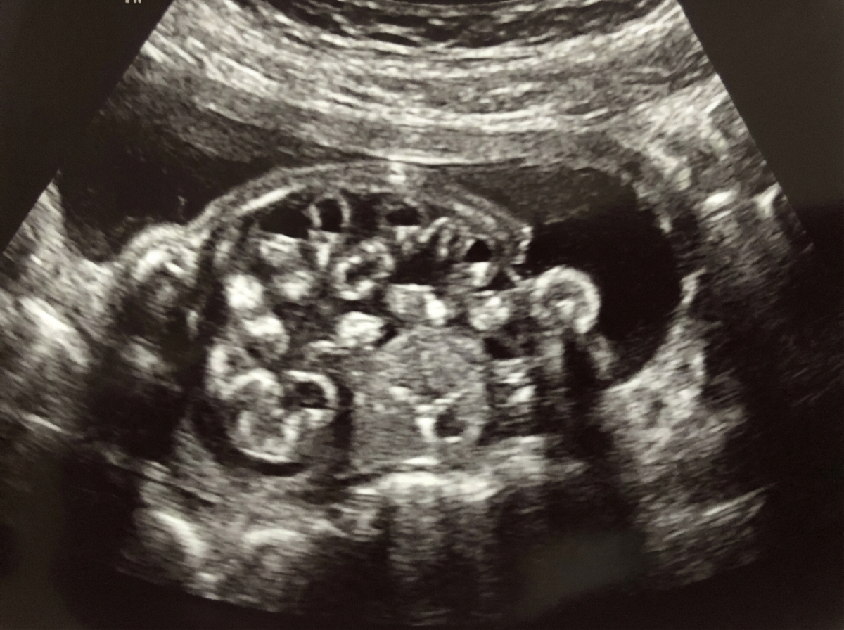
A 26-year-old gravida 2 para 1 presents to her physician at 12 weeks gestation. She has no complaints. Her previous pregnancy 5 years ago had an uncomplicated course with vaginal delivery of a healthy boy at 39 + 1 weeks gestation. Her weight is 75 kg (165 lb) and the height is 168 cm (5 ft 6 in). On presentation, the blood pressure is 110/70 mm Hg, the heart rate is 83/min, the respiratory rate is 14/min, and the temperature is 36.6℃ (97.9℉). The physical examination is within normal limits. The gynecologic examination demonstrates a fetal heart rate of 180/min. The uterus cannot be palpated and the ultrasound exam is benign. Blood testing showed the following: RBC count 3.9 million/mm3 Leukocyte count 11,100/mm3 Hb 11.6 g/dL Hct 32% MCV 87 fl Reticulocyte count 0.4% The patient’s blood type is A neg. Which testing is indicated in this patient?
A 24-year-old woman with a missed menstrual cycle has a positive pregnancy test. The estimated gestational age is 4 weeks. The patient questions the pregnancy test results and mentions that a urinary pregnancy test she took 3 weeks ago was negative. What is the explanation for the patient’s first negative pregnancy test result?
Hormone balance is essential for maintaining a normal pregnancy. Early on, elevated progesterone levels are needed to maintain pregnancy and progesterone is produced in excess by the corpus luteum. In the normal menstrual cycle the corpus luteum involutes, but this process is impeded during pregnancy because of the presence of which hormone?
A 25-year-old G1P0000 presents to her obstetrician’s office for a routine prenatal visit at 32 weeks gestation. At this visit, she feels well and has no complaints. Her pregnancy has been uncomplicated, aside from her Rh negative status, for which she received Rhogam at 28 weeks gestation. The patient has a past medical history of mild intermittent asthma and migraine headaches. She currently uses her albuterol inhaler once a week and takes a prenatal vitamin. Her temperature is 98.6°F (37.0°C), pulse is 70/min, blood pressure is 117/68 mmHg, and respirations are 13/min. Cardiopulmonary exam is unremarkable, and abdominal exam reveals a gravid uterus with fundal height at 30 centimeters. Bedside ultrasound reveals that the fetus is in transverse lie. The patient states that she prefers to have a vaginal delivery. Which of the following is the best next step in management?
A 29-year-old G1P0 woman, at 12 weeks estimated gestational age, presents for her first prenatal visit. Past medical history reveals the patient has type O+ blood and that her husband has type A+ blood. The patient is worried about the risk of her baby having hemolytic disease. Which of the following is correct regarding fetomaternal incompatibility in this patient?
A 19-year-old woman, gravida 1, para 0, at 21 weeks’ gestation comes to the physician for a follow-up prenatal visit. At her previous appointment, her serum α-fetoprotein concentration was elevated. She had smoked 1 pack of cigarettes daily for 3 years but quit at 6 weeks' gestation. Examination shows a uterus consistent in size with a 21-week gestation. Ultrasonography shows fetal viscera suspended freely into the amniotic cavity. Which of the following is the most likely diagnosis?
Three weeks after delivering a healthy boy, a 28-year-old woman, gravida 1, para 1, comes to the physician for a postpartum check-up. Labor and delivery were uncomplicated. Two days after delivery she was diagnosed with postpartum endometritis and received intravenous clindamycin plus gentamicin for 2 days. She had painful swelling of the breasts at the beginning of lactation, but frequent breastfeeding and warm compresses prior to breastfeeding improved her symptoms. Physical examination shows no abnormalities. The patient asks about a reliable contraceptive method. Which of the following is the most appropriate recommendation?
Practice by Chapter
Routine prenatal visit schedule
Practice Questions
Nutrition in pregnancy
Practice Questions
Weight gain recommendations
Practice Questions
Exercise in pregnancy
Practice Questions
Medication safety in pregnancy
Practice Questions
Immunizations in pregnancy
Practice Questions
Management of common pregnancy complaints
Practice Questions
Fetal growth assessment
Practice Questions
Fetal movement monitoring
Practice Questions
Antepartum fetal surveillance (NST, BPP)
Practice Questions
Anemia in pregnancy
Practice Questions
Travel during pregnancy
Practice Questions
Patient education topics
Practice Questions
Want unlimited practice?
Get full access to all questions, explanations, and performance tracking.
Scan to download app