A 27-year-old dental radiographer presented to a clinic with red lesions on his palate, right lower and mid-upper lip, as well as one of his fingers. These lesions were accompanied by slight pain, and the patient had a low-grade fever 1 week before the appearance of the lesions. The patient touched the affected area repeatedly, which resulted in bleeding. Two days prior to his visit, he observed a small vesicular eruption on his right index finger, which merged with other eruptions and became cloudy on the day of the visit. He has not had similar symptoms previously. He did not report drug usage. A Tzanck smear was prepared from scrapings of the aforementioned lesions by the attending physician, and multinucleated epithelial giant cells were observed microscopically. According to the clinical presentation and histologic finding, which viral infection should be suspected in this case?
A 16-year-old male presents to his pediatrician with a sore throat. He reports a severely painful throat preceded by several days of malaise and fatigue. He has a history of seasonal allergies and asthma. The patient is a high school student and is on the school wrestling team. He takes cetirizine and albuterol. His temperature is 100.9°F (38.3°C), blood pressure is 100/70 mmHg, pulse is 100/min, and respirations are 20/min. Physical examination reveals splenomegaly and posterior cervical lymphadenopathy. Laboratory analysis reveals the following: Serum: Na+: 145 mEq/L K+: 4.0 mEq/L Cl-: 100 mEq/L HCO3-: 24 mEq/L BUN: 12 mg/dL Ca2+: 10.2 mg/dL Mg2+: 2.0 mEq/L Creatinine: 1.0 mg/dL Glucose: 77 mg/dL Hemoglobin: 17 g/dL Hematocrit: 47% Mean corpuscular volume: 90 µm3 Reticulocyte count: 1.0% Platelet count: 250,000/mm3 Leukocyte count: 13,000/mm3 Neutrophil: 45% Lymphocyte: 42% Monocyte: 12% Eosinophil: 1% Basophil: 0% Which of the following cell surface markers is bound by the pathogen responsible for this patient’s condition?
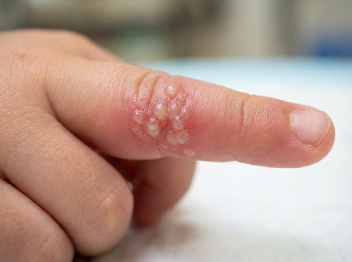
A previously healthy 16-year-old girl comes to the physician because of fever, fatigue, and a sore throat for 8 days. She also has a diffuse rash that started yesterday. Three days ago, she took amoxicillin that she had at home. She is sexually active with two male partners and uses condoms inconsistently. Her temperature is 38.4°C (101.1°F), pulse 99/min, blood pressure 106/70 mm Hg. Examination shows a morbilliform rash over her trunk and extremities. Oropharyngeal examination shows tonsillar enlargement and erythema with exudates. Tender cervical and inguinal lymphadenopathy are present. Abdominal examination shows mild splenomegaly. A peripheral blood smear shows lymphocytosis with > 10% atypical lymphocytes. Which of the following is most likely to be positive in this patient?
A 36-year-old woman presents to the emergency department with a 2-day history of conjunctivitis, sensitivity to bright light, and decreased visual acuity. She denies a history of ocular trauma. She wears contact lenses and thought that the contact lenses may be the cause of the symptoms, although she has always used proper hygiene. Fluorescein staining showed a corneal dendritic branching ulcer with terminal bulbs that stained with rose bengal. Giemsa staining revealed multinucleated giant cells. What is the most likely causative agent?
A 35-year-old male nurse presents to the emergency room complaining of fever and malaise. He recently returned from a medical trip to Liberia to help with a deadly outbreak of a highly infectious disease. He reports severe generalized muscle pain, malaise, fatigue, and a sore throat. He has recently developed some difficulty breathing and a nonproductive cough. His past medical history is notable for asthma. He drinks alcohol socially and does not smoke. His temperature is 102.1°F (38.9°C), blood pressure is 115/70 mmHg, pulse is 115/min, and respirations are 24/min. On examination, a generalized maculopapular rash and bilateral conjunctival injection are noted. Laboratory testing reveals the presence of negative sense, singled-stranded linear genetic material with filaments of varying lengths. The pathogen responsible for this patient’s symptoms is most similar to which of the following?
An investigator is studying the structural characteristics of pathogenic viruses. Cell cultures infected by different viruses are observed under a scanning electron microscope. One of the cell samples is infected by a virus that has an envelope composed of nuclear membrane molecules. The most likely virus that has infected this cell sample can cause which of the following conditions?
A 25-year-old G1P0 gives birth to a male infant at 33 weeks’ gestation. The mother immigrated from Sudan one month prior to giving birth. She had no prenatal care and took no prenatal vitamins. She does not speak English and is unable to provide a medical history. The child’s temperature is 101.0°F (38.3°C), blood pressure is 90/50 mmHg, pulse is 140/min, and respirations are 30/min. Physical examination reveals flexed upper and lower extremities, minimal response to stimulation, and slow and irregular respirations. A murmur is best heard over the left second intercostal space. The child’s lenses appear pearly white. Which of the following classes of pathogens is most likely responsible for this patient’s condition?
A 15-year-old high school rugby player presents to your clinic with a sore throat. He reports that he started feeling fatigued along with body aches about a week ago. His vitals and physical are normal except for an exudative pharynx and an enlarged spleen. Monospot test comes back positive and the student is told not to participate in contact sports for a month. What is the most likely causative agent and which immune cell does it affect?
A 14-year-old boy is brought to the physician with fever, malaise, and bilateral facial pain and swelling that began 2 days ago. He has no history of serious illness and takes no medications. He was born in India, and his mother received no prenatal care. She is unsure of his childhood vaccination history. He returned from a trip to India 3 weeks ago, where he was visiting his family. His temperature is 38.2°C (100.8°F). There is erythema, edema, and tenderness of the right and left parotid glands. The remainder of the examination shows no abnormalities. Laboratory studies show: Leukocyte count 13,000/mm3 Hemoglobin 13.0 g/dL Hematocrit 38% Platelet count 180,000/mm3 This patient is at greatest risk for which of the following complications?
A 6-year-old female from a rural village in Afghanistan presents with her mother to a local health center complaining of leg weakness. Her mother also reports that the patient had a fever, fatigue, and headache a week prior that resolved. The patient has not received any immunizations since being born. Her temperature is 98.6°F (37°C), blood pressure is 110/70 mmHg, pulse is 90/min, and respirations are 18/min. Physical examination reveals 1/5 strength in right hip and knee actions and 0/5 strength in left hip and knee actions. Tone is notably decreased in both lower extremities. Sensation to touch, temperature, and vibration is intact. Patellar and Achilles reflexes are absent bilaterally. The most likely cause of this patient’s condition has which of the following characteristics?
Explanation: ***Herpes simplex infection*** - The presence of **red lesions on the palate, lips, and finger**, along with **vesicular eruptions** that become cloudy and are accompanied by **pain** and a preceding **low-grade fever**, are classic signs of **herpes simplex virus (HSV) infection**. - The histological finding of **multinucleated epithelial giant cells** on a **Tzanck smear** is highly characteristic of herpes virus infections, including HSV. *Varicella-zoster infection* - While **varicella-zoster virus (VZV)** also causes vesicular lesions and produces multinucleated giant cells, it typically presents with a **widespread rash** (chickenpox) or a **dermatomal distribution** (shingles), which is not described here. - The patient's initial symptoms are more consistent with **primary herpes simplex infection**, particularly with the localized presentation. *Measles* - **Measles** presents with a characteristic **maculopapular rash** that starts on the face and spreads downwards, often preceded by **Koplik spots** in the mouth, and significant **catarrhal symptoms** (cough, coryza, conjunctivitis). - It does not typically involve vesicular lesions or the presence of multinucleated giant cells on a Tzanck smear. *Herpangina* - **Herpangina** is characterized by painful **vesicles and ulcers localized to the posterior oropharynx** (soft palate, tonsillar pillars, uvula), typically caused by Coxsackieviruses. - It does not usually affect the lips or fingers, and a Tzanck smear would not show multinucleated giant cells. *Hand-foot-and-mouth disease* - **Hand-foot-and-mouth disease (HFMD)** is caused by coxsackieviruses and presents with **vesicular lesions** on the **hands, feet, and oral cavity**, particularly the tongue and buccal mucosa. - While oral lesions are present, the specific involvement of the palate and the characteristic Tzanck smear findings point away from HFMD.
Explanation: ***CD21*** - The patient's symptoms (sore throat, fatigue, splenomegaly, posterior cervical lymphadenopathy, and increased lymphocytes) are consistent with **infectious mononucleosis**, which is caused by the **Epstein-Barr virus (EBV)**. - EBV primarily infects **B lymphocytes** by binding to the **CD21 receptor** (also known as the C3d complement receptor or CR2) on their surface. *CD3* - **CD3** is a protein complex found on the surface of **T lymphocytes**, associated with the T-cell receptor. - EBV primarily targets B cells, not T cells, for initial infection and replication. *CD4* - **CD4** is a glycoprotein found on the surface of **helper T cells, monocytes, macrophages, and dendritic cells**. - It is primarily known as the co-receptor for HIV entry into T cells. *CD20* - **CD20** is a transmembrane protein expressed on the surface of **B lymphocytes** from the pre-B cell stage through differentiated B cells, but not on plasma cells. - While involved in B cell activation and a target for certain immunotherapies, it is not the primary receptor used by EBV for entry. *CD19* - **CD19** is another transmembrane glycoprotein expressed on the surface of **B lymphocytes** and is involved in B cell development, activation, and signaling. - Although it's a B cell marker, **CD21** is the specific receptor that EBV uses for attachment and entry into B cells.
Explanation: ***Heterophile antibody test*** - The patient's symptoms (fever, fatigue, sore throat with exudates, generalized rash after amoxicillin, lymphadenopathy, splenomegaly, and **atypical lymphocytes** on blood smear) are classic for **infectious mononucleosis**. - **Infectious mononucleosis** is most commonly caused by the **Epstein-Barr virus (EBV)**, which is diagnosed by a positive **heterophile antibody test** (Monospot test). *Anti-CMV IgM* - While **cytomegalovirus (CMV)** can cause a mononucleosis-like syndrome, it is a less common cause than EBV, and other classic features like diffusely enlarged tender lymph nodes and splenomegaly are more suggestive of EBV. - CMV mononucleosis typically lacks the prominent **pharyngitis and exudative tonsillitis** seen in EBV infection. *Flow cytometry* - **Flow cytometry** is used to identify and quantify specific cell populations, primarily in the diagnosis and monitoring of **hematologic malignancies** (e.g., leukemia, lymphoma). - It is not a primary diagnostic test for infectious diseases like mononucleosis, though it could rule out other conditions if there was diagnostic uncertainty regarding atypical lymphocytes. *ELISA for HIV* - Acute **HIV infection** can present with a mononucleosis-like syndrome and a rash. However, the prominent **exudative tonsillitis** and **atypical lymphocytosis** with definite splenomegaly point more strongly towards EBV. - While considering the patient's sexual activity, the constellation of symptoms is more directly explained by EBV infection. *Throat swab culture* - A **throat swab culture** is primarily used to diagnose bacterial pharyngitis, such as **Streptococcus pyogenes (Group A Strep)**. - While Group A Strep can cause exudative tonsillitis, it would not typically cause diffuse lymphadenopathy, splenomegaly, a generalized rash (especially after amoxicillin), or atypical lymphocytes.
Explanation: ***Herpes simplex virus (HSV)-1*** - The classic **dendritic corneal ulcer with terminal bulbs** revealed by fluorescein staining, coupled with **multinucleated giant cells** on Giemsa staining, is pathognomonic for **herpes simplex keratitis**. - Though contact lens wear is mentioned, the specific corneal findings point away from typical bacterial keratitis and strongly towards viral etiology. *Candida albicans* - Ocular infections with *Candida* typically present as **fungal keratitis**, often with feathery-edged infiltrates and satellite lesions, which differ from the dendritic pattern. - While contact lens wear can be a risk factor for fungal infections, the specific branching ulcer described is not characteristic of *Candida*. *Acanthamoeba* - *Acanthamoeba* keratitis is a severe infection often associated with **contact lens use** and presents with severe pain and a **ring-shaped infiltrate (ring infiltrate)**, rather than a dendritic ulcer. - *Acanthamoeba* organisms would be identified on microscopy, not multinucleated giant cells. *Varicella zoster virus* - While VZV can cause **dendritic ulcers** (similar to HSV), it is typically associated with a history of **shingles (herpes zoster ophthalmicus)**, often with a vesicular rash in the distribution of the trigeminal nerve. - The absence of a painful vesicular rash and the specific morphology of the dendrite with true terminal bulbs make HSV-1 more likely. *Pseudomonas* - **Pseudomonas aeruginosa** is a common cause of bacterial keratitis, particularly in contact lens wearers, and is known for its rapid progression and severe pain. - It typically causes a **dense, central corneal infiltrate** with a stromal abscess and often hypopyon, which is distinct from a dendritic ulcer.
Explanation: ***Marburg virus*** - The history of travel to **Liberia** during a deadly outbreak, prolonged fever, malaise, myalgia, respiratory symptoms, and the presence of a **maculopapular rash** and **conjunctival injection** are all highly suggestive of a **viral hemorrhagic fever**. - The finding of **negative-sense, single-stranded linear genetic material with filaments of varying lengths** points to a **filovirus**, such as the Marburg virus, which causes severe hemorrhagic fever and fits the clinical picture. *Lassa fever virus* - While Lassa fever is a **viral hemorrhagic fever endemic to West Africa**, including Liberia, its causative agent, the **Lassa virus**, is an **arenavirus** with a **circular segmented RNA genome**, not filamentous. - The clinical presentation can be similar, but the genomic characteristic described in the question rules out Lassa virus. *Hantavirus* - **Hantaviruses** cause **hemorrhagic fever with renal syndrome** or **hantavirus pulmonary syndrome**, and are primarily found in the **Americas and Asia**, not West Africa. - They are **bunyaviruses** with a **segmented, tripartite RNA genome**, which does not match the filamentous morphology described. *Dengue virus* - **Dengue fever** is a mosquito-borne illness common in **tropical and subtropical regions**, but less likely to be described as a "deadly outbreak of a highly infectious disease" in Liberia of the severity implied here. - The **dengue virus** is a **flavivirus** with a **single-stranded, positive-sense RNA genome** and an **icosahedral shape**, which differs from the filamentous negative-sense RNA found. *Lymphocytic choriomeningitis virus* - **Lymphocytic choriomeningitis virus (LCMV)** is an **arenavirus** that typically causes **aseptic meningitis or meningoencephalitis** and is transmitted via rodents. - Its genomic characteristics (circular segmented RNA) and typical clinical presentation do not align with the patient's severe, systemic hemorrhagic fever symptoms and the filamentous nature of the pathogen.
Explanation: ***Shingles*** - The virus causing shingles, **Varicella Zoster Virus (VZV)**, is a **herpesvirus** that acquires its envelope from the **nuclear membrane of the host cell**. - Herpesviruses bud through the inner nuclear membrane, picking up host nuclear membrane components to form their envelope. *Ebola* - Ebola virus is a **filovirus** that acquires its envelope from the **plasma membrane** of the host cell as it buds off. - Its envelope is not derived from the nuclear membrane. *Yellow fever* - Yellow fever is caused by a **flavivirus**, which is an **enveloped RNA virus** that acquires its envelope from the **endoplasmic reticulum** and Golgi apparatus. - Its envelope is not derived from the nuclear membrane. *Erythema infectiosum* - Erythema infectiosum is caused by **Parvovirus B19**, which is a **non-enveloped DNA virus**. - As it lacks an envelope, it does not bud from any host membrane. *Condylomata acuminata* - Condylomata acuminata (genital warts) are caused by **Human Papillomavirus (HPV)**, which is a **non-enveloped DNA virus**. - Like Parvovirus B19, HPV does not possess an envelope derived from host cell membranes.
Explanation: ***Togavirus*** - The combination of **congenital cataracts** (pearly white lenses), **PDA murmur** (left second intercostal space), **prematurity**, and generalized distress in an infant whose mother had no prenatal care points strongly to **congenital rubella syndrome**, caused by the **rubella virus (a Togavirus)**. - Exposure to rubella during pregnancy, especially in the first trimester, can lead to a triad of **cataracts**, **cardiac defects** (like PDA), and **sensorineural hearing loss**. *Retrovirus* - **Retroviruses**, such as HIV, can cause congenital infections but typically present with different symptom complexes, like **immunodeficiency**, **failure to thrive**, and **opportunistic infections**, rather than the classic congenital rubella triad. - While HIV can cause cardiomyopathy, it doesn't typically cause **cataracts** as a primary congenital defect in the same manner as rubella. *Protozoan* - **Protozoans** like **Toxoplasma gondii** can cause congenital infections (**toxoplasmosis**), which typically manifest as **chorioretinitis**, **hydrocephalus**, and **intracranial calcifications**, a different set of symptoms from those described. - **Toxoplasmosis** does not commonly cause **cataracts** or **patent ductus arteriosus**. *Spirochete* - **Spirochetes**, particularly **Treponema pallidum** (causing **congenital syphilis**), can lead to prematurity and other congenital anomalies. - Symptoms of congenital syphilis often include **maculopapular rash**, **hepatosplenomegaly**, **bone abnormalities**, and **rhinitis**, distinct from the findings in this case. *Herpesvirus* - **Herpesviruses** (e.g., CMV, HSV) can cause congenital infections with varied presentations. **Congenital CMV** is associated with **periventricular calcifications**, **microcephaly**, and **sensorineural hearing loss**, while **congenital HSV** typically presents with **skin lesions**, **neurological deficits**, and **ocular manifestations** that are different from the described cataracts. - While CMV can cause **cataracts**, it's less commonly associated with **PDA** compared to rubella.
Explanation: ***Epstein-Barr virus; B-cells*** - This clinical picture of **fatigue, sore throat (exudative pharyngitis)**, and **splenomegaly** in an adolescent, along with a **positive Monospot test**, is classic for infectious mononucleosis caused by the **Epstein-Barr virus (EBV)**. - EBV primarily infects **B lymphocytes** by binding to the CD21 receptor, leading to their proliferation and activation. *Group A Streptococcus; Neutrophils* - While **Group A Streptococcus (GAS)** can cause exudative pharyngitis, it is not typically associated with **prolonged fatigue** and **splenomegaly**, nor would it result in a **positive Monospot test**. - GAS is a bacterium and primarily causes an immune response involving **neutrophils** and other innate immune cells, rather than primarily affecting neutrophils directly in the context of persistent infection. *Epstein-Barr virus; T-cells* - While EBV infection does lead to a robust **T-cell response** (especially cytotoxic T-cells) to control infected B-cells, its **primary infection and latency site are B-cells**, not T-cells. - The atypical lymphocytes observed in mononucleosis are largely activated cytotoxic T-cells, but they are responding to B-cell infection. *Streptococcus; Macrophages* - **Streptococcus** (a broad term) causes various infections, but the constellation of symptoms including pronounced **fatigue, splenomegaly**, and a **positive Monospot test** is not characteristic of typical streptococcal infections. - Macrophages are significant in the immune response to bacteria, but they are not the primary cell type directly targeted or dysfunctional in streptococcal infections in the way B-cells are in mono. *Cytomegalovirus; T-cells* - **Cytomegalovirus (CMV)** can cause a mononucleosis-like syndrome with symptoms like fatigue and splenomegaly, but the **Monospot test would be negative** in CMV infection. - CMV primarily infects various cell types including **epithelial cells, fibroblasts, and macrophages**, and can also have tropism for T-cells, but it's not the primary causative agent when a Monospot is positive.
Explanation: ***Impaired fertility*** - The patient's symptoms (fever, malaise, **bilateral parotid swelling**) are highly suggestive of **mumps**, especially given his uncertain vaccination status and recent travel. - In post-pubertal males, mumps can cause **orchitis** (testicular inflammation), a significant risk factor for **impaired fertility** or even sterility due to testicular atrophy. *Facial nerve palsy* - While facial nerve issues can theoretically occur with severe parotitis, **facial nerve palsy** is not a common or direct complication of mumps itself. - More often, facial nerve palsy is associated with conditions like Bell's palsy or tumors of the parotid gland, not typically the acute viral infection seen here. *Osteomyelitis of facial bone* - **Osteomyelitis** is an infection of the bone, usually bacterial, and is not a typical complication of mumps. - There is no clinical evidence (e.g., severe localized pain, skin breakdown, or signs of deep soft tissue infection) to suggest bone involvement. *Glomerulonephritis* - **Glomerulonephritis** is an inflammatory kidney condition, sometimes seen post-streptococcal infection or in systemic autoimmune diseases. - It is not a recognized and common complication of mumps virus infection. *Diabetes mellitus* - Mumps can, in rare cases, cause **pancreatitis**, which theoretically could lead to damage to the insulin-producing cells and increase the risk of diabetes. - However, **impaired fertility** due to orchitis is a much more common and direct complication in post-pubertal males with mumps than diabetes mellitus.
Explanation: ***Non-enveloped (+) ssRNA virus*** - This describes **Poliovirus**, the causative agent of polio, which is characterized by **acute flaccid paralysis** and absent reflexes due to damage to **anterior horn cells**. - The patient's presentation with **unvaccinated status**, **acute onset of asymmetric flaccid paralysis** following a febrile illness, and **areflexia** is highly suggestive of poliomyelitis. *Enveloped (-) ssRNA virus* - This describes viruses such as **Measles**, **Mumps**, or **Rabies**, which cause different clinical syndromes and are not typically associated with acute flaccid paralysis. - While these can cause neurological symptoms, they manifest differently (e.g., encephalitis, specific rashes) and are not the primary cause of the described motor deficits. *Enveloped (+) ssRNA virus* - This describes viruses like **Dengue**, **Zika**, or **Coronaviruses (SARS-CoV-2)**, which are associated with various fever syndromes, rashes, or respiratory illness, but not typically the specific flaccid paralysis seen here. - While some can cause neurological complications (e.g., Guillain-Barré syndrome with Zika), the direct neuronal damage leading to areflexic flaccid paralysis as seen in polio is not characteristic. *dsRNA virus* - This describes viruses like **Rotavirus**, which primarily cause **gastroenteritis** (nausea, vomiting, diarrhea), or **Reoviruses**. - They are not known to cause acute flaccid paralysis or the specific neurological findings described in the patient. *Non-enveloped (-) ssRNA virus* - This type of virus is less common, but the description does not fit the typical etiologies for acute flaccid paralysis. Most common medically relevant (-) ssRNA viruses are enveloped. - This classification does not align with any known human pathogen that presents with the classic symptoms of poliomyelitis.
Explanation: ***Epstein-Barr virus*** - The symptoms of fever, sore throat, **cervical lymphadenopathy**, **hepatosplenomegaly**, and **tonsillar exudates** in an adolescent, coupled with a **positive Monospot test**, are highly characteristic of **infectious mononucleosis** caused by the Epstein-Barr virus (EBV) - The positive Monospot test detects **heterophile antibodies**, which are a hallmark of acute EBV infection and essentially confirms the diagnosis *Varicella virus* - Varicella virus (chickenpox) typically presents with a **pruritic vesicular rash** that progresses through different stages (macules → papules → vesicles → crusts), which is not described in this patient - While fever can be present, the distinctive rash, rather than lymphadenopathy and tonsillar exudates, is the defining feature *Cytomegalovirus* - Cytomegalovirus (CMV) can cause a **mononucleosis-like syndrome** with similar clinical features - However, CMV mononucleosis typically presents with **negative heterophile antibodies** (negative Monospot test), which distinguishes it from EBV - This patient's positive Monospot test makes CMV unlikely as the primary cause *Herpes simplex virus* - Herpes simplex virus (HSV) infections typically cause **oral ulcers** (cold sores), **gingivostomatitis**, or **genital lesions** - While HSV can cause fever and sore throat, it would not typically lead to the diffuse lymphadenopathy, hepatosplenomegaly, and positive Monospot test seen in this case *Variola virus* - Variola virus (smallpox) is characterized by a **distinctive rash** of deep-seated pustules that begin on the face and extremities, often with systemic symptoms like high fever and malaise - Smallpox has been **eradicated worldwide since 1980**, making this diagnosis impossible in contemporary practice - The presentation is distinctly different from the symptoms described, especially with the absence of the characteristic rash
Explanation: ***Mosquito repellent*** - The patient's symptoms (high fever, severe body pains, retro-orbital pain, rash, leukopenia, thrombocytopenia, and recent travel to Taiwan) are highly suggestive of **Dengue fever**, which is a **mosquito-borne** illness. - **Mosquito repellents** are the most effective method for preventing mosquito bites and, consequently, Dengue infection in endemic areas. *Frequent hand washing* - Frequent hand washing is crucial for preventing the spread of **fecal-oral pathogens** and **respiratory viruses**, but it does not protect against mosquito-borne diseases like Dengue. - This practice is important for general hygiene but irrelevant to the transmission mechanism of the patient's likely condition. *Prophylaxis with doxycycline* - **Doxycycline** is used for prophylaxis against certain bacterial infections, such as **malaria** (though other drugs are preferred for some regions) and some rickettsial diseases. - It is **ineffective** against viral infections like Dengue fever, which is the likely diagnosis in this case. *Vaccination* - While a **Dengue vaccine (Dengvaxia)** exists, it is approved for specific populations (individuals with confirmed prior Dengue infection) and has limitations. - The most reliable and widely recommended preventive measure for travelers to Dengue-endemic areas remains **mosquito bite prevention**, which includes repellents. *Safe sexual practices* - Safe sexual practices, such as consistent condom use, prevent the transmission of **sexually transmitted infections (STIs)**, including HIV, syphilis, and gonorrhea. - The patient's symptoms and travel history do not align with common STIs, and his HIV test was negative, making this measure irrelevant to his current condition.
Explanation: ***Translocation of the antigen into the endoplasmic reticulum via TAP proteins*** - The **endogenous pathway** processes **intracellular antigens**, such as those from viruses, presenting them on **MHC class I** molecules. - After proteolytic degradation of viral proteins in the **cytosol** by the **proteasome**, the resulting peptides are transported into the **endoplasmic reticulum (ER)** by **TAP (transporter associated with antigen processing) proteins** to bind with MHC class I molecules. *Degradation of the antigen by the proteases in the phagolysosome* - This process describes the degradation of **extracellular antigens** within the **phagolysosome** for presentation via the **exogenous pathway (MHC class II)**. - Viral antigens, being replicated *intracellularly*, are primarily handled by the endogenous pathway. *Binding of the peptide to MHC class II* - This step is characteristic of the **exogenous pathway** of antigen presentation, which primarily handles **extracellular antigens**. - **MHC class II molecules** are typically loaded with peptides derived from endocytosed or phagocytosed antigens in **endosomal/lysosomal compartments.** *Translocation of the antigen into endosome after phagocytosis* - **Endosomes** and **phagocytosis** are key components of the **exogenous pathway** for presenting extracellular antigens. - This mechanism is not typically associated with the processing and presentation of **intracellular viral antigens** for **MHC class I**. *Interaction of the MHC class II complex with its target CD4+ T cell* - This describes a downstream event of antigen presentation, specifically the interaction involving **MHC class II** and **CD4+ T cells**, which is part of the **exogenous pathway**. - The question asks for a critical step in the *endogenous pathway* of antigen presentation.
Explanation: ***Infection with herpes simplex virus*** - The patient's presentation with **altered mental status**, focal neurological deficits (left hand twitching, speech difficulties), and **bizarre behavior** (mania-like symptoms) suggests **herpes simplex encephalitis**. - The CSF analysis showing **lymphocytic pleocytosis**, normal glucose, and a slightly elevated protein are consistent with **viral encephalitis**. The presence of **RBCs in the CSF** is also highly suggestive of HSV encephalitis due to its propensity to cause **hemorrhagic necrosis** in the temporal lobes. *Brain abscess* - A brain abscess typically presents with **fever**, **headache**, and **focal neurological deficits**, similar to viral encephalitis. - However, brain abscesses often show **neutrophilic pleocytosis** in the CSF and may have a **ring-enhancing lesion** on CT/MRI, which was not described here (unremarkable CT). *Infection with Streptococcus pneumoniae* - Infection with *Streptococcus pneumoniae* would cause **bacterial meningitis**, which is characterized by a **high fever**, **neck stiffness**, and altered mental status. - The CSF analysis for bacterial meningitis typically shows **neutrophilic predominance**, **low glucose**, and **significantly elevated protein**, which is not seen in this patient's CSF. *Neurosyphilis* - While the patient has a history of treated syphilis, **neurosyphilis** can present with myriad neurological and psychiatric symptoms, including altered mental status and behavioral changes. - However, the rapid onset of focal neurological symptoms and the CSF findings (particularly the high cell count with relatively normal protein and glucose, and presence of RBCs) are *less typical* for neurosyphilis and more indicative of an acute viral encephalitis. *Undiagnosed bipolar disorder* - The patient's initial "bizarre behavior" and grandiosity (elaborate plans, "genius project") could suggest a **manic episode** consistent with bipolar disorder. - However, the subsequent rapid decline in mental status, development of **focal neurological deficits** (twitching, speech difficulties), **fever**, and **abnormal CSF findings** rule out bipolar disorder as the sole explanation for her current presentation.
Explanation: ***Anti-HEV IgM*** - The patient's presentation with **acute liver failure** (jaundice, encephalopathy, coagulopathy, elevated aminotransferases) following a trip to South Asia, especially during pregnancy, is highly suggestive of **hepatitis E virus (HEV) infection**. - **IgM antibodies** indicate an **acute infection**, and HEV can have a particularly severe course in pregnant women, leading to high mortality rates. *Anti-HAV IgM* - This would indicate an **acute hepatitis A virus (HAV) infection**, which is transmitted feco-orally and endemic in regions like South Asia. - While HAV can cause acute hepatitis, it typically has a **milder course in adults** and is **not associated with the severe liver failure and high mortality** seen in pregnant women that is characteristic of HEV. *Anti-HCV IgG* - **IgG antibodies** to hepatitis C virus (HCV) indicate **past exposure or chronic infection**, not acute hepatitis. - Acute HCV infection is often asymptomatic or mild and rarely causes the fulminant liver failure described here. *Anti-HBc IgM* - **IgM antibodies to hepatitis B core antigen (HBcAg)** indicate **acute hepatitis B virus (HBV) infection**. - While acute HBV can cause severe hepatitis, the combination of **pregnancy, travel to South Asia, and the rapid progression to liver failure** makes HEV a more likely culprit in this specific clinical context. *HBsAg* - **HBsAg (Hepatitis B surface antigen)** is a marker of active HBV infection, either acute or chronic. - While present in acute HBV, the clinical picture, particularly the travel history and pregnancy, points more strongly towards **HEV as the cause of fulminant hepatic failure** in this demographic.
Explanation: ***Rotavirus*** - The patient's age (4-year-old), symptoms of **fever, vomiting, and non-bloody diarrhea**, and being **unvaccinated** against rotavirus strongly point to this diagnosis. - Rotavirus is a common cause of **severe gastroenteritis** in unvaccinated infants and young children, often leading to dehydration as evidenced by **dry mucous membranes**. - The rotavirus vaccine has dramatically reduced cases in vaccinated populations, making unvaccinated children particularly vulnerable. *Campylobacter jejuni* - This typically causes **bloody diarrhea**, which is not seen in this patient's presentation. - **Campylobacter jejuni** is often associated with consumption of raw poultry or contaminated water, and usually presents with more severe abdominal pain. *Norovirus* - While Norovirus can cause vomiting and non-bloody diarrhea, it typically affects individuals of **all ages** and often occurs in outbreaks in close-knit communities. - Though possible, **rotavirus** is a more common cause of severe gastroenteritis in unvaccinated young children. *Vibrio cholerae* - **Cholera** is characterized by **profuse watery diarrhea** (rice-water stools) leading to rapid dehydration. - It is typically seen in endemic areas or after exposure to contaminated water sources, and the patient's symptoms are not as extreme to suggest cholera. *Adenovirus* - While adenovirus can cause gastroenteritis in children, it typically presents with **respiratory symptoms** (pharyngitis, conjunctivitis) in addition to gastrointestinal symptoms. - Adenovirus gastroenteritis is generally **less severe** than rotavirus and lacks the classic vaccine-preventable epidemiology that makes rotavirus more likely in this unvaccinated child.
Explanation: ***Antigenic shift*** - **Antigenic shift** in influenza viruses refers to the process where two different influenza strains **coinfect** the same host cell, leading to a **reassortment** of their segmented genomes. - This reassortment creates a novel viral strain with a new combination of hemagglutinin (H) and neuraminidase (N) antigens (e.g., H7N1 + H3N8 → H7N8), which can lead to pandemics. *Transduction* - **Transduction** is a process where foreign DNA is introduced into a cell by a virus or viral vector, primarily seen in bacteria. - It involves the transfer of genetic material via **bacteriophages** and does not describe the reassortment of influenza virus segments. *Antigenic drift* - **Antigenic drift** involves small, gradual changes in the H and N antigens of influenza viruses due to **point mutations** during replication. - These slow mutations lead to seasonal epidemics, but not the creation of a completely new subtype as described. *Transformation* - **Transformation** is the process by which a cell takes up naked DNA from its environment, incorporating it into its own genome. - This mechanism is common in bacteria for acquiring new genetic traits and is not applicable to the reassortment of viral segments within a coinfected host cell. *Conjugation* - **Conjugation** is a process of genetic material transfer between bacteria through direct cell-to-cell contact, typically via a **pilus**. - This mechanism is distinct from viral genetic reassortment and does not involve the coinfection of a host cell by different viral strains.
Explanation: ***CD4+ and CD8+*** - In the **thymic cortex**, T cells undergo **positive selection**, where only T cells that can bind dimly to self-MHC molecules survive. - At this stage, cortical thymocytes are typically **double-positive**, expressing both **CD4** and **CD8** co-receptors. *CD4+* - While CD4+ T cells are a mature T cell subset, **positive selection** in the cortex involves cells that are still expressing both co-receptors before lineage commitment. - T cells that exclusively express CD4 have already undergone **lineage commitment** and generally exit the cortex for the medulla if they pass positive selection. *T cell precursor* - **T cell precursors** (prothymocytes and early thymocytes) enter the thymus and develop in the subcapsular region before migrating to the cortex. - They are typically **double-negative** (CD4-CD8-) and have not yet rearranged their TCR genes or expressed CD4/CD8. *CD8+* - Similar to CD4+, CD8+ T cells represent a **mature T cell subset** that has already committed to the cytotoxic lineage. - During positive selection in the cortex, T cells are still in the **double-positive** stage (CD4+CD8+) before differentiating into single-positive cells. *Th2* - **Th2 cells** (T helper type 2 cells) are a subset of mature CD4+ T cells that differentiate in the periphery after activation. - They are not found in the thymus differentiating during the **positive selection** phase in the cortex.
Explanation: ***A rhinovirus-specific, cell-mediated immune response*** - The **interferon response** is part of the **innate immune system** and acts as a first line of defense against viral infections. - While interferons can modulate adaptive immunity, they do not directly trigger a **pathogen-specific B or T cell-mediated immune response**; that function belongs to antigen-presenting cells and lymphocytes. *Interferon binding to nearby uninfected epithelial cells* - This is a primary function of interferons, which are secreted by infected cells to warn neighboring cells. - Upon binding through a **receptor-ligand interaction**, interferons induce an antiviral state in these uninfected cells, making them resistant to viral replication. *Decreased viral replication within the cell* - Interferons induce the expression of **antiviral proteins (AVPs)** like PKR and 2',5'-OAS, which inhibit viral protein synthesis and degrade viral RNA, respectively. - This leads to a significant reduction in the virus's ability to replicate within the infected cell and subsequently in neighboring cells. *Activation of NK cells* - Interferons, particularly **Type I interferons (IFN-α/β)**, can directly activate **natural killer (NK) cells**. - Activated NK cells then play a crucial role in the **innate immune response** by recognizing and killing virus-infected cells. *Upregulation of NK cell ligands on the infected cell* - Viruses often downregulate MHC class I molecules to evade cytotoxic T cells, but this can make infected cells more susceptible to NK cell mediated killing (missing self hypothesis). - Interferons can induce the expression of ligands that are recognized by NK cells, thereby enhancing the ability of NK cells to detect and eliminate infected cells.
Explanation: ***Interleukin 2*** - The description of the lymphocyte binding the **constant portion of MHC class I** and requiring a signaling molecule for activation, proliferation, and survival points to a **T cell**. - **Interleukin-2 (IL-2)** is a crucial cytokine for the proliferation, differentiation, and survival of T lymphocytes, acting in an autocrine or paracrine fashion after T cell activation. *Interleukin 1* - **Interleukin-1 (IL-1)** is primarily involved in inflammation and fever, produced by macrophages and other innate immune cells. - While it can act as a costimulator for T cells, it is not the primary cytokine required for their sustained proliferation and survival after initial activation. *Interleukin 4* - **Interleukin-4 (IL-4)** is a key cytokine in humoral immunity, promoting B cell proliferation and differentiation, and inducing IgE class switching. - It also plays a role in the differentiation of naive T cells into **Th2 cells**, but it is not the main cytokine for general T cell proliferation and survival. *Interleukin 8* - **Interleukin-8 (IL-8)**, also known as CXCL8, is a chemokine primarily responsible for attracting and activating neutrophils to sites of infection or inflammation. - It does not have a direct role in the sustained proliferation and survival of activated lymphocytes. *Interleukin 6* - **Interleukin-6 (IL-6)** is a pleiotropic cytokine involved in acute phase reactions, hematopoiesis, and the immune response, particularly B cell differentiation and antibody production. - Although it can influence T cell responses, it is not the primary growth factor for activated T lymphocytes as IL-2 is.
Explanation: ***Measles-specific IgM antibodies*** - The clinical presentation with **fever**, **cough**, **coryza** (runny nose), and a **maculopapular rash** spreading from the face downward in an unvaccinated child from an endemic area (Philippines) is classic for **measles (rubeola)**. - The **3 C's** (cough, coryza, conjunctivitis) along with high fever precede the rash by 2-4 days. **Koplik spots** (white spots on buccal mucosa) may appear before the rash but are often transient. - Detecting **measles-specific IgM antibodies** in serum is the **gold standard** for confirming acute measles infection, with IgM appearing within days of rash onset and persisting for weeks. - Generalized lymphadenopathy is also consistent with measles. *Monospot test* - The Monospot test detects **heterophile antibodies** associated with **infectious mononucleosis** caused by **Epstein-Barr virus (EBV)**. - EBV typically presents with **fatigue**, **pharyngitis**, **posterior cervical lymphadenopathy**, and **splenomegaly**; the rash (if present) usually occurs after ampicillin administration. - The cephalocaudal spread of rash and prominent **respiratory prodrome** are more characteristic of measles. *Tzanck smear* - A Tzanck smear identifies **multinucleated giant cells** characteristic of **herpes simplex virus (HSV)** or **varicella-zoster virus (VZV)** infections. - These conditions present with **vesicular or blistering lesions**, not the **maculopapular, blanching rash** described in this patient. - Varicella (chickenpox) has a vesicular rash in different stages, not confluent maculopapular lesions. *Rapid antigen detection testing* - Rapid antigen tests are typically used for diagnosing **streptococcal pharyngitis** or **influenza**. - While respiratory symptoms are present, the **prominent maculopapular exanthem** with cephalocaudal spread and **generalized lymphadenopathy** are not consistent with these diagnoses. - Measles rapid antigen tests exist but are not standard first-line confirmatory tests; **serology (IgM)** is preferred. *Rapid plasma reagin* - Rapid Plasma Reagin (RPR) is a non-treponemal test used to screen for **syphilis**. - Congenital syphilis can present with rash, but this patient's **age (6 years)**, **acute febrile illness with respiratory prodrome**, and **classic rash progression** make measles far more likely. - Secondary syphilis (which causes rash) requires sexual transmission, making it highly unlikely in a 6-year-old child.
Explanation: ***Vesicle*** - A **vesicle** is defined as a **circumscribed, elevated lesion** (macule/papule) containing **clear fluid** and measuring less than 1 cm in diameter. - The patient's lesions, which are 5-6 mm in diameter and contain clear fluid, perfectly fit the description of vesicles, characteristic of **herpes simplex virus (HSV)** infection. *Pustule* - A **pustule** is a small, elevated lesion similar to a vesicle but filled with **pus**, not clear fluid. - Examples include acne or folliculitis, which are typically opaque and yellowish, unlike the described lesions. *Ulcer* - An **ulcer** is a defect or excavation of the skin past the **epidermis**, resulting in the loss of tissue. - The patient's lesions are described as fluid-filled and elevated, not as an open wound with tissue loss. *Papule* - A **papule** is a **solid, elevated lesion** measuring less than 1 cm in diameter. - While elevated and small, a papule does **not contain fluid**, which is a key characteristic of the described lesions. *Bulla* - A **bulla** is a **fluid-filled lesion** that is **larger than 1 cm** in diameter. - The lesions described are 5-6 mm, making them smaller than the definition of a bulla.
Explanation: ***Specific autoclave sterilization*** - The patient's symptoms (rapidly progressive dementia, behavioral changes, fatigue, depression, and myoclonus in the left foot) coupled with a history of **corneal transplant** are highly suggestive of **Creutzfeldt-Jakob Disease (CJD)**, which can be iatrogenically transmitted. - **Inadequate sterilization** of surgical instruments, particularly for procedures involving neural tissue or corneas, can transmit prions. **Specific autoclave sterilization** or other prion-deactivating methods are essential to prevent iatrogenic CJD. *Antidepressant therapy* - While the patient exhibits symptoms of depression, this is likely a manifestation of the underlying neurodegenerative process (CJD), not the primary cause. - Antidepressants would only address the symptomatic depression and would not prevent the progression of the underlying CJD. *Early screening for depression* - Early screening might identify the patient's depressed mood sooner, but it would not prevent the onset or progression of CJD, which is caused by infectious prions. - The dementia and other neurological symptoms indicate a much broader and more severe condition than primary depression. *Good social and familial support system* - While a strong support system is beneficial for managing chronic illnesses, it does not prevent the development of a prion disease like CJD. - Emotional and social support can improve quality of life but has no bearing on the pathological process of prion accumulation. *Statin therapy* - Statins are primarily used to treat **dyslipidemia** and prevent cardiovascular disease. - There is no evidence to suggest that statin therapy protects against or prevents prion diseases such as Creutzfeldt-Jakob Disease.
Explanation: ***Overflow incontinence*** - The patient's symptoms of **difficulty initiating a urine stream**, **intermittent and slow stream**, **needing extra effort to pass urine**, and **feeling the need to urinate again immediately after** are classic signs of **urinary retention** and **overflow incontinence**. - The presence of an **anterior vaginal wall herniation beyond the hymen** suggests a significant cystocele, which can obstruct the bladder outlet, leading to chronic incomplete bladder emptying and overflow. *Functional incontinence* - This type of incontinence occurs when a person has **normal bladder control** but is unable to reach the toilet in time due to **physical or cognitive impairments**, such as mobility issues, dementia, or severe depression. - The patient's presentation does not describe any such physical or cognitive limitations preventing her from accessing the toilet. *Mixed incontinence* - Mixed incontinence is characterized by the presence of **both stress and urgency incontinence symptoms**. - While it's a common type, the patient's primary symptoms are more indicative of outlet obstruction and incomplete emptying, rather than involuntary leakage with exertion or a sudden, strong urge. *Stress incontinence* - **Stress incontinence** is defined by involuntary urine leakage that occurs with **physical exertion** such as coughing, sneezing, laughing, or lifting. - The patient's symptoms are primarily related to difficulty voiding and incomplete emptying, not leakage during physical activity. *Urgency incontinence* - **Urgency incontinence** is characterized by a **sudden, strong, and uncontrollable urge to urinate** that leads to involuntary urine leakage. - While the patient mentions needing to go to the restroom again, her main symptoms revolve around obstructive voiding and incomplete emptying, rather than uncontrollable urges.
Explanation: ***Adenovirus*** - **Adenovirus** has a **DNA genome**, which can be directly amplified by **DNA-dependent Taq polymerase** in a standard PCR reaction. - The other viruses listed are **RNA viruses**, requiring **reverse transcriptase** to convert their RNA into DNA before amplification by Taq polymerase. *Rhinovirus* - **Rhinovirus** is an **RNA virus** (Picornaviridae), and its RNA genome cannot be directly amplified by **DNA-dependent Taq polymerase**. - A successful PCR would require **reverse transcription** of its RNA into cDNA first. *Poliovirus* - **Poliovirus** is a **RNA virus** (Picornaviridae), meaning its genome is RNA and not DNA. - **DNA-dependent Taq polymerase** can only amplify DNA templates; hence, a **reverse transcriptase** step is necessary. *Yellow Fever virus* - **Yellow Fever virus** is an **RNA virus** (Flaviviridae) with an RNA genome. - It would require a **reverse transcriptase** enzyme to synthesize a complementary DNA (cDNA) strand before **Taq polymerase** could perform PCR. *Rubella virus* - **Rubella virus** is an **RNA virus** (Togaviridae), and its genetic material is RNA. - Standard PCR with **DNA-dependent Taq polymerase** would not yield a product without an initial **reverse transcription** step.
Explanation: ***Flavivirus*** - The clinical presentation, including acute onset of **high fever**, headache, nausea, vomiting (**dark vomitus**), **scleral icterus**, and **hepatomegaly** following travel to Nigeria, is highly suggestive of **yellow fever**. - Yellow fever is caused by the **yellow fever virus**, which is a **flavivirus** transmitted by mosquitoes, primarily *Aedes aegypti*. *Togavirus* - The Togavirus family includes viruses like **rubella virus** and **alphaviruses** (e.g., Eastern equine encephalitis virus). - While some alphaviruses can cause fever and encephalitis, they typically do not present with the characteristic **hemorrhagic fever** and severe liver involvement seen in this case. *Calicivirus* - The Calicivirus family includes **Norovirus**, which is a common cause of **gastroenteritis** with vomiting and diarrhea. - Norovirus infections are typically self-limiting and do not usually lead to the severe systemic symptoms, **jaundice**, or fatal outcome described here. *Hepevirus* - The Hepevirus family includes the **hepatitis E virus (HEV)**. - HEV causes **acute viral hepatitis**, characterized by jaundice, nausea, and vomiting, but it rarely progresses to the rapid, severe, and fatal hemorrhagic form seen in this patient. *Bunyavirus* - The Bunyavirus family (now split into several families) includes viruses like Hantavirus and Rift Valley fever virus, which can cause **hemorrhagic fevers**. - While some bunyaviruses are found in Africa, the specific constellation of symptoms, particularly the prominent **scleral icterus** and rapid progression to severe liver failure and death, is most consistent with **yellow fever**, a flavivirus.
Explanation: ***Reassortment*** - This patient presents with **influenza**, confirmed by a positive nasopharyngeal viral culture for an **orthomyxovirus**. The seasonal nature of his illness, despite having it before, points to antigenic changes. - **Reassortment** (also known as **antigenic shift**) is the primary mechanism responsible for **influenza pandemics**. It involves the exchange of entire gene segments between different influenza strains (e.g., human and avian or swine strains) when a host cell is co-infected with two distinct viral strains, leading to a new subtype with novel hemagglutinin (HA) or neuraminidase (NA) proteins that human populations have little to no immunity against. *Complementation* - **Complementation** occurs when two viruses infect the same cell, and one virus provides a **necessary gene product** that the other mutated or defective virus lacks, allowing the latter to replicate. - This mechanism does not involve genetic exchange leading to new viral subtypes and is therefore not responsible for the emergence of pandemic strains. *Transduction* - **Transduction** is a process by which **bacteriophages** (viruses that infect bacteria) transfer bacterial DNA from one bacterium to another. - This is a mechanism of gene transfer in bacteria and is not relevant to the genetic changes in influenza viruses. *Phenotypic mixing* - **Phenotypic mixing** occurs when the genome of one virus is packaged into the **capsid** of another virus, or when genetic material from two viruses is packaged into a mixed capsid. - While it can alter the tropism or antigenicity of progeny viruses temporarily, it does not involve a change in the viral genome itself and is therefore not responsible for permanent shifts leading to pandemics. *Recombination* - **Recombination** involves the exchange of genetic material between two homologous DNA or RNA molecules, leading to new combinations of genes within the same gene segment. - While recombination can occur in viruses, **reassortment** of entire gene segments (antigenic shift) is the specific and most significant mechanism for creating novel influenza strains capable of causing pandemics, rather than recombination within gene segments.
Explanation: ***Prions*** - Prions are **abnormally folded proteins** that are highly resistant to standard sterilization methods like steam autoclaving at 121°C, making them a risk for transmission through reused surgical instruments. - They cause transmissible spongiform encephalopathies (TSEs) like **Creutzfeldt-Jakob disease**, where even trace amounts can be highly infectious. *Non-enveloped viruses* - Non-enveloped viruses are generally **more resistant to heat and disinfectants** than enveloped viruses but are typically inactivated by recommended steam sterilization protocols. - Standard autoclaving conditions are effective in destroying most non-enveloped viruses. *Sporulating bacteria* - **Bacterial spores**, such as those from *Clostridium* or *Bacillus*, are known for their high resistance to heat and chemicals, but are usually **inactivated by steam sterilization at 121°C** for 15 minutes. - This method is specifically designed to kill bacterial spores effectively. *Enveloped viruses* - Enveloped viruses are the **least resistant to heat and chemical disinfectants** due to their lipid envelope. - They are readily **inactivated by standard steam sterilization** at 121°C. *Yeasts* - **Yeasts** are eukaryotic microorganisms that are typically **susceptible to heat sterilization**. - They are effectively killed by typical steam autoclaving conditions used for surgical instruments.
Explanation: ***Morbillivirus*** - The constellation of symptoms—**elevated temperature**, **sore throat**, **runny nose**, **lacrimation**, a **maculopapular erythematous rash** that began on the face and spread downward, and especially the **irregularly-shaped red dots on the mucosa of the lower lip** (likely **Koplik spots**)—are classic for **measles**, caused by Morbillivirus. - The patient's **unvaccinated status** against MMR further supports measles as the most probable diagnosis, as it is a highly contagious disease prevented by vaccination. *Group A Streptococcus* - This bacterium causes **scarlet fever**, characterized by a **sandpaper-like rash** and **strawberry tongue**, not a maculopapular rash spreading from face to extremities with Koplik spots. - While it can cause pharyngitis and fever, the specific rash progression and oral lesions rule out Group A Streptococcus. *Rubulavirus* - Rubulavirus causes **mumps**, which primarily presents with **parotitis** (swelling of parotid glands), fever, and headache. - It does not typically cause a generalized maculopapular rash or Koplik spots, making it an unlikely cause for the described symptoms. *Influenzavirus* - Influenzavirus causes **influenza**, characterized by sudden onset of high fever, cough, myalgia, and headache. - While it can cause fever and respiratory symptoms, it does not typically present with a widespread maculopapular rash or Koplik spots. *Herpesvirus* - Herpesviruses cause a variety of conditions, including **chickenpox** (Varicella-zoster virus), which presents with **vesicular lesions** that crust over, and **roseola infantum** (HHV-6/7), which primarily causes a high fever followed by a non-pruritic rash appearing *after* the fever subsides. - Neither of these typically presents with Koplik spots or the specific maculopapular rash progression described.
Explanation: ***Herpesvirus*** - The presence of an **outer coating high in phospholipids** indicates an **enveloped virus**. Herpesviruses are large, enveloped DNA viruses. - The detection of surface proteins expressed around the **nucleus** is a key finding that suggests the virus **buds from the nuclear membrane**, a characteristic feature of **herpesviruses**. - Many herpesviruses cause dermatitis, including **HSV-1, HSV-2, VZV**, and **HHV-6**. *Adenovirus* - Adenoviruses are **non-enveloped viruses**, meaning they lack a lipid outer coating, which contradicts the phospholipid-rich coating described. - Their replication and assembly occur exclusively in the **nucleus**, but they do not acquire an envelope by budding from the nuclear membrane. *Papillomavirus* - Papillomaviruses are also **non-enveloped viruses** with an **icosahedral capsid**, lacking the phospholipid envelope described. - They replicate and assemble in the nucleus but do not possess the phospholipid-rich outer coating. *Poxvirus* - While poxviruses are **enveloped**, they acquire their envelope from the **Golgi apparatus** or **plasma membrane**, not the nuclear membrane. - They are also distinct from other DNA viruses in that they replicate entirely in the **cytoplasm**, not the nucleus, which does not explain the perinuclear protein expression. *Hepadnavirus* - Hepadnaviruses (e.g., **Hepatitis B virus**) are enveloped DNA viruses, but their envelope is acquired by budding through the **endoplasmic reticulum** and **Golgi apparatus**, not the nuclear membrane. - The perinuclear localization of surface proteins in immunofluorescence is not a characteristic feature of hepadnaviruses.
Explanation: ***Correct: Picornavirus*** - **Coxsackievirus B** and other enteroviruses (Picornaviruses) are the **most common viral causes of acute pericarditis**, making this the best answer for USMLE. - The classic presentation includes a **preceding upper respiratory infection** followed by **pleuritic chest pain**, **fever**, and a **pericardial friction rub** - exactly as described in this case. - Coxsackievirus B can also cause myocarditis and is the prototypical viral pathogen associated with both pericarditis and myocarditis in medical education. *Incorrect: Coronavirus* - While coronaviruses (including SARS-CoV-2) can cause pericarditis, this is **not the classic or most common viral cause** taught in USMLE content. - Coronaviruses are more commonly associated with respiratory symptoms, and pericarditis as a complication is less typical than with Picornaviruses. *Incorrect: Paramyxovirus* - Paramyxoviruses (e.g., **measles**, **mumps**, RSV) primarily cause respiratory infections but are **less commonly associated with pericarditis** compared to Picornaviruses. - Mumps can rarely cause pericarditis but typically presents with parotitis, which is absent in this case. *Incorrect: Flavivirus* - Flaviviruses (e.g., **West Nile virus**, **dengue virus**) are **arthropod-borne** viruses and not associated with typical URI prodrome. - Their presentations typically include **fever**, **rash**, and potential neurological complications, not the classic pericarditis presentation described here. *Incorrect: Togavirus* - Togaviruses (e.g., **rubella**, **chikungunya**) typically cause **rash** and **arthralgias** rather than pericarditis. - These are not commonly associated with the URI-to-pericarditis progression seen in this patient.
Explanation: ***Isotype switching*** - **Isotype switching** (or class switch recombination) is the process by which a B cell changes the class of antibody it produces from IgM to other classes like IgE, IgG, or IgA, while maintaining the same antigen specificity. - This process is essential for diversifying the effector functions of antibodies, allowing **IgE** to mediate allergic reactions like the one described. *Junctional diversity* - **Junctional diversity** refers to the addition or subtraction of nucleotides at the junctions between V, D, and J gene segments during V(D)J recombination, thereby increasing antibody diversity. - While it contributes to antigen-binding diversity, it does not explain the change in the **antibody class** (e.g., from IgM to IgE). *Allelic exclusion* - **Allelic exclusion** is a mechanism that ensures each B cell produces only one functional heavy chain and one functional light chain of a specific antibody. - This process prevents the production of multiple antibody specificities by a single B cell, but it does not account for the change in **antibody class**. *Somatic hypermutation* - **Somatic hypermutation** introduces point mutations into the variable regions of antibody genes after antigen encounter, leading to changes in antigen-binding affinity. - This process is crucial for **affinity maturation** but does not directly cause the switch from IgM to IgE production. *Affinity maturation* - **Affinity maturation** is the process by which the affinity of antibodies for their specific antigen increases over time due to repeated exposure to the antigen, driven by somatic hypermutation and selection. - While important for a strong immune response, it describes the refinement of antibody binding, not the change in the **antibody class** itself.
Explanation: ***Norovirus*** - The constellation of **vomiting**, **watery diarrhea**, **abdominal cramps**, and a **recent cruise travel history** in a young adult is highly suggestive of norovirus. Norovirus outbreaks are common in semi-closed communities like cruise ships. - Its high infectivity and rapid onset (1-2 days) align with the patient's symptoms and timeline. *Helicobacter pylori* - This bacterium is primarily associated with **peptic ulcers**, **gastritis**, and gastric cancer, not acute gastroenteritis with watery diarrhea and vomiting. - Symptoms would typically be chronic abdominal pain, bloating, and possibly GI bleeding. *Staphylococcus aureus* - Causes **food poisoning** with a very rapid onset (1-6 hours) of severe nausea, vomiting, and abdominal cramps due to preformed toxins. - While vomiting is present, the 2-day history is longer than typical for *S. aureus* toxin-mediated illness, and watery diarrhea is less prominent. *Rotavirus* - Primarily affects **infants and young children**, causing severe watery diarrhea, vomiting, and fever. - While it can occur in adults, it's less common and the cruise ship setting points more towards norovirus in this age group. *Enterotoxigenic Escherichia coli* - This is a common cause of **traveler's diarrhea**, characterized by watery diarrhea and abdominal cramps, often acquired through contaminated food or water. - While possible, the strong association with cruise ships and the prominent vomiting make norovirus a more characteristic diagnosis.
Explanation: ***Poliomyelitis*** - The isolation of **purified viral genetic material** directly leading to viral protein production (polymerase) and genome replication indicates the virus has an **RNA genome that can directly serve as mRNA**. - **Poliovirus** is a **positive-sense single-stranded RNA (+ssRNA) virus**, meaning its genome can immediately be translated by host ribosomes upon entry, acting like mRNA. *Rotavirus infection* - Rotavirus is a **double-stranded RNA (dsRNA) virus** and requires its own **RNA-dependent RNA polymerase** to synthesize mRNA before protein production and genome replication can occur. - Its purified genetic material alone would not directly lead to viral protein synthesis in the absence of viral enzymes. *Hepatitis B* - Hepatitis B virus (HBV) is a **DNA virus** and replicates through an **RNA intermediate** via **reverse transcriptase**. - Its genetic material cannot directly initiate the production of viral polymerase or genome replication without complex cellular machinery and viral enzymes. *Rabies* - Rabies virus is a **negative-sense single-stranded RNA (-ssRNA) virus**, which means its genome cannot be directly translated into protein. - It requires its own **RNA-dependent RNA polymerase** to first synthesize complementary positive-sense mRNA strands. *Influenza* - Influenza virus is also a **negative-sense single-stranded RNA (-ssRNA) virus**. - Like rabies, it carries its own **RNA-dependent RNA polymerase** to transcribe its genome into mRNA before protein synthesis can begin.
Explanation: ***Double-stranded, icosahedral, non-enveloped*** - The clinical presentation of severe diarrhea, vomiting, and fever in a young child, especially in a daycare setting with a history of vaccine exemption, is highly suggestive of **Rotavirus infection**. - **Rotavirus** is a member of the Reoviridae family and is characterized by its **double-stranded RNA (dsRNA) genome**, **icosahedral capsid**, and **lack of an envelope**. *Single-stranded, icosahedral, non-enveloped* - This describes viruses like **Picornaviruses** (e.g., Poliovirus, Rhinovirus) or **Caliciviruses** (e.g., Norovirus), which can cause gastrointestinal symptoms but lack the dsRNA genome of Rotavirus. - While Norovirus can cause outbreaks of gastroenteritis, the structural features provided do not align with the most likely pathogen given the severity and typical age group for Rotavirus. *Single-stranded, helical, enveloped* - This describes viruses such as **Paramyxoviruses** (e.g., Measles, Mumps) or **Orthomyxoviruses** (e.g., Influenza), which typically cause respiratory or systemic infections, not primarily severe gastroenteritis in this manner. - The presence of an **envelope** and **helical symmetry** rule out Rotavirus. *Single-stranded, icosahedral, enveloped* - This describes viruses such as **Togaviruses** (e.g., Rubella) or **Flaviviruses** (e.g., Dengue), which cause a variety of systemic diseases but are not common causes of severe diarrheal illness in this demographic. - The combination of **enveloped** and an **icosahedral capsid** does not match Rotavirus structure. *Double-stranded, helical, non-enveloped* - While some viruses have **double-stranded RNA**, none of the medically significant viruses are known to be **helical** and **non-enveloped** simultaneously. - **Helical symmetry** is usually associated with enveloped viruses in RNA viruses; therefore, this combination is not characteristic of common human viral pathogens causing gastroenteritis.
Explanation: ***Herpesvirus*** - The loss of infectivity with nonionic detergents indicates the presence of a **lipid envelope**, a characteristic of herpesviruses. - The genetic material survived **RNase treatment**, indicating it is **DNA** (not RNA), which is consistent with herpesviruses being DNA viruses. - Under experimental conditions with **direct intracellular injection**, purified herpesvirus DNA can initiate viral replication by utilizing host cell transcription machinery, ultimately producing viral particles. *Togavirus* - Togaviruses are **enveloped RNA viruses**; they would lose infectivity with detergent treatment. - However, their **RNA genome** would have been destroyed by RNase treatment, preventing any subsequent viral particle production. *Hepevirus* - Hepeviruses are **non-enveloped RNA viruses**; they would **not** lose infectivity with nonionic detergent, which contradicts the experimental observation. - Additionally, their **RNA genome** would be destroyed by RNase, preventing viral replication. *Calicivirus* - Caliciviruses are **non-enveloped RNA viruses**, so they would not be inactivated by nonionic detergents. - Their **RNA genome** would be susceptible to degradation by RNase, precluding viral production. *Adenovirus* - Adenoviruses are **non-enveloped DNA viruses**, meaning they would **not lose infectivity** when treated with nonionic detergent, which contradicts the first experimental result. - Although they have a DNA genome that would survive RNase treatment, the lack of envelope rules them out.
Explanation: ***Aerosol inhalation*** - This scenario describes prophylaxis for **Respiratory Syncytial Virus (RSV)** with palivizumab, a monoclonal antibody typically given to high-risk infants (like those with HIV). **RSV** is primarily transmitted via **aerosol inhalation** of respiratory droplets. - RSV infection can lead to severe **bronchiolitis** and **pneumonia** in immunocompromised infants, making prophylaxis crucial. *Fecal-oral route* - Pathogens transmitted via the **fecal-oral route** (e.g., rotavirus, poliovirus) typically cause gastrointestinal or systemic infections but are not primarily targeted by monthly monoclonal antibody injections in this context. - While immune-compromised infants are susceptible to various infections, **RSV** is specifically associated with aerosol transmission and prophylaxis with palivizumab. *Skin inoculation* - **Skin inoculation** is the route for infections like tetanus, human papillomavirus (HPV), or some arboviruses, which are less likely to be the target of monthly monoclonal antibody prophylaxis in a 3-month-old HIV-positive infant. - This route is not characteristic of the common severe respiratory infections that disproportionately affect immunocompromised neonates. *Blood transfusion* - Infections transmitted via **blood transfusion** (e.g., HIV, hepatitis B, hepatitis C) are typically not prevented with monthly intramuscular monoclonal antibody injections in infants. - The risk of acquiring new infections via transfusion is usually addressed through proper screening of blood products. *Breast feeding* - While **breastfeeding** can transmit certain infections, including HIV itself, it is not the primary transmission route for respiratory viruses like RSV, which is targeted by the described prophylaxis. - Recommendations for breastfeeding in mothers with HIV vary based on resource availability and local guidelines.
Explanation: ***Phenotypic mixing*** - Involves the **packaging of the genome of one virus type into a capsid** that contains proteins from another virus type or a mosaic of proteins from both types. - The new virion (Virus X) has the **genome of virus A** but a **coat with components from both A and B**, which is the hallmark of phenotypic mixing. *Complementation* - Occurs when one virus provides a **missing gene product** (e.g., an enzyme or structural protein) that allows a co-infecting, functionally deficient virus to replicate. - In complementation, the progeny viruses typically retain their original genetic material and coat proteins, unlike the described scenario where the coat is mixed. *Genetic reassortment* - Applies to viruses with **segmented genomes**, where entire gene segments are exchanged between different viral strains co-infecting the same cell. - This results in progeny viruses with a **novel combination of gene segments**, altering both the genome and potentially the viral proteins; the scenario described involves a mixed coat, not a whole genome segment exchange. *Genetic recombination* - Involves the **physical exchange of genetic material between two different viruses** during co-infection, leading to progeny viruses with a new combination of genes within a single genome. - This creates permanent changes in the viral genome, whereas **phenotypic mixing is typically a transient phenomenon** affecting only the virion's physical structure, not its inherited genetic information. *Antigenic shift* - Refers to a **major change in the influenza virus genome** due to genetic reassortment, resulting in a novel hemagglutinin or neuraminidase protein. - It leads to a **new subtype of the virus** with high pandemic potential, but it is a specific example of genetic reassortment, not the general phenomenon described.
Explanation: ***Eosinophilic intranuclear inclusions*** - The patient's symptoms (painful genital sores, fever, malaise, tender lymphadenopathy, and punched-out ulcers) are highly suggestive of **herpes simplex virus (HSV) infection**, specifically HSV-2, which causes genital herpes. - Microscopic examination of cells from HSV lesions often reveals **Cowdry type A intranuclear inclusions**, which are **eosinophilic** and represent viral replication within the nucleus, as well as **multinucleated giant cells**. *Bipolar-staining intracytoplasmic inclusions* - This description typically refers to the **safety pin appearance** of *Yersinia pestis* (the causative agent of plague) on Giemsa stain, which is a bacterial infection and has a completely different clinical presentation. - This finding is not associated with genital ulcer disease. *Eosinophilic intracytoplasmic inclusions* - While *eosinophilic inclusions* can be seen in some viral infections, they are typically **cytoplasmic** in diseases like **rabies** (Negri bodies) or some poxvirus infections, not HSV. - HSV characteristically forms intranuclear inclusions. *Basophilic intranuclear inclusions* - **Basophilic intranuclear inclusions** are characteristic findings in infections caused by **cytomegalovirus (CMV)**, often described as an "owl's eye" appearance. - CMV can cause genital ulcers but HSV is a more common cause of acute, painful, recurrent genital lesions and the inclusions are distinctly eosinophilic. *Basophilic intracytoplasmic inclusions* - **Basophilic intracytoplasmic inclusions** are seen in conditions such as **Chlamydia trachomatis infections** (e.g., in cells from conjunctivitis or cervical smears, known as elementary or reticulate bodies). - While *Chlamydia* can cause genital ulcers (lymphogranuloma venereum), the inclusions are not intranuclear and the characteristic HSV inclusions are distinct.
Explanation: ***CD3*** - As double-positive T cells are actively differentiating in the thymus, they express **CD3** as a crucial component of the **TCR complex**, which is essential for signal transduction. - The presence of **CD3**, along with CD4 and CD8, indicates that the cells are progressing through positive and negative selection stages, preparing to recognize antigen. *CD44* - **CD44** is a cell surface glycoprotein involved in **cell adhesion** and **migration**, particularly expressed on **hematopoietic stem cells** and early thymocytes. - While present earlier in T-cell development (double-negative stage), its expression often *decreases* as T cells mature into the double-positive stage. *CD32* - **CD32** is an **Fcγ receptor (FcγRII)**, primarily found on **B cells**, macrophages, and neutrophils, mediating **antibody-dependent cellular cytotoxicity** and antigen presentation. - It is *not* a characteristic marker of developing T cells in the thymus. *CD10* - **CD10** (also known as **CALLA**) is a **neutral endopeptidase** primarily expressed on **early B-cell precursors** and **lymphoid progenitor cells** (pre-B cells). - Its presence on T cells is *not* typical, especially at the double-positive stage. *CD14* - **CD14** is a **monocyte/macrophage differentiation antigen** and a **co-receptor for Toll-like receptors**, involved in innate immunity. - It is *not* expressed on T cells and is unrelated to T-lymphocyte development.
Explanation: ***Th2 lymphocytes*** - **Th2 lymphocytes** play a central role in the pathogenesis of **extrinsic (allergic) asthma** by promoting allergic inflammation. - They produce cytokines like **IL-4, IL-5, and IL-13**, which drive **IgE production**, **eosinophil activation**, and **mucus hypersecretion**, all characteristic features of asthma. *Kupffer cells* - **Kupffer cells** are specialized macrophages found in the liver, primarily involved in clearing pathogens and debris from the portal circulation. - They are not directly involved in the pathogenesis of airway obstruction or allergic asthma. *Th17 lymphocytes* - **Th17 lymphocytes** are involved in host defense against extracellular bacteria and fungi and contribute to inflammation in autoimmune diseases. - While they can be involved in some forms of severe asthma, they are not the primary drivers of **extrinsic, allergic asthma** characterized by IgE and eosinophilia. *Th1 lymphocytes* - **Th1 lymphocytes** primarily mediate cellular immunity against intracellular pathogens and are involved in delayed-type hypersensitivity reactions. - They produce **IFN-γ**, which typically *suppresses* Th2 responses, and are therefore generally considered protective against allergic asthma. *Treg lymphocytes* - **Treg lymphocytes** (regulatory T cells) are crucial for maintaining immune tolerance and suppressing excessive immune responses. - Their primary role is to *prevent* allergic and autoimmune diseases; therefore, an *increase* in their numbers would typically *reduce* the likelihood of asthma, not predispose to it.
Explanation: ***Interleukin-10*** - The description points to **regulatory T cells (Tregs)**, which are CD4+, CD25+, and Foxp3+. A key function of Tregs in maintaining **immunologic tolerance** is the secretion of **IL-10** and TGF-β. - **IL-10** is a potent **anti-inflammatory cytokine** that suppresses the activation and proliferation of various immune cells, including T cells, macrophages, and dendritic cells, thereby preventing immune responses against self-antigens. *Interleukin-6* - **IL-6** is a **pro-inflammatory cytokine** primarily involved in the acute phase response, hematopoiesis, and differentiation of Th17 cells, which is contrary to the immunosuppressive role of Tregs. - It promotes inflammation and is secreted by various cells, including macrophages, T cells, and B cells, but not typically by Tregs as part of their suppressive function. *Interleukin-2* - **IL-2** is an important **T cell growth factor**, crucial for the proliferation and differentiation of T cells, including Tregs themselves, but it is primarily secreted by activated helper T cells (Th1). - While Tregs express the **CD25 (IL-2 receptor alpha chain)** and require IL-2 for their survival and function, they do not typically secrete IL-2 as their primary immunomodulatory cytokine. *Interleukin-12* - **IL-12** is a cytokine mainly produced by antigen-presenting cells (APCs) like dendritic cells and macrophages, and plays a critical role in promoting **Th1 differentiation** and cell-mediated immunity. - It is a **pro-inflammatory cytokine** that drives immune responses, which is opposite to the suppressive function described for these cells. *Interleukin-17* - **IL-17** is the signature cytokine of **Th17 cells**, which are primarily involved in host defense against extracellular bacteria and fungi, but also play a significant role in mediating autoimmune diseases. - It is a **pro-inflammatory cytokine** and its production is antagonistic to the immunosuppressive function of regulatory T cells.
Explanation: ***Herpes simplex virus*** - Herpes simplex virus is an **enveloped virus**, meaning it has a lipid outer layer that is easily disrupted by disinfectants like **70% ethanol**. - The disruption of its envelope renders the virus inactive and unable to infect host cells, making this a highly effective prevention strategy. *Hepatitis A virus* - Hepatitis A virus is a **non-enveloped virus**, making it relatively **resistant to many common disinfectants**, including alcohol-based ones. - Its robust protein capsid protects its genetic material, requiring stronger disinfection methods than 70% ethanol for inactivation. *Poliovirus* - Poliovirus is another **non-enveloped virus** that exhibits significant **resistance to alcohol-based disinfectants** due to its stable protein capsid. - Effective inactivation typically requires disinfectants with greater germicidal activity, such as chlorine-based solutions. *Parvovirus* - Parvovirus is one of the **most resistant non-enveloped viruses** to disinfection, including inactivation by 70% ethanol. - Its small size and extremely stable capsid make it challenging to eliminate from surfaces, often necessitating harsh chemical treatments. *Polyomavirus* - Polyomaviruses are **non-enveloped DNA viruses** that are generally more **resistant to alcohol-based disinfectants** than enveloped viruses. - Their lack of a lipid envelope provides protection against agents like ethanol that target lipid bilayers.
Explanation: ***Poliomyelitis*** - The patient's presentation with **acute flaccid paralysis** (lower limb weakness with absent reflexes), recent emigration from a region with potential **endemic polio** (Pakistan), and **lack of vaccination** are highly suggestive of poliomyelitis. - The **CSF findings** (mild pleocytosis with lymphocytic predominance and normal glucose) are consistent with a viral infection of the central nervous system, which is characteristic of polio. *Tetanus* - Tetanus typically presents with **spastic paralysis**, muscle rigidity, and **lockjaw**, not flaccid paralysis. - The onset of symptoms is also usually preceded by a wound contamination, which is not mentioned in this case. *Guillain-Barre syndrome* - While GBS can cause **flaccid paralysis** and is often preceded by a viral illness, it typically presents with **ascending paralysis** and the CSF classically shows **albumino-cytological dissociation** (high protein with normal or low cell count), which is not fully consistent with the CSF findings here. - The rapid onset of significant asymmetry in reflexes is also less typical for GBS. *Botulism* - Botulism causes **descending flaccid paralysis**, often starting with cranial nerve palsies (e.g., ptosis, diplopia), and is typically associated with ingestion of contaminated food or honey in infants. - The patient's symptoms are more focused on lower limb weakness without initial cranial nerve involvement, and the CSF findings are usually normal in botulism. *HSV encephalitis* - HSV encephalitis typically presents with **fever, seizures, altered mental status, and focal neurological deficits**, not primarily acute flaccid paralysis. - While it is a viral encephalitis, the predominant symptom pattern and the specific lower limb weakness are not characteristic of HSV encephalitis.
Explanation: ***Enveloped, SS + nonsegmented RNA*** - The clinical presentation with a **low-grade fever**, a **rash** that starts on the face and spreads downwards while fading from the face, and **post-auricular lymphadenopathy** is highly characteristic of **Rubella** (German measles). - Rubella virus is an **enveloped**, **single-stranded (SS)**, **positive-sense (+)**, **nonsegmented RNA virus** belonging to the *Togaviridae* family. *Nonenveloped, DS segmented RNA* - This description matches **Rotavirus** (a cause of gastroenteritis) or **Reoviruses**, which are **nonenveloped** and have **double-stranded (DS) segmented RNA** genomes. - These viruses do not cause the described rubella-like symptoms with rash and lymphadenopathy. *Enveloped, SS - nonsegmented RNA* - This describes viruses like **measles, mumps, influenza, and rabies viruses**, which are **enveloped, single-stranded (SS) negative-sense (-) nonsegmented RNA viruses**. - While measles causes a rash, it typically presents with a **high fever**, **Koplik spots**, and a rash that does not fade from the face as it spreads. *Nonenveloped, SS linear DNA* - This description is incorrect as DNA viruses are typically double-stranded. Single-stranded DNA viruses are rare, such as **Parvovirus B19**, which causes **Fifth disease** (erythema infectiosum). - Parvovirus B19 causes a "slapped cheek" rash, which is distinct from the rubella rash described. *Enveloped, DS linear DNA* - This describes viruses such as **Herpesviruses** (e.g., Varicella-Zoster virus causing chickenpox, Herpes Simplex virus) or **Poxviruses**. - While chickenpox involves an enveloped, DS linear DNA virus and a rash, the rash typically presents as **vesicles** and does not have the classic head-to-toe progression with fading on the face.
Explanation: ***Parvovirus B19*** - The patient's history of **low-grade fever**, **runny nose**, **painful joints**, and **sore throat** followed by fetal hydrops (fluid accumulation in the scalp and pleural effusions) strongly suggests a **Parvovirus B19 infection**. - Parvovirus B19 is a common cause of **aplastic crisis** in the fetus, leading to severe **anemia** and subsequent **hydrops fetalis**. *Listeria monocytogenes* - **Listeria infection** typically presents with flu-like symptoms and can cause **preterm labor**, **stillbirth**, or **neonatal sepsis**, but it is not typically associated with fetal hydrops. - While it can cross the placenta, the classic presentation of fetal hydrops is not its primary manifestation. *Toxoplasma gondii* - **Toxoplasmosis** can cause **hydrocephalus**, **chorioretinitis**, and **intracranial calcifications** in the fetus, but it usually doesn't present with diffuse fetal hydrops as seen here. - The maternal symptoms would also be more non-specific and often asymptomatic. *Rubella virus* - **Congenital rubella syndrome** is characterized by **cataracts**, **cardiac defects** (e.g., patent ductus arteriosus), **sensorineural hearing loss**, and **blueberry muffin rash**. - While it can cause fetal compromise, **hydrops fetalis** is not a typical or prominent feature of rubella infection. *Herpes simplex virus* - **HSV infection** in pregnancy usually leads to **neonatal herpes**, which is acquired during delivery and presents with **skin lesions**, **encephalitis**, or **disseminated disease**. - **In utero transmission** is rare and does not typically cause the generalized fetal hydrops described.
Explanation: ***Mature cytotoxic T lymphocytes*** - **CD8** is a characteristic surface marker for **cytotoxic T lymphocytes**, indicating their immune function in directly killing infected or cancerous cells. - The "mature" designation implies they have fully developed and are ready to exert their effector functions. *Activated regulatory T lymphocytes* - **Regulatory T lymphocytes** are typically identified by the expression of **CD4** and **CD25**, along with the intracellular transcription factor **FOXP3**, not CD8. - Their primary role is immune suppression, not direct cytotoxicity. *Inactive B lymphocytes* - **B lymphocytes** are characterized by the expression of **CD19**, **CD20**, and surface immunoglobulins, which are distinct from the CD8 marker. - Their main function is antibody production. *Dendritic cells* - **Dendritic cells** are primarily **antigen-presenting cells** and are identified by markers such as **CD11c** and **MHC class II molecules**, not CD8. - While some rare subsets of dendritic cells can express CD8α, it is not their predominant or defining marker. *Mature helper T lymphocytes* - **Helper T lymphocytes** are defined by the expression of **CD4** and play a crucial role in coordinating the immune response. - They do not express CD8, which is characteristic of cytotoxic T cells.
Explanation: ***Non-enveloped virus with single-stranded DNA*** - Erythema infectiosum (fifth disease) is caused by **Parvovirus B19**, which is a **non-enveloped virus** with a **single-stranded DNA** genome. - Its unique genomic structure makes it distinct from many other common human viruses. *Enveloped virus with single-stranded RNA* - This description typically applies to viruses like **influenza virus** or **measles virus**, which are not the cause of erythema infectiosum. - The presence of an **envelope** and an **RNA genome** differentiates them from parvoviruses. *Non-enveloped virus with double-stranded DNA* - Viruses such as **adenoviruses** and some **papillomaviruses** fit this description, but they cause different clinical syndromes. - Parvovirus B19 specifically has a **single-stranded DNA** genome. *Enveloped virus with single-stranded DNA* - This is a rare combination for human viruses, as most DNA viruses are **double-stranded** and many enveloped viruses are **RNA viruses**. - No major human pathogen causing erythema infectiosum fits this description. *Enveloped virus with double-stranded DNA* - This describes viruses like **herpesviruses** or **hepadnaviruses**, which cause diseases such as cold sores or hepatitis, respectively. - These are distinct in both their **envelope** and **DNA structure** from Parvovirus B19.
Explanation: ***Rhinovirus*** - **Rhinovirus** is a **positive-sense single-stranded RNA virus**. Its genetic material can directly serve as mRNA in the host cell cytoplasm, leading to immediate protein synthesis and viral replication without needing DNA intermediates or a nuclear phase. - This direct translation allows for the production of viable, infectious virions upon cytoplasmic injection of the genetic material. *Lassa fever virus* - **Lassa fever virus** is an **ambisense RNA virus** and requires an RNA-dependent RNA polymerase (RdRp) to transcribe its genome into mRNA. - This RdRp is packaged within the virion, meaning the injected genetic material alone is not sufficient to initiate replication without the viral proteins. *Rabies virus* - **Rabies virus** is a **negative-sense single-stranded RNA virus**. Its genome cannot directly act as mRNA. - It requires a virion-associated **RNA-dependent RNA polymerase (RdRp)** to transcribe its negative-sense RNA into positive-sense mRNA, which is essential for protein synthesis. *Mumps virus* - **Mumps virus** is a **negative-sense single-stranded RNA virus** and, like rabies virus, cannot directly translate its genome into proteins. - It also requires its own **virion-associated RNA-dependent RNA polymerase** to synthesize mRNA from its negative-sense genome. *Influenza virus* - **Influenza virus** is a **negative-sense segmented RNA virus**. Its replication cycle involves the nucleus, where its RNA genome is transcribed into mRNA. - This process requires the viral **RNA-dependent RNA polymerase**, which is brought into the cell by the virion, and interaction with host nuclear machinery.
Explanation: ***Natural killer cell-induced lysis of infected cells*** - In a **primary viral infection**, before the adaptive immune response is fully developed, natural killer (NK) cells play a crucial role by recognizing and lysing **virally infected cells** that downregulate MHC class I molecules. - The patient's presentation with acute influenza symptoms, indicating a primary infection without prior exposure, points to the activation of the **innate immune system**, where NK cells are key early responders. *Eosinophil-mediated lysis of infected cells* - **Eosinophils** are primarily involved in the immune response to **parasitic infections** and allergic reactions, not typically in viral infections like influenza. - Their mechanism involves releasing toxic granules, mainly effective against large pathogens, which is not the primary defense against intracellular viruses. *Virus-specific immunoglobulins to remove free virus* - **Virus-specific immunoglobulins (antibodies)** are part of the **adaptive immune response**, which takes several days to develop during a primary infection. - While antibodies are crucial for neutralizing free virus and preventing reinfection, they are not the immediate host defense mechanism in the very early stages of a first exposure to influenza. *Presentation of viral peptides on MHC-II of CD4+ T cells* - The **presentation of viral peptides on MHC-II** molecules occurs on **antigen-presenting cells (APCs)** such as dendritic cells, macrophages, and B cells, which then activate **CD4+ T helper cells**. - While this is a critical step in initiating adaptive immunity, it occurs after initial antigen processing and presentation, making it a later event in the immune response compared to innate mechanisms in a primary infection. *Complement-mediated lysis of infected cells* - **Complement-mediated lysis** is more effective against **extracellular bacteria** and involves the formation of the **membrane attack complex (MAC)**. - While complement can be activated by viruses, directly lysing infected host cells is not its primary function in viral infections, especially given that many viruses have mechanisms to evade complement.
Explanation: ***Impaired production of secretory immunoglobulins*** - This patient likely has **selective IgA deficiency**, suggested by recurrent infections (especially in childhood) and the development of severe **anaphylaxis** (hypotension, wheezing, pruritus, urticaria) after receiving IgA-containing blood products. - In individuals with **IgA deficiency**, antibodies (IgE) can form against IgA, leading to a severe **allergic reaction** when IgA is transfused. *Impaired development of the third and fourth pharyngeal pouches* - This describes **DiGeorge syndrome**, which leads to **T-cell deficiency** due to thymic hypoplasia and can cause hypocalcemia due to parathyroid hypoplasia. - This patient's presentation with an acute transfusion reaction and recurrent mild infections does not fit the typical profound immune deficiencies and congenital anomalies seen in DiGeorge syndrome. *Absence of neutrophilic reactive oxygen species* - This is characteristic of **chronic granulomatous disease (CGD)**, where phagocytes cannot kill certain bacteria and fungi due to defective **NADPH oxidase**. - Patients with CGD suffer from severe, recurrent bacterial and fungal infections, often forming granulomas, which is a different clinical picture than presented. *Absence of mature circulating B cells* - This describes severe **B-cell immunodeficiencies** like **X-linked agammaglobulinemia (Bruton's agammaglobulinemia)**, leading to recurrent bacterial infections beginning in infancy. - While recurrent infections are present, the **anaphylactic transfusion reaction** points specifically to IgA deficiency rather than a global lack of B cells. *Dysfunction of phagosome-lysosome fusion* - This is seen in conditions like **Chédiak-Higashi syndrome**, characterized by recurrent pyogenic infections, partial albinism, and neurological abnormalities. - The patient's symptoms are not consistent with this severe, multi-system disorder.
Explanation: ***Enveloped (+) ssRNA virus*** - The symptoms of **sore throat**, **runny nose**, **cough**, and **mild fevers** are characteristic of the **common cold**. - While **rhinoviruses** (nonenveloped picornaviruses) are the most common cause overall, **coronaviruses** are **enveloped, positive-sense single-stranded RNA viruses** that frequently cause the common cold, accounting for approximately 15-20% of cases. - Among the given viral classifications, **coronaviruses** best match this presentation of a self-limited upper respiratory infection in an otherwise healthy adult. *Nonenveloped dsRNA virus* - This describes **Rotavirus**, which causes **gastroenteritis** with severe diarrhea and vomiting, not upper respiratory symptoms. - The patient's respiratory symptoms and lack of gastrointestinal complaints rule out this viral type. *Nonsegmented, enveloped (-) ssRNA virus* - This describes viruses like **Respiratory Syncytial Virus (RSV)** or **parainfluenza virus**. - While these cause respiratory infections, they more commonly cause **bronchiolitis** in infants and young children, or **croup** in children. - In adults, they typically cause more severe lower respiratory symptoms than the mild cold presented here. *Segmented, enveloped (-) ssRNA* - This describes **influenza viruses**, which typically present with **abrupt onset**, **high fevers**, **severe myalgias**, **headache**, and **profound fatigue**. - Influenza is generally **more severe** than the mild, self-limited illness described here. - The gradual onset over 2 days and reassurance of self-limited disease argue against influenza. *Nonenveloped dsDNA virus* - This describes **adenoviruses**, which can cause upper respiratory infections, **conjunctivitis**, **pharyngitis**, and sometimes **gastroenteritis**. - While adenoviruses can cause cold-like symptoms, the typical mild common cold presentation is more consistent with **coronaviruses** among the enveloped RNA virus options.
Explanation: ***Dengue fever*** - This patient presents with a classic constellation of symptoms including **high fever**, **myalgia**, **arthralgia** (break-bone fever), **nausea**, and **petechial rash**, along with **thrombocytopenia** and evidence of **hemorrhagic manifestations** (mild gingival bleeding, petechiae, and prolonged PTT due to thrombocytopenia). Recent travel to Southeast Asia, an endemic region, further supports this diagnosis. - The combination of **fever**, **leukopenia**, **thrombocytopenia**, and **hemorrhagic signs** in a patient returning from an endemic area is highly suggestive of dengue fever. *Leptospirosis* - While leptospirosis can cause **fever**, **myalgia**, and **nausea**, it is typically associated with contact with **contaminated water** or animal urine and often presents with **conjunctival suffusion** and sometimes **renal or hepatic involvement**, which are not prominent here. - **Thrombocytopenia** and **hemorrhagic manifestations** are less common or severe in typical leptospirosis compared to dengue. *Typhoid fever* - Typhoid fever is characterized by a **gradually escalating fever**, **relative bradycardia**, and often a **"rose spot" rash**, along with **gastrointestinal symptoms** like constipation or diarrhea. - While leukopenia can occur, **thrombocytopenia** and **hemorrhagic signs** like petechiae and gingival bleeding are not typical features. *Yellow fever* - Yellow fever, though mosquito-borne and endemic in some tropical regions, typically presents with **jaundice** (hence "yellow" fever), **renal failure**, and more severe **hemorrhage** (black vomitus) in its toxic phase. - The patient's symptoms are more consistent with dengue's milder hemorrhagic picture and lack the prominent liver and kidney involvement seen in yellow fever. *Ebola virus* - Ebola virus disease causes a severe **hemorrhagic fever** with rapid onset and high mortality, characterized by profound **multi-organ failure**, widespread **hemorrhage** (internal and external), and severe **gastrointestinal symptoms** (vomiting, diarrhea). - The clinical presentation, while including fever and some hemorrhagic signs, is not as severe or rapidly progressing as typical Ebola, nor does it fit the travel epidemiology for this patient (Ebola is endemic to Central and West Africa, not Southeast Asia).
Explanation: ***Parvovirus*** - The presented symptoms of **symmetric polyarthralgia** (hands, knees, ankles), **morning stiffness**, and preceding **low-grade fever with malaise** in an otherwise healthy young adult, followed by **spontaneous resolution**, are highly characteristic of a **Parvovirus B19 infection**. - Adults with Parvovirus B19 infection typically experience an **arthropathy** that can mimic rheumatoid arthritis, but it is usually self-limiting and does not cause chronic erosive joint damage. *Osteoarthritis* - This condition is characterized by **degenerative joint disease**, often affecting weight-bearing joints and typically worsening with activity, not improving. - While it can cause morning stiffness, it usually progresses slowly over years and does not typically resolve spontaneously within weeks. *Reactive arthritis* - Reactive arthritis usually follows a **genitourinary or gastrointestinal infection** and often presents with an **asymmetric oligoarthritis** (affecting a few joints) with enthesitis. - The patient's presentation of symmetric polyarthralgia and spontaneous resolution without specific infectious triggers for reactive arthritis makes it less likely. *Rheumatoid arthritis* - Although rheumatoid arthritis presents with **symmetric polyarthralgia** and **morning stiffness**, it is a **chronic inflammatory disease** that typically progresses and causes persistent joint pain, swelling, and potential joint destruction if untreated. - The patient's complete resolution of symptoms within a month without requiring ongoing medication makes rheumatoid arthritis highly unlikely. *Transient synovitis* - Transient synovitis primarily affects the **hip joint** in **young children**, causing acute hip pain and limping, and typically resolves within a week. - It does not present as widespread symmetric polyarthralgia in an adult.
Explanation: ***Single-stranded positive-sense RNA virus*** - The clinical presentation of **fever, sore throat, runny nose, maculopapular rash** spreading from the face downwards, **lymphadenopathy**, and **palatal petechiae** (Forchheimer spots) with an incomplete vaccination history is highly suggestive of **rubella (German measles)**. - Rubella is caused by the **rubella virus**, which is a **single-stranded, positive-sense RNA virus** belonging to the *Maviridae* family (genus Rubivirus). *Double-stranded RNA virus* - **Double-stranded RNA viruses** include **rotaviruses**, which cause **gastroenteritis**, or reoviruses, which are not typically associated with this clinical picture of rash and lymphadenopathy. - Their clinical manifestations are distinct from rubella, primarily involving severe diarrhea and vomiting rather than a systemic rash. *Cocci in chains* - **Cocci in chains**, such as *Streptococcus pyogenes*, typically cause **strep throat**, **scarlet fever** (erythematous rash with sandpaper texture, not maculopapular), or **erysipelas**. - While they can cause fever, sore throat, and rash, the rash characteristics (fine, sandpaper-like, sparing the face) and the absence of a diffuse maculopapular rash spreading from the face differentiate it from rubella. *Double-stranded DNA virus* - **Double-stranded DNA viruses** such as **adenoviruses** can cause respiratory infections and fevers but do not typically present with the classic rubella rash. - Other DNA viruses like **Varicella-Zoster virus** cause **chickenpox** with vesicular rash, and **parvovirus B19** causes **erythema infectiosum** ("slapped cheek" rash), which have different rash distributions and characteristics. *Single-stranded RNA retrovirus* - **Single-stranded RNA retroviruses** (e.g., **HIV**) typically cause **immunodeficiency** and do not present with an acute, self-limiting febrile illness with a characteristic maculopapular rash like rubella in a previously healthy child. - The clinical course and symptoms described are inconsistent with a retroviral infection.
Explanation: ***Human T-lymphotropic virus 1*** - The patient's presentation with **chronic fatigue**, **generalized lymphadenopathy**, **abnormal lymphocytes**, and **hypercalcemia** is highly suggestive of **Adult T-cell Leukemia/Lymphoma (ATLL)**. - The history of being sexually promiscuous and frequent visits to **Japan**, an endemic area for **HTLV-1**, further supports this diagnosis, as HTLV-1 is the causative agent of ATLL. *Hepatitis B virus* - While **Hepatitis B virus (HBV)** can be sexually transmitted, it is primarily associated with **liver disease**, such as **hepatitis**, **cirrhosis**, and **hepatocellular carcinoma**. - It does not cause **lymphadenopathy**, **atypical lymphocytes**, or **hypercalcemia** as seen in this patient. *Human T-lymphotropic virus 2* - **HTLV-2** is endemic in specific populations (e.g., Native Americans, intravenous drug users) and is less clearly linked to severe diseases compared to HTLV-1. - While it can cause some neurological disorders, it is **not associated with ATLL** or the specific constellation of symptoms presented by this patient, particularly hypercalcemia. *Hepatitis C virus* - **Hepatitis C virus (HCV)** is also sexually transmitted but is mainly known for causing **chronic hepatitis**, **cirrhosis**, and some extrahepatic manifestations like **cryoglobulinemia** and **non-Hodgkin lymphoma**. - It does not typically present with the **generalized lymphadenopathy**, **atypical lymphocytes**, or **hypercalcemia** that are characteristic of ATLL. *Human immunodeficiency virus* - **Human immunodeficiency virus (HIV)** causes a range of symptoms including **lymphadenopathy** and **fatigue**, progressing to AIDS. - However, the presence of **hypercalcemia with abnormal lymphocytes** pointing to a specific T-cell malignancy, especially with the epidemiological link to Japan, makes **HTLV-1** a more specific diagnosis than HIV.
Explanation: ***Herpes simplex virus*** - The presence of **vesicles with an erythematous base** on the eyelids is highly characteristic of a **herpes simplex virus (HSV)** infection. - In a child with a history of **atopic dermatitis**, there is an increased risk for severe or disseminated HSV infections, including **eczema herpeticum**. *Adenovirus* - Adenovirus typically causes **epidemic keratoconjunctivitis**, characterized by **follicular conjunctivitis** and preauricular lymphadenopathy, not vesicular lesions. - While it can cause watery discharge and conjunctival injection, the absence of vesicles makes it less likely than HSV. *Molluscum contagiosum virus* - Molluscum contagiosum causes **dome-shaped, pearly papules with central umbilication**, which are distinct from the vesicles described. - Lesions are usually chronic and can lead to follicular conjunctivitis through viral shedding into the conjunctival sac, but not acute vesicular eruptions. *Chlamydia trachomatis* - **Chlamydia trachomatis** is a common cause of **bacterial conjunctivitis** in newborns and sexually active adults, presenting as a **mucopurulent discharge**. - While chronic forms can cause scarring and pannus, it does not typically cause acute vesicular lesions on the eyelids. *Staphylococcus aureus* - **Staphylococcus aureus** is a common cause of **bacterial conjunctivitis**, characterized by **purulent discharge** and crusting, and can cause cellulitis or abscesses. - However, it does not typically cause the distinct **vesicular lesions** described on the eyelids.
Explanation: ***Single-stranded, positive-sense RNA*** - The constellation of a runny nose, mild cough, sore throat, and lethargy in a 3-year-old child strongly suggests a **common cold** (viral upper respiratory infection). - The most frequent causes of the common cold are **rhinoviruses** and **coronaviruses**, both of which possess **single-stranded, positive-sense RNA genomes**. *Double-stranded DNA* - Viruses with double-stranded DNA genomes include **adenoviruses** and **herpesviruses**, which can cause respiratory infections but often present with more severe or distinct symptoms (e.g., adenoviral conjunctivitis, herpetic stomatitis). - While adenoviruses can cause common cold-like symptoms, rhinoviruses and coronaviruses are statistically more prevalent in this clinical picture. *Single-stranded, negative-sense RNA* - Viruses with single-stranded, negative-sense RNA genomes include **influenza viruses**, **respiratory syncytial virus (RSV)**, and **paramyxoviruses**. - While these can cause respiratory symptoms, influenza often presents with more significant **fever** and myalgia, and RSV more commonly causes **bronchiolitis** in young children. *Double-stranded RNA* - **Rotaviruses**, which have double-stranded RNA genomes, are primarily associated with **gastroenteritis**, causing severe diarrhea and vomiting, not typical respiratory symptoms. - Though some reoviruses (a family of double-stranded RNA viruses) can cause respiratory illness, it is less common in this context than the positive-sense RNA viruses. *Single-stranded DNA* - **Parvovirus B19**, a single-stranded DNA virus, is known for causing **erythema infectiosum** (fifth disease) and aplastic crises, not general upper respiratory symptoms associated with the common cold. - There are very few human pathogens with single-stranded DNA genomes that would cause this specific set of symptoms.
Explanation: ***Norovirus infection*** - The combination of **vomiting** and **diarrhea** along with a history of **cruise ship travel** is highly suggestive of norovirus. Norovirus is a common cause of gastroenteritis outbreaks in crowded settings like cruise ships, schools, and nursing homes. - While fever, headaches, and muscle aches can be present, the absence of **bloody diarrhea** and **tenesmus** points away from bacterial dysentery. *Traveler’s diarrhea due to ETEC* - **Enterotoxigenic *E. coli* (ETEC)** is a common cause of traveler's diarrhea, but the primary symptom is typically **watery diarrhea** often without significant vomiting. - While travel is a risk factor, **cruise ship outbreaks** are more characteristic of norovirus due to its highly contagious nature and short incubation period. *Irritable bowel syndrome* - **Irritable bowel syndrome (IBS)** is a chronic functional gastrointestinal disorder characterized by recurrent abdominal pain associated with altered bowel habits. - It does not typically present with an acute onset of **vomiting, diarrhea, fever, and muscle aches** following a specific exposure like a cruise. *Giardiasis* - **Giardiasis** is caused by the parasite *Giardia lamblia* and typically presents with **prolonged watery diarrhea**, abdominal cramps, bloating, and malabsorption. - The acute, self-limiting nature and prominent **vomiting** in this case are less typical for giardiasis, and stool microscopy was negative for ova or parasites. *C. difficile colitis* - ***C. difficile* colitis** is primarily associated with **recent antibiotic use** and symptoms include **severe watery diarrhea**, abdominal pain, and fever. - There is no history of antibiotic use, and the cruise travel context and prominent vomiting are not typical for *C. difficile* infection.
Explanation: ***Epstein-Barr virus (EBV)*** - EBV primarily infects B cells by binding to **CD21**, also known as the **C3d receptor** or CR2. - Absence of CD21 would prevent EBV from entering B cells, thereby disrupting its **pathogenesis** and replication cycle. *Human papillomavirus* - HPV primarily infects **epithelial cells** and uses entry receptors other than CD21, such as alpha-6 integrins and heparan sulfate proteoglycans. - Its pathogenesis is not directly dependent on B cell CD21 expression. *Human immunodeficiency virus (HIV)* - HIV primarily infects **CD4+ T cells and macrophages** by binding to CD4 and chemokine co-receptors (CCR5 or CXCR4). - CD21 on B cells is not a primary receptor for HIV entry or infection. *Parvovirus B19* - Parvovirus B19 primarily targets **erythroid progenitor cells** by binding to the **P antigen** (globoside) on their surface. - Its infection pathway does not involve CD21 on B cells. *Measles virus* - Measles virus primarily uses **CD150 (SLAM)** as its receptor on immune cells (including B cells and T cells) and nectin-4 on epithelial cells. - While B cells can be infected, CD21 is not the primary receptor for measles virus entry.
Explanation: ***Hydrops fetalis*** - The description of a nonenveloped virus with a **single-stranded DNA genome** is characteristic of **Parvovirus B19**. This virus commonly causes hydrops fetalis due to **fetal anemia** and subsequent heart failure. - Parvovirus B19 infection in pregnant women can lead to severe complications for the fetus, primarily due to tropism for **erythroid progenitor cells**, resulting in anemia. *Notched teeth* - **Hutchinson's teeth**, characterized by notches, are a classic manifestation of **congenital syphilis**, caused by the bacterium *Treponema pallidum*, not a virus. - Syphilis is a spirochete and not a single-stranded DNA virus. *Microcephaly* - **Microcephaly** is a severe neurological abnormality often associated with congenital infections like **Zika virus** or **cytomegalovirus (CMV)**, which are RNA and double-stranded DNA viruses, respectively. - While viral infections can cause microcephaly, Parvovirus B19 is primarily known for causing fetal anemia and hydrops, not typically microcephaly. *Chorioretinitis* - **Chorioretinitis** is a common ocular manifestation of congenital infections such as **toxoplasmosis**, **CMV**, and **rubella**, but it is not a hallmark of Parvovirus B19 infection. - These pathogens have different genomic structures and disease presentations. *Vesicular rash* - A **vesicular rash** is characteristic of infections caused by **herpesviruses**, such as **varicella-zoster virus (VZV)** or herpes simplex virus. - These are **double-stranded DNA viruses**, not single-stranded DNA viruses like Parvovirus B19.
Explanation: ***HPV (types 6 & 11)*** - These types of **Human Papillomavirus** are responsible for the vast majority of **genital warts (condyloma acuminata)**, which typically present as **painless, skin-colored, flattened, and papilliform lesions**. - The lesions turning white upon application of **5% acetic acid solution (acetowhitening)** is a characteristic finding that helps visualize and identify HPV-related lesions. *HSV (type 2)* - **Herpes Simplex Virus type 2** causes **genital herpes**, which presents as painful, vesicular lesions that typically rupture to form ulcers. - The lesions described in the question are painless and papilliform, not vesicular or ulcerated. *Neisseria gonorrhoeae* - **Neisseria gonorrhoeae** causes **gonorrhea**, a bacterial infection that typically presents as urethritis with purulent discharge in men, or can be asymptomatic. - It does not cause wart-like lesions on the skin. *HPV (types 16 & 18)* - While these are high-risk types of **Human Papillomavirus**, they are primarily associated with **cervical, anal, and other anogenital cancers**, rather than benign genital warts. - The lesions described are characteristic of condyloma acuminata, which are typically caused by low-risk HPV types. *Molluscum contagiosum* - **Molluscum contagiosum** manifests as **umbilicated papules**, meaning they have a central indention, which is distinct from the papilliform lesions described. - These lesions are typically small, flesh-colored to pearly, and dome-shaped.
Explanation: ***Phenotypic mixing*** - **Phenotypic mixing** occurs when a virus genome is packaged into a capsid or envelope proteins derived from a *different* but co-infecting virus. In this case, the influenza strain's genome lacks the **neuraminidase gene**, but it expresses neuraminidase on its surface because it acquired the protein from another co-infecting strain. - This process is temporary, as the progeny of this mixed virus will replicate normally according to its own **genome**, and thus would not express neuraminidase unless it also inherited the gene. *Complementation* - **Complementation** occurs when two viruses infect the same cell, and one virus provides a **gene product** (a protein) that the other virus, which has a defective gene, needs to replicate. - In complementation, the defective virus *replicates its own genome*, but uses the protein supplied by the other virus; it would not express a protein derived directly from another virus's genome on its surface if its own genome did not encode it. *Recombination* - **Recombination** involves the exchange of genetic material between two different but related viruses, resulting in a **hybrid genome** containing genes from both parental viruses. - If recombination had occurred, the virus's genome *would* contain the neuraminidase gene, which is contradicted by the finding that the genome *lacks* it. *Transduction* - **Transduction** is a process where **bacteriophages** (viruses that infect bacteria) transfer bacterial DNA from one bacterium to another. This mechanism is specific to bacteria and their phages, and is not applicable to human influenza viruses. - This process involves the transfer of genetic material between bacteria via a viral vector, which is unrelated to the expression of a surface protein from a *non-encoded gene* in influenza. *Reassortment* - **Reassortment** is a unique characteristic of viruses with **segmented genomes**, such as influenza, where segments can be exchanged between different strains during co-infection. - If reassortment had occurred, the influenza strain's genome *would have acquired* the neuraminidase gene segment, which contradicts the finding that the genome *lacks* the neuraminidase gene.
Explanation: ***Orthomyxoviruses*** - The description of a virus with a **segmented RNA genome** undergoing reassortment (mixing segments from coinfecting strains) is characteristic of **Orthomyxoviruses**, which notably include the influenza virus. - Influenza virus is well-known for its ability to **reassort** its **8 segmented RNA genome**, leading to antigenic shifts responsible for pandemics. *Caliciviruses* - **Caliciviruses** (e.g., Norovirus) have a **single-stranded, non-segmented RNA genome** and do not undergo genomic reassortment. - They are a common cause of **gastroenteritis** but their genetic features differ. *Flaviviruses* - **Flaviviruses** (e.g., Dengue, Yellow Fever, Zika) possess a **single-stranded, non-segmented RNA genome**. - They replicate via a polyprotein cleavage mechanism and do not exhibit genomic reassortment. *Retroviruses* - **Retroviruses** (e.g., HIV) have a **diploid, positive-sense, single-stranded RNA genome** that is reverse transcribed into DNA. - While they can undergo **recombination** during reverse transcription, this is distinct from the described reassortment of segmented genomes. *Picornaviruses* - **Picornaviruses** (e.g., Poliovirus, Rhinovirus) have a **single-stranded, non-segmented RNA genome**. - They are known for their rapid replication but do not exhibit genomic segmentation or reassortment.
Explanation: ***Deficiency of C1 esterase inhibitor*** - The patient's history of **recurrent angioedema** since childhood, nonpitting edema, and the current presentation with **facial and airway swelling** after starting an ACE inhibitor (lisinopril) are classic signs of **hereditary angioedema (HAE)**, which is caused by a C1 esterase inhibitor deficiency. - C1 esterase inhibitor normally regulates the **bradykinin pathway** and the complement system; a deficiency leads to uncontrolled bradykinin production, resulting in increased vascular permeability and angioedema. *Lack of NADPH oxidase* - This deficiency causes **chronic granulomatous disease**, characterized by recurrent severe infections, particularly with catalase-positive organisms. - It does not present with isolated angioedema. *MHC class I deficiency* - This is a rare primary immunodeficiency presenting with recurrent viral infections, particularly respiratory viruses, and **CD8 T-cell lymphopenia**. - It does not typically cause angioedema. *Defect in cytoskeletal glycoprotein* - Defects in cytoskeletal glycoproteins are associated with various disorders, such as certain types of **muscular dystrophy** or **junctional epidermolysis bullosa**, depending on the specific protein involved. - These conditions do not manifest as isolated angioedema. *Defective lysosomal storage proteins* - Defective lysosomal storage proteins lead to **lysosomal storage disorders**, which are a group of metabolic diseases characterized by the accumulation of undigested or partially digested macromolecules within lysosomes. - These disorders present with a wide range of symptoms, including neurological dysfunction, organomegaly, and skeletal abnormalities, but not angioedema.
Explanation: ***Enterovirus meningitis*** - The patient's symptoms of **headache**, **photophobia**, **fever**, and a **fine erythematous rash**, combined with CSF findings of **lymphocytic pleocytosis (75%)**, **elevated protein**, and **normal glucose**, are classic for **viral (aseptic) meningitis**. - Enteroviruses (including coxsackievirus and echovirus) are the **most common cause** of viral meningitis in immunocompetent adults, particularly in summer and fall. - The **normal glucose level** (70 mg/dL) effectively rules out bacterial meningitis, while the **lymphocytic predominance** with mildly elevated protein is pathognomonic for viral etiology. - The fine erythematous rash is consistent with enteroviral exanthem. *Lymphocytic choriomeningitis virus* - While LCMV can cause aseptic meningitis with similar CSF findings (lymphocytic pleocytosis, normal glucose), it is typically acquired through contact with **rodent urine or feces**, particularly from pet hamsters or mice. - The patient **denies animal contact**, making this diagnosis less likely than the more prevalent enterovirus infection. *Ehrlichiosis* - Ehrlichiosis is a **tick-borne illness** (from *Ehrlichia* species) that can cause fever, headache, and rash, but typically presents with **leukopenia**, **thrombocytopenia**, and elevated liver enzymes. - The rash in ehrlichiosis is often petechial or absent entirely. CNS involvement is uncommon and would more likely present as meningoencephalitis rather than isolated meningitis. - The CSF profile with prominent lymphocytic pleocytosis and normal glucose fits viral meningitis better than ehrlichiosis. *Brucellosis* - Brucellosis is a **zoonotic infection** acquired through contact with **unpasteurized dairy products** or infected livestock (cattle, goats, pigs), which the patient denies. - While *Brucella* can cause chronic meningitis with lymphocytic pleocytosis, it classically presents with **undulating fever**, hepatosplenomegaly, and a more protracted course (weeks to months). - The acute 6-day presentation without exposure history makes this unlikely. *Mumps meningitis* - Mumps virus can cause aseptic meningitis with a similar CSF profile (lymphocytic pleocytosis, normal glucose). - However, mumps meningitis typically occurs in association with or following **parotitis (parotid gland swelling)**, which is not mentioned in this case. - With widespread MMR vaccination, mumps is now rare in immunized populations, making enterovirus a more likely diagnosis.
Explanation: ***Adenovirus*** - **Adenovirus** commonly causes outbreaks of **conjunctivitis** and **pharyngitis**, often referred to as **pharyngoconjunctival fever**, especially in crowded settings like summer camps. - The acute onset of symptoms, including itchy red eyes, low-grade fever, and sore throat, in a child who was previously healthy and recently attended a camp with ill children, strongly points to an adenoviral infection. *Influenza virus* - Influenza typically presents with more prominent **systemic symptoms** such as high fever, body aches, chills, and productive cough, with conjunctivitis being less common or severe. - While respiratory symptoms like sore throat can occur, the combination with prominent bilateral conjunctivitis less strongly suggests influenza. *Metapneumovirus* - **Metapneumovirus** primarily causes **lower respiratory tract infections**, such as bronchiolitis, pneumonia, and croup, especially in young children and the elderly. - Conjunctivitis and pharyngitis as primary presenting symptoms are not typical of metapneumovirus infection. *Rhinovirus* - **Rhinovirus** is the most common cause of the **common cold**, characterized primarily by rhinorrhea, sneezing, and sore throat. - While it can cause pharyngitis, conjunctivitis is a less common and less prominent feature compared to what is described in this case. *Enterovirus* - **Enteroviruses** can cause a variety of syndromes, including **hand, foot, and mouth disease**, **herpangina**, and aseptic meningitis, which often have specific dermatological or neurological manifestations. - While some enteroviruses can cause non-specific febrile illness, the classic constellation of bilateral conjunctivitis and pharyngitis as prominently described is less typical of a primary enteroviral infection.
Explanation: ***Subacute sclerosing panencephalitis*** - This patient's symptoms (malaise, pink eyes, cough, runny nose, high fever, conjunctival injection, and **Koplik spots** on the buccal mucosa) are highly indicative of **measles** (rubeola) in its prodromal phase. - **Subacute sclerosing panencephalitis (SSPE)** is a rare, fatal, progressive neurodegenerative disease that occurs years after an initial measles infection due to persistent measles virus infection in the brain. *Glomerular immune complex deposition* - While some viral infections can lead to **glomerulonephritis** via immune complex deposition, it is not a direct or common complication of measles itself. - Measles primarily causes a **rash**, respiratory symptoms, and can lead to complications such as pneumonia or otitis media, but not typically glomerular immune complex deposition. *Immune thrombocytopenic purpura* - **ITP** involves the destruction of platelets by autoantibodies and can be triggered by various viral infections (e.g., rubella, varicella, Epstein-Barr virus) in children. - While theoretically possible with any severe viral infection, ITP is not a particularly common or characteristic complication of measles compared to other well-established sequelae. *Non-Hodgkin lymphoma* - Non-Hodgkin lymphoma is a type of cancer that affects lymphocytes and is more commonly associated with conditions like **Epstein-Barr virus**, HIV, or immunosuppression. - There is **no direct or well-established link** between measles infection and an increased risk of developing Non-Hodgkin lymphoma. *Transient arrest of erythropoiesis* - **Transient aplastic crisis** (transient arrest of erythropoiesis) is most commonly associated with **Parvovirus B19 infection**, especially in patients with underlying chronic hemolytic anemias (e.g., sickle cell disease). - Measles is not typically associated with either significant anemia or a direct arrest of erythropoiesis.
Explanation: ***Yellow fever vaccine*** - The Yellow fever vaccine is a **live-attenuated vaccine**, which mimics natural infection and effectively stimulates both **cellular and humoral immune responses**, leading to strong and long-lasting immunity. - Live-attenuated vaccines contain a weakened form of the pathogen, allowing for replication within the host and robust immune system activation. *Hepatitis A vaccine* - The Hepatitis A vaccine is an **inactivated vaccine**, which primarily induces a **humoral (antibody-mediated) immune response**. - Inactivated vaccines generally do not stimulate a strong cellular immune response and often require booster doses to maintain protective immunity. *Polio vaccine (Salk)* - The Salk polio vaccine is an **inactivated polio vaccine (IPV)**, meaning it contains killed viral particles. - As an inactivated vaccine, it mainly elicits a **humoral immune response** producing circulating antibodies but less mucosal or cellular immunity. *Rabies vaccine* - The Rabies vaccine is an **inactivated vaccine** given after exposure or for pre-exposure prophylaxis. - It primarily induces a **humoral antibody response** rather than a strong cellular immune response. *Hepatitis B vaccine* - The Hepatitis B vaccine is a **recombinant vaccine**, containing only a portion of the viral antigen (HBsAg). - This type of vaccine primarily stimulates a **humoral immune response** leading to antibody production, which is effective but does not typically induce a strong cellular response like live vaccines.
Explanation: ***An enveloped, double-stranded DNA virus*** - This description refers to **Epstein-Barr virus (EBV)**, which is a common cause of **infectious mononucleosis** in children and adolescents. - Symptoms like **sore throat**, **fever**, **fatigue**, **cervical lymphadenopathy**, and **erythematous tonsils with exudates** are classic for mononucleosis; a negative strep test helps rule out bacterial pharyngitis. *A gram-negative, pleomorphic, obligate intracellular bacteria* - This describes organisms like **Chlamydia** or **Rickettsia**, which typically cause different sets of symptoms, such as sexually transmitted infections or tick-borne diseases. - These are **not common causes of pharyngitis** with exudates and cervical lymphadenopathy in children. *An enveloped, single-stranded, negative sense RNA virus* - This describes viruses such as **influenza virus** or **respiratory syncytial virus (RSV)**. - While these can cause pharyngitis, the overall clinical picture, particularly the prominent **lymphadenopathy** and **exudative tonsillitis** in the presence of a negative strep test, is less typical for these viruses compared to EBV. *A gram-positive, beta-hemolytic cocci in chains* - This describes **Group A Streptococcus (GAS)**, which is the causative agent of **streptococcal pharyngitis (strep throat)**. - Although the symptoms are consistent with strep throat, the information states that the **rapid antigen detection test for streptococcus was negative**, making this diagnosis highly unlikely. *A naked, double-stranded DNA virus* - This describes viruses such as **adenoviruses** or **human papillomaviruses**. - While adenoviruses can cause pharyngitis, the detailed clinical presentation of **prominent exudative tonsillitis** and **cervical lymphadenopathy** in the context of a negative strep test points more strongly to infectious mononucleosis caused by EBV.
Explanation: ***Arthrocentesis aspirate showing abundant, purulent joint effusion with negative culture*** - This finding is characteristic of **disseminated gonococcal infection (DGI)**, where joint cultures are often negative due to the fastidious nature of *Neisseria gonorrhoeae*, despite a **purulent** aspirate. - The patient's presentation with **migratory polyarthralgia**, **tenosynovitis (wrist pain extending to fingers)**, **fever**, **petechial rash**, and **risk factors for STIs** (multiple partners, inconsistent condom use) strongly points towards DGI. *Arthrocentesis aspirate showing gram-positive cocci in clusters* - This typically indicates a **staphylococcal septic arthritis**, often *Staphylococcus aureus*, which usually presents as a **monoarticular arthritis** and lacks the migratory polyarthralgia and characteristic rash seen here. - While septic arthritis can cause purulent effusion, the specific pattern of joint involvement and the patient's risk factors make gonococcal infection more likely. *Positive serum ASO titer* - A positive **antistreptolysin O (ASO) titer** is indicative of a recent **Streptococcus pyogenes** infection, often associated with **rheumatic fever**, which can cause migratory polyarthritis. - However, rheumatic fever typically does not cause a purulent joint effusion, tenosynovitis, or the specific rash described, and the patient's sexual history is not a relevant risk factor for this condition. *Radiographs of right wrist and left knee showing osteopenia and joint space narrowing* - **Osteopenia** and **joint space narrowing** are chronic changes seen in long-standing inflammatory or degenerative arthritis (e.g., rheumatoid arthritis, osteoarthritis). - These findings would not be expected in an acute presentation of only 2 days, where imaging is typically normal or shows only soft tissue swelling. *Arthrocentesis aspirate showing negatively birefringent crystals under polarised light* - This finding is pathognomonic for **gout**, caused by **monosodium urate crystals**. - While gout can present with acute, severe arthritis, it is typically monoarticular (though polyarticular gout can occur), and the patient's risk factors, rash, and tenosynovitis are inconsistent with a primary diagnosis of gout.
Explanation: Both the clinical presentation (runny nose, headache, cough, viral spread) and the electron microscopy finding of a **non-enveloped RNA virus with an icosahedral capsid** are characteristic of a **rhinovirus** (a member of the Picornaviridae family). ***Correct: ICAM-1*** - **Rhinoviruses** primarily infect the upper respiratory tract and bind to **ICAM-1 (intercellular adhesion molecule-1)** on respiratory epithelial cells. - Approximately **90% of rhinovirus serotypes** use ICAM-1 as their primary cellular receptor. - This binding facilitates viral entry and is the key factor in the **virulence** of rhinovirus, leading to common cold symptoms. *Incorrect: CD21* - **CD21** is the receptor for **Epstein-Barr virus (EBV)**, which causes infectious mononucleosis, a condition distinct from the common cold. - EBV is an **enveloped DNA virus**, unlike the non-enveloped RNA virus described in this case. *Incorrect: Integrin* - **Integrins** are cell adhesion receptors used by some viruses for entry (e.g., certain adenoviruses and enteroviruses). - However, they are **not the primary receptor** for rhinovirus, which predominantly uses ICAM-1. - The description of an **icosahedral non-enveloped RNA virus** with upper respiratory symptoms specifically points to rhinovirus and ICAM-1. *Incorrect: P antigen* - The **P antigen** (globoside) is the receptor for **parvovirus B19**, which causes Fifth disease (erythema infectiosum) and aplastic crisis. - Parvovirus B19 is a **non-enveloped DNA virus** (not RNA), which is distinct from the virus described in this question. *Incorrect: Sialic acid residues* - **Sialic acid residues** are the receptors for **influenza viruses** (via hemagglutinin) for viral attachment and entry. - Influenza viruses are **enveloped RNA viruses** and typically cause more severe systemic symptoms (fever, myalgias) than the mild upper respiratory symptoms described here.
Explanation: ***False negative*** - The **heterophile antibody test (Monospot test)** has a sensitivity of only **70-92%** for infectious mononucleosis, meaning false negatives occur in **10-25% of cases**. - Heterophile antibodies typically appear **1-2 weeks after symptom onset**, and this patient has been symptomatic for only **7 days**, making it likely the heterophile antibodies have not yet developed to detectable levels. - The positive **EBV IgM and IgG for capsid antigen** confirm acute EBV infection, so the negative heterophile test is a **false negative** result. - False negatives are especially common **early in the course of illness**. *Age of the patient* - Age 20 years is actually within the **peak sensitivity range** for heterophile antibody testing (adolescents and young adults 15-25 years have 85-90% sensitivity). - The heterophile test is **less sensitive in young children (<12 years)**, with sensitivity as low as 30-50% in children under 4 years. - This patient's age would not explain the negative result. *Concurrent viral hepatitis A infection* - Serologic testing for **hepatitis A is negative**, ruling out co-infection. - Hepatitis A co-infection would not cause a false negative heterophile test. *CMV infection* - Serologic testing for **CMV is negative**, and the patient has **positive EBV-specific serology**. - While CMV can cause heterophile-negative mononucleosis syndrome, the confirmed EBV infection makes this irrelevant. *Low specificity* - The heterophile antibody test has **high specificity (95-100%)** for infectious mononucleosis, meaning false positives are rare. - The limitation of the test is its **low sensitivity**, not low specificity, which explains false negatives but doesn't directly answer why this specific test is negative.
Explanation: ***IgM*** - A lack of **CD40 ligand** on T-cells prevents proper **T-cell dependent B-cell activation** and subsequent **class switching**. - This leads to an inability to produce other immunoglobulin isotopes, resulting in an accumulation of **IgM** and a deficiency of IgG, IgA, and IgE. *IgA* - **IgA** production requires **T-cell help** and functioning CD40-CD40L interaction for class switching. - In the absence of CD40 ligand, **IgA production** will be significantly impaired, not in excess. *IgE* - **IgE** class switching is also a **T-cell dependent process** that relies on the CD40-CD40L pathway. - A deficiency in CD40 ligand would therefore lead to **reduced IgE levels**, not elevated ones. *IgG* - **IgG** is the most abundant immunoglobulin and its production is highly dependent on **T-cell signaling** via CD40-CD40L interaction for class switching. - A lack of CD40 ligand would result in a severe **deficiency of IgG**, predisposing the patient to recurrent infections. *IgD* - **IgD** is primarily found on the surface of naïve B cells and its production is **not typically regulated** by class switching mechanisms involving the CD40-CD40L pathway in the same way as other isotypes. - While its role is less understood, its levels are generally **not significantly elevated** in conditions affecting class switching.
Explanation: ***α3 domain in class I molecules and β2 domain in class II molecules*** - The **α3 domain of MHC Class I** molecules and the **β2 domain of MHC Class II** molecules are invariant (non-polymorphic) and interact with the co-receptors **CD8** and **CD4**, respectively. - These domains are crucial for the stability and proper function of the MHC molecules, as well as for T cell recognition, making their conserved nature essential. *β2-microglobulin in class I molecules and β1 domain in class II molecules* - **β2-microglobulin** is a non-polymorphic component of MHC Class I molecules, but the **β1 domain in Class II MHC** is **highly polymorphic** and forms part of the peptide-binding groove. - Thus, this option incorrectly states that β1 domain of Class II is non-polymorphic. *α1 domain in class I molecules and α1 domain in class II molecules* - Both the **α1 domain of Class I MHC** and the **α1 domain of Class II MHC** are **polymorphic**, contributing to the diversity of the peptide-binding groove. - This option incorrectly identifies these polymorphic domains as non-polymorphic. *α2 domain in class I molecules and β2 domain in class II molecules* - While the **β2 domain of MHC Class II** is non-polymorphic, the **α2 domain of MHC Class I** is **highly polymorphic** and forms part of the peptide-binding groove. - This option incorrectly states that α2 domain of Class I is non-polymorphic. *α1-α2 domains in class I molecules and α1-β1 domains in class II molecules* - The **α1 and α2 domains of Class I MHC** molecules and the **α1 and β1 domains of Class II MHC** molecules all contain the **polymorphic residues** that form the peptide-binding groove. - Therefore, these are examples of 'P' (polymorphic) domains, not 'N' (non-polymorphic) domains.
Explanation: ***Respiratory syncytial virus*** - The patient's age (6 weeks), symptoms of **bronchiolitis** (wheezing, retractions, tachypnea), and **hypoxia** are highly characteristic of a **Respiratory Syncytial Virus (RSV)** infection. - RSV is the most common cause of **bronchiolitis** in infants and young children, especially during colder months. *Coronavirus* - While coronaviruses can cause respiratory infections, they are a less common cause of classic **bronchiolitis** in this age group compared to RSV. - The constellation of severe bronchiolitis symptoms makes RSV a more likely primary pathogen in this specific case. *Listeria monocytogenes* - *Listeria* typically causes **meningitis** or **sepsis** in neonates and young infants, often presenting with fever, poor feeding, and lethargy, but not typically with severe respiratory symptoms like wheezing and retractions. - This pathogen is usually acquired perinatally and would present earlier than 6 weeks with more systemic signs of infection. *Streptococcus pneumoniae* - *S. pneumoniae* is a common cause of **pneumonia** and **otitis media** in infants, but it rarely causes acute **bronchiolitis** with prominent wheezing and retractions as the primary presentation. - While pneumonia can cause respiratory distress, the expiratory wheezes point more towards airway obstruction characteristic of viral bronchiolitis. *Rhinovirus* - Rhinovirus can cause upper and lower respiratory tract infections in infants, including some cases of **bronchiolitis**. - However, RSV is statistically the most common and severe cause of bronchiolitis with significant hypoxia and respiratory distress in this age group.
Explanation: ***Infectious mononucleosis*** - The classic triad of **fever**, **pharyngitis (sore throat with exudates)**, and **lymphadenopathy** is highly characteristic of infectious mononucleosis, often caused by the **Epstein-Barr virus (EBV)**. - The presence of **splenomegaly**, sometimes hepatomegaly, and **atypical lymphocytes** on a blood smear (large lymphocytes with blue-gray cytoplasm and irregular nuclei) further supports this diagnosis. *Toxoplasmosis* - While it can cause **lymphadenopathy** and sometimes **hepatosplenomegaly**, toxoplasmosis typically does not present with prominent **exudative pharyngitis** as described. - The specific morphology of atypical lymphocytes on the blood smear is more indicative of a viral infection like EBV. *Cytomegalovirus infection* - **CMV can cause a mononucleosis-like syndrome** with fever, malaise, and atypical lymphocytes, but **exudative tonsillitis is less common** and often less severe than in EBV mononucleosis. - While hepatosplenomegaly can occur, the clinical picture strongly favors EBV given the pharyngeal findings. *Viral hepatitis* - Viral hepatitis primarily manifests with **liver inflammation**, leading to symptoms like **jaundice**, dark urine, and elevated liver enzymes, which are not mentioned here. - While some forms can cause generalized malaise and mild hepatomegaly, exudative tonsillitis and distinctive atypical lymphocytes are not typical features. *Graves' disease* - Graves' disease is an **autoimmune disorder** causing **hyperthyroidism**, characterized by symptoms like **tachycardia**, weight loss, tremor, and exophthalmos. - It does not present with fever, sore throat, splenomegaly, or atypical lymphocytes on a blood smear.
Explanation: ***Coxsackie A virus*** - The presentation of **fever**, **painful papules** on the trunk, knees, palms, and soles, and **reddish macules on the hard palate** (enanthem) is classic for **hand-foot-and-mouth disease (HFMD)**, which is primarily caused by **Coxsackie A virus**. - The child's reports of **severe pain in lower extremities** and **difficulty eating** are consistent with the painful lesions (enanthem and exanthem) associated with HFMD. *Measles virus* - Measles typically presents with a **prodrome of cough, coryza, and conjunctivitis**, followed by a **maculopapular rash that starts on the face** and spreads downwards. - While measles can cause an enanthem (**Koplik spots**), these are usually white spots on the buccal mucosa, not reddish macules on the hard palate. The rash distribution described is also atypical for measles. *Rubella virus* - Rubella (German measles) causes a **milder maculopapular rash** that originates on the face and spreads rapidly, often accompanied by **postauricular and occipital lymphadenopathy**. - The described **tender papules** on palms and soles and **mucosal lesions** are not characteristic features of rubella. *Human herpesvirus 6* - Human herpesvirus 6 (HHV-6) is the primary cause of **roseola infantum**, characterized by a **high fever followed by a blanching macular or maculopapular rash** that appears as the fever subsides. - The rash of roseola typically does not involve distinct papules on the palms and soles or painful oral lesions. *Herpes simplex virus 1* - HSV-1 primarily causes **oral herpes (cold sores)**, **gingivostomatitis**, or **herpetic whitlow**. - While it can cause oral lesions, the widespread **flesh-colored papules** on the trunk and extremities, particularly the palms and soles, are not typical for a primary HSV-1 infection in this age group.
Explanation: ***Herpes simplex virus (HSV)*** - The combination of **bitemporal hyperintensities** on MRI, **agitation**, **uncooperativeness**, **fever**, and **lymphocytic pleocytosis** with elevated protein in the CSF is highly suggestive of **HSV encephalitis**. The history of a camping trip and insect bites is a distractor, but the MRI and CSF findings are classic for HSV. - HSV encephalitis often presents with **focal neurologic deficits**, such as diffuse hyperreflexia and an extensor plantar response, and behavioral changes, reflecting the **temporal lobe involvement**. *Naegleria fowleri* - Causes **primary amebic meningoencephalitis**, which is rapidly fatal and typically associated with recent exposure to **warm freshwater**. While she went swimming in a freshwater lake, the MRI findings and CSF profile (lymphocytic predominance, moderate protein elevation) are not typical for Naegleria, which usually causes a **hemorrhagic necrosis** and often a **neutrophilic pleocytosis**. - The disease progression with Naegleria fowleri is much more aggressive and rapid, usually leading to death within 3-7 days, and the MRI findings would often be more widespread. *West Nile virus (WNV)* - Transmitted by **mosquitoes** and can cause West Nile encephalitis, characterized by fever, headache, body aches, and sometimes a rash. However, WNV encephalitis typically does not show the characteristic **bitemporal hyperintensities** on MRI and often presents with more generalized encephalopathy or flaccid paralysis rather than focal temporal lobe findings. - While CSF can show lymphocytic pleocytosis, the specific MRI findings are less consistent with WNV compared to HSV. *Tick-borne encephalitis virus (TBEV)* - Transmitted by **ticks** and is common in parts of Europe and Asia. It can cause encephalitis with fever, headache, and neurologic symptoms. However, it typically does not present with the specific **bitemporal hyperintensities** seen in this patient's MRI. - The clinical picture, particularly the MRI findings and behavioral changes, is not a classic presentation for TBEV, which often has a more diffuse cerebral involvement. *La Crosse virus (LACV)* - Transmitted by **mosquitoes** and primarily affects children, causing encephalitis with fever, headache, nausea, and seizures. While it can cause an acute encephalitis, it typically does not feature the distinct **bitemporal hyperintensities** on MRI that are characteristic of HSV encephalitis. - The geographic distribution of LACV is typically in the Midwestern and eastern United States, and while possible, the specific MRI pattern strongly points away from LACV.
Explanation: ***Herpes simplex virus*** - The combination of **acute severe headache**, **fever**, **confusion**, **bitemporal hyperintensities** on MRI, and CSF showing a **lymphocytic pleocytosis** with some red blood cells (erythrocytes) is highly suggestive of **herpes simplex encephalitis (HSV encephalitis)**. - HSV encephalitis characteristically causes **temporal lobe involvement**, leading to focal neurological deficits and neuropsychiatric symptoms, and CSF often has **lymphocytic predominance** with a slightly elevated protein. *La Crosse virus* - This virus causes **encephalitis** primarily in children and adolescents, often preceded by fever, headache, and nausea; seizures are common. - While it can cause encephalitis, the **bitemporal hyperintensities** and specific CSF findings (lymphocytic pleocytosis, erythrocytes) are more classic for HSV. *Rabies virus* - Rabies presents as **encephalitis** with symptoms escalating from non-specific flu-like illness to neurological symptoms like hydrophobia, hallucinations, and paralysis. - The patient's presentation with **acute severe headache** and rapid onset of confusion, without a history of animal bite, makes rabies less likely. *Enterovirus* - Enteroviruses are a common cause of **aseptic meningitis** and can cause **encephalitis**, especially in children. - While they can cause fever, headache, and CSF lymphocytic pleocytosis, **bitemporal hyperintensities** and significant confusion pointing to focal neurological involvement are less characteristic. *Naegleria fowleri* - *Naegleria fowleri* causes **primary amoebic meningoencephalitis (PAM)**, typically in individuals with recent freshwater exposure, leading to rapid onset of severe headache, fever, and altered mental status. - CSF analysis in PAM usually shows a **neutrophilic pleocytosis** rather than lymphocytic predominance, differentiating it from HSV encephalitis.
Explanation: ***Patient A has a higher level of duodenal IgA antibodies*** - The **Sabin oral vaccine** (live attenuated virus) replicates in the gut, stimulating a strong **mucosal IgA response** in the duodenum and elsewhere within the gastrointestinal tract. This provides excellent local immunity against future re-infection by wild-type polio. - This **mucosal immunity** is crucial for preventing the shedding of poliovirus and thus contributes to **herd immunity** by reducing transmission. *Patient A has a lower level of serum IgA antibodies* - While the **oral vaccine** primarily stimulates mucosal immunity (IgA), it does not necessarily lead to a *lower* level of IgA in the serum compared to the Salk vaccine, as both can induce systemic responses to varying degrees. - The most significant difference is in the *mucosal* IgA, not serum IgA, and the oral vaccine is designed to *increase* mucosal IgA. *Patient A has a higher level of serum IgG antibodies* - Both Sabin and Salk vaccines induce systemic IgG antibodies, but the **Salk vaccine** (inactivated virus given intramuscularly) is generally better at stimulating **high levels of serum IgG antibodies**, as its primary site of action is systemic rather than mucosal. - Sabin does induce serum IgG, but the Salk vaccine often results in a more robust and sustained systemic IgG response providing protection against viremia. *Patient B has a higher level of duodenal IgA antibodies* - The **Salk vaccine** is an **inactivated vaccine** administered intramuscularly, meaning it primarily stimulates a **systemic immune response** (IgG and IgM) and does not replicate in the gut. - Therefore, it provokes a **minimal, if any, duodenal IgA response**, which is a key difference from the Sabin vaccine. *Patient B has a lower level of serum IgM antibodies* - Both vaccine types would induce a **primary immune response** including IgM antibodies in the serum after initial vaccination. - There is no specific reason why the Salk vaccine would lead to a *lower* level of serum IgM compared to Sabin; typically, both would show an IgM rise followed by an IgG class switch.
Explanation: ***Coxsackievirus A*** - The presentation of **fever**, **throat pain**, **oral lesions** (fewer than 10, on tonsillar pillars and soft palate), and **spontaneous resolution within 4 days** is classic for **herpangina**, which is most commonly caused by **Coxsackievirus A**. - **Herpangina** typically affects young children during summer/fall and is characterized by discrete, small lesions in the posterior oropharynx that resolve without specific treatment. *Varicella-zoster* - **Varicella-zoster virus** causes **chickenpox**, which presents with a widespread **vesicular rash** primarily on the trunk and face, rather than isolated oral lesions in the posterior oropharynx. - While oral lesions can occur in chickenpox, they would generally be accompanied by the characteristic diffuse skin rash and would not be limited to the posterior pharynx as described. *Staphylococcus aureus* - **Staphylococcus aureus** can cause various infections, but it is not a typical cause of **viral-like oral vesicular lesions** such as those described. - Bacterial pharyngitis caused by *S. aureus* is uncommon and would more likely present with bacterial tonsillitis, **abscess formation**, or **impetigo**-like skin lesions, not discrete oral vesicles. *Herpes simplex virus type 1* - **Herpes simplex virus type 1 (HSV-1)** typically causes **gingivostomatitis** in young children, characterized by numerous, painful **vesicles and ulcers on the gingiva, tongue, buccal mucosa, lips, and hard palate**. - This presentation differs from the described lesions, which are fewer in number and localized to the **tonsillar pillars and soft palate**, as seen in herpangina. *Epstein-Barr virus* - **Epstein-Barr virus (EBV)** causes **infectious mononucleosis**, which presents with **fever**, **fatigue**, **pharyngitis**, and **lymphadenopathy**, often with **splenomegaly**. - While pharyngitis can be severe, EBV does not typically cause the **discrete vesicular or ulcerative lesions limited to the tonsillar pillars and soft palate** as described in this case.
Explanation: ***Transfer of saliva*** - The clinical presentation with headaches, facial numbness, epistaxis, weight loss, cervical lymphadenopathy, and an exophytic nasopharyngeal mass, particularly in a patient from Hong Kong, is highly suggestive of **Nasopharyngeal Carcinoma (NPC)**. - NPC is strongly associated with the **Epstein-Barr virus (EBV)**, which is commonly transmitted via **saliva** (e.g., through kissing or sharing utensils), leading to infectious mononucleosis in most cases but can cause NPC in susceptible individuals. *Tick bite* - Transmission via a **tick bite** is characteristic of diseases like **Lyme disease**, **Rocky Mountain spotted fever**, or **ehrlichiosis**, which present with different symptoms (e.g., rash, fever, widespread arthralgia) and are not associated with nasopharyngeal masses or EBV infection. - This route is not relevant to the development of **nasopharyngeal carcinoma**. *Fecal-oral* - The **fecal-oral route** is responsible for transmitting pathogens like **Hepatitis A**, **poliovirus**, and **norovirus**, which primarily cause gastrointestinal or systemic infections distinct from nasopharyngeal carcinoma. - This mode of transmission is not associated with **EBV** or the development of NPC. *Sexual contact* - **Sexual contact** is the primary mode of transmission for sexually transmitted infections (STIs) such as **HPV**, **HIV**, and **herpes simplex virus (HSV)**. While some of these (e.g., HPV) can cause cancers, they are generally not associated with nasopharyngeal carcinoma linked to EBV. - The symptoms presented are not characteristic of a sexually transmitted disease. *Mother to baby* - **Mother-to-baby** (vertical) transmission occurs with infections like **HIV**, **syphilis**, or **CMV**, which cause congenital abnormalities or early childhood diseases. This route is not typically associated with the acquisition of EBV leading to nasopharyngeal carcinoma in a 55-year-old adult. - While EBV can be transmitted vertically, it's not the primary route for the development of NPC in adulthood.
Explanation: ***Dengue virus*** - The patient's presentation with **acute onset of high fever**, severe **muscle and joint pains** ("breakbone fever"), headache, and a **blanching rash** after recent travel to **South Asia** is highly characteristic of dengue fever. - The description of the pathogen as an **enveloped virus** with an **icosahedral capsid** and **positive-sense, single-stranded linear RNA** perfectly matches the **Flaviviridae family** to which the dengue virus belongs. - Among the options, only dengue virus and Saint Louis encephalitis virus have these exact structural characteristics (both are flaviviruses), but the **clinical presentation** with severe myalgia/arthralgia and travel to South Asia clearly points to dengue. *Norovirus* - Norovirus typically causes **gastroenteritis**, characterized primarily by **vomiting, diarrhea**, and abdominal cramps, which are not the dominant symptoms in this patient. - While fever can occur, it's usually **mild** and not as prominent as the high fever and severe myalgia/arthralgia seen in dengue. - **Structurally**, norovirus is **non-enveloped** (naked capsid), which does not match the pathogen description. *Coronavirus* - Coronaviruses are associated with **respiratory illnesses** (e.g., common cold, SARS, MERS, COVID-19) causing symptoms like cough, shortness of breath, and sore throat. - While fever and body aches can occur, the **severe joint pains** and typical rash are not hallmarks of coronavirus infections. - **Structurally**, coronaviruses have **helical nucleocapsid symmetry**, not icosahedral, which excludes this option based on the pathogen description. *Marburg virus* - Marburg virus causes a severe **hemorrhagic fever** with symptoms including high fever, severe headache, malaise, followed by gastrointestinal symptoms, and eventually **hemorrhagic manifestations** (e.g., bleeding from orifices, petechiae, purpura). - The patient's presentation does not describe any hemorrhagic signs, and the rash is blanching, not petechial or purpuric. - **Structurally**, Marburg is a filovirus with **helical symmetry** and **negative-sense ssRNA**, not positive-sense with icosahedral capsid, which excludes this option. *Saint Louis encephalitis virus* - Saint Louis encephalitis virus causes a **neuroinvasive disease** characterized by encephalitis, presenting with altered mental status, seizures, and focal neurological deficits, although some patients may have milder fever and headache. - While it shares the **same viral structure** as dengue (both are flaviviruses with enveloped, icosahedral, (+)ssRNA), the **clinical presentation** differs significantly—this patient lacks neurological symptoms. - The prominent **severe myalgia, arthralgia**, typical blanching rash, and **travel history to dengue-endemic South Asia** distinguish dengue from Saint Louis encephalitis.
Explanation: ***Coxsackie virus infection*** - The patient's symptoms (fatigue, dyspnea, chest pain, S3 gallop, jugular venous distention, edema, crackles, and cardiac enlargement) suggest **myocarditis** and **congestive heart failure**. - **Coxsackie B virus** is the most common viral cause of myocarditis, often following an upper respiratory infection. - The **temporal relationship** (symptoms developing 2-3 weeks after URI) is classic for viral myocarditis. *Rhinovirus infection* - **Rhinovirus** typically causes the common cold and does not usually lead to severe cardiac complications like myocarditis and heart failure. - While it can cause an upper respiratory infection, it lacks the typical association with later development of **myocardial inflammation**. *Borrelia burgdorferi infection* - **Borrelia burgdorferi** (Lyme disease) can cause **Lyme carditis**, but Lyme disease is uncommon in Colorado (endemic in Northeast and Upper Midwest US). - Lyme carditis typically presents as **AV block or pericarditis**, not diffuse myocarditis with dilated cardiomyopathy and heart failure. - The patient lacks other key features of Lyme disease, such as a **bull's-eye rash (erythema migrans)**, joint pain, or neurological symptoms. *Giant cell myocarditis* - **Giant cell myocarditis** is a rare and aggressive form of myocarditis, often presenting with rapid onset heart failure and arrhythmias. - While it causes severe cardiac dysfunction, it's less common in this age group and usually has a more fulminant course requiring urgent diagnosis and immunosuppressive therapy. *Acute rheumatic fever* - **Acute rheumatic fever** typically follows a **Streptococcal pharyngitis** and can cause pancarditis (myocarditis, pericarditis, endocarditis). - However, the absence of fever, migratory arthritis, skin rash (erythema marginatum), or chorea makes acute rheumatic fever less likely in this case.
Explanation: ***Adenovirus*** - The constellation of **fever**, **sore throat**, **conjunctivitis** (itchy, red eyes), and **preauricular lymphadenopathy** is classic for **pharyngoconjunctival fever**, which is most commonly caused by adenovirus. - Adenoviruses are **non-enveloped DNA viruses** that frequently cause respiratory and ocular infections, especially in children and in crowded settings such as summer camps. - The **preauricular lymphadenopathy** is a key finding that distinguishes adenoviral conjunctivitis from other causes. *Picornavirus* - Picornaviruses (e.g., rhinoviruses, enteroviruses) are **RNA viruses**, not DNA viruses, which contradicts the information given in the prompt. - While enteroviruses can cause hand-foot-and-mouth disease or herpangina, they typically do not present with prominent **pharyngoconjunctival fever** symptoms like those described. *Human herpes virus 4* - Human herpesvirus 4, also known as **Epstein-Barr virus (EBV)**, causes **infectious mononucleosis**, characterized by fatigue, fever, **lymphadenopathy** (often cervical), and **pharyngitis**, but typically not conjunctivitis. - While EBV is a DNA virus, the clinical picture with prominent conjunctivitis and preauricular lymphadenopathy is less characteristic of mononucleosis. *Paramyxovirus* - Paramyxoviruses (e.g., measles, mumps) are **RNA viruses**, which contradicts the prompt's finding of a DNA virus. - While measles can cause conjunctivitis, it presents with a characteristic **rash**, **Koplik spots**, and more severe systemic symptoms not described here. *Parvovirus* - Parvovirus B19 is a **DNA virus** that causes **erythema infectiosum** (fifth disease), characterized by a "slapped cheek" rash and lacy rash on the extremities, often preceded by mild flu-like symptoms. - It does not typically cause the combination of severe pharyngitis, conjunctivitis, and preauricular lymphadenopathy seen in this case.
Explanation: ***Picornavirus*** - Picornaviruses are **non-enveloped viruses**, meaning they lack a lipid outer membrane. - As they do not possess a lipid membrane, they would not be susceptible to disruption by a detergent like Lipidator, which targets lipid bilayers. *Flavivirus* - Flaviviruses are **enveloped viruses**, possessing an outer lipid membrane derived from the host cell. - This lipid envelope would be disrupted by a nonionic surfactant, leading to the inactivation of the virus. *Herpesvirus* - Herpesviruses are also **enveloped viruses**, characterized by a complex lipid-containing outer membrane. - Their envelopes are susceptible to disruption by detergents, which would compromise viral integrity and infectivity. *HIV* - HIV is an **enveloped retrovirus**, meaning it has a lipid bilayer that surrounds its capsid. - This lipid envelope is essential for its infectious cycle and would be easily destroyed by a detergent, rendering the virus non-functional. *Hepadnavirus* - Hepadnaviruses, such as Hepatitis B virus, are **enveloped viruses** that contain an outer lipid membrane crucial for their structure and entry into host cells. - A detergent like Lipidator would disrupt this lipid envelope, thereby inactivating the virus.
Explanation: ***After somatic hypermutation, only a small amount of B cells antigen receptors have increased affinity for the antigen.*** * **Somatic hypermutation** introduces random mutations in the **variable regions** of immunoglobulin genes, leading to a spectrum of B cells with varying affinities for the antigen; only a small fraction will have significantly **increased affinity**. * B cells with higher-affinity receptors are preferentially selected to proliferate and differentiate through a process called **affinity maturation**, ensuring a more effective immune response. *Deletions are the most common form of mutations that occur during somatic hypermutation in this patient’s B cells.* * **Point mutations**, specifically **substitutions**, are the predominant type of mutations introduced during **somatic hypermutation**, not deletions. * These point mutations lead to subtle changes in the **antibody binding site**, enabling refinement of antigen affinity. *V(D)J recombination results in the formation of a B cell clone, which produces specific antibodies against influenza virus antigens.* * **V(D)J recombination** occurs *before* antigen exposure and is responsible for generating the diverse repertoire of B cell receptors; it does not directly "form a B cell clone" after antigen binding. * **Clonal selection**—the proliferation of a specific B cell upon antigen recognition—is what forms the clone responsible for producing specific antibodies. *During antibody class switching, variable region of antibody heavy chain changes, and the constant one stays the same.* * During **antibody class switching**, the **constant region** of the heavy chain gene changes (e.g., from IgM to IgG), while the **variable region** remains the same. * This allows the B cell to produce antibodies with different effector functions (e.g., complement activation, opsonization) but with the same **antigen specificity**. *The first event that occurs after B lymphocyte activation is V(D)J recombination.* * **V(D)J recombination** is a process that occurs during **B cell development** in the bone marrow, *before* the B cell encounters an antigen and becomes activated. * After **B lymphocyte activation** by an antigen, subsequent events include proliferation, differentiation into plasma cells and memory cells, **somatic hypermutation**, and **class switching**.
Explanation: ***Zika virus disease*** - The patient's travel to **Brazil**, **mosquito exposure**, fever, malaise, headache, arthralgia, and **maculopapular rash** (without palm/sole involvement) are classic symptoms of Zika virus. - Additional findings like **conjunctival suffusion**, **hematospermia**, and **hearing impairment** are consistent with atypical presentations or complications of Zika. *Rocky Mountain spotted fever* - While it presents with fever, headache, and rash, the rash typically involves the **palms and soles** and can be **petechial**, which is not described. - This disease is common in the **southeastern and south-central US**, not typically Brazil, and is transmitted by ticks, not mosquitoes. *Chagas disease* - This is a parasitic disease (Trypanosoma cruzi) transmitted by **reduviid bugs** (kissing bugs) in Central and South America. - Acute symptoms often include a **chagoma** (local swelling at the bite site) or **Romaña's sign** (periorbital swelling), followed by chronic cardiac or gastrointestinal involvement, which don't fit the current presentation. *Whipple’s disease* - This rare systemic bacterial infection (Tropheryma whipplei) primarily affects the **gastrointestinal tract**, leading to malabsorption, diarrhea, and weight loss. - While it can cause arthralgia, fever, and neurological symptoms, the prominent rash, conjunctival suffusion, and recent travel/mosquito exposure are not characteristic. *Malaria* - Malaria presents with cyclical **fevers** and **chills**, headache, and myalgia, often with **anemia** and **splenomegaly**. - A **rash is uncommon** in malaria, and the other specific symptoms like conjunctival suffusion, hematospermia, and hearing impairment are not typical.
Explanation: ***Adenovirus and rhinovirus*** - **Adenovirus** and **rhinovirus** are both **non-enveloped viruses**, meaning they lack a lipid envelope. - Non-enveloped viruses are generally **more resistant** to inactivation by alcohol-based disinfectants because alcohol primarily acts by dissolving lipid envelopes. *Coronavirus and herpesvirus* - Both **coronavirus** and **herpesvirus** are **enveloped viruses**. - **Enveloped viruses** are highly susceptible to destruction by alcohol, which disrupts their lipid envelope. *Adenovirus and coronavirus* - While **adenovirus** is non-enveloped and resistant, **coronavirus** is enveloped and thus susceptible to alcohol. - Therefore, the coronavirus component of this pair would likely be inactivated. *Coronavirus and rhinovirus* - While **rhinovirus** is non-enveloped and resistant, **coronavirus** is enveloped and thus susceptible to alcohol. - As a result, the coronavirus would likely be destroyed by the alcohol. *Adenovirus and herpesvirus* - While **adenovirus** is non-enveloped and resistant, **herpesvirus** is enveloped and susceptible to alcohol. - The herpesvirus would likely be inactivated by the alcohol cleaning.
Explanation: ***Reassortment*** - **Reassortment** occurs in viruses with **segmented genomes**, like influenza, when a host cell is co-infected with two different viral strains. - During replication, the progeny viruses can package segments from either parent, leading to novel combinations of surface proteins (**H** and **N** antigens), as seen with the emergence of H1N2 and H2N1. *Phenotypic mixing* - **Phenotypic mixing** involves the packaging of the genome of one virus into the capsid or envelope proteins derived from another virus, without genetic exchange. - While progeny viruses might temporarily display characteristics of both parents, their genetic material remains unchanged, so subsequent generations would revert to the original serotype unless continuous co-infection occurs. *Complementation* - **Complementation** happens when one virus provides a functional protein that is deficient in another co-infecting virus, allowing the deficient virus to replicate. - This process does not lead to the formation of new hybrid serotypes but rather allows a 'defective' virus to replicate alongside a 'helper' virus. *Transformation* - **Transformation** in microbiology typically refers to the uptake of foreign genetic material (DNA) by a bacterial cell, altering its genetic makeup. - In virology, it can also refer to the process by which some viruses induce uncontrolled cell growth in eukaryotic cells, leading to oncogenesis, which is unrelated to the formation of hybrid serotypes. *Recombination* - **Recombination** involves the exchange of genetic material between two homologous chromosomes or DNA molecules, leading to a new genetic sequence. - While it results in new genetic combinations, the term **reassortment** is specifically used for the exchange of entire genome segments characteristic of viruses like influenza.
Explanation: ***Picornavirus*** - This patient presents with **myopericarditis** (concurrent myocarditis and pericarditis), which is particularly characteristic of **picornavirus** infection, specifically **coxsackievirus B**. - **Pericarditis features:** Pleuritic chest pain, pericardial friction rub (scratchy sound), diffuse ST elevations with PR depression on EKG. - **Myocarditis features:** Left ventricular enlargement with contractile dysfunction, signs of heart failure (pitting edema, tachycardia, weakness). - **Coxsackievirus B** is the **most common viral cause** of acute myocarditis and pericarditis in previously healthy young adults, often following a prodrome of respiratory or gastrointestinal symptoms. *Orthomyxovirus* - **Orthomyxoviruses** (influenza virus) can cause myocarditis, but the **negative rapid influenza test** makes this diagnosis unlikely in this case. - While influenza commonly causes respiratory symptoms and fever, the combination of classic pericarditis findings with severe myocarditis points more strongly toward coxsackievirus infection. *Flavivirus* - **Flaviviruses** (e.g., Dengue, Zika, West Nile) typically present with **fever, arthralgias, rash**, or **neurological symptoms**, which are not described here. - Cardiac complications from flaviviruses are rare and would typically occur in the context of their characteristic systemic manifestations. They are not a common cause of acute myopericarditis. *Togavirus* - **Togaviruses** include **rubella virus** and **alphaviruses** (e.g., Chikungunya). Rubella presents with diffuse rash and lymphadenopathy, while alphaviruses cause prominent arthralgias. - While rubella can rarely cause myocarditis, it is not a common cause of acute myopericarditis, and the characteristic rash and joint symptoms are absent in this patient. *Paramyxovirus* - **Paramyxoviruses** include measles, mumps, RSV, and parainfluenza. **Mumps virus** is the paramyxovirus most associated with myocarditis. - However, mumps typically presents with characteristic **parotitis** (parotid gland swelling), which is absent in this patient, making it less likely than coxsackievirus as the cause of this myopericarditis presentation.
Explanation: ***Weakened live microorganisms*** - **Live-attenuated vaccines** (weakened live microorganisms) mimic a natural infection most closely, eliciting strong, long-lasting immunity involving both **humoral (antibody)** and **cellular immune responses**. - They induce a more robust immune response because the pathogen can replicate to a limited extent, presenting a broader range of **antigens** and stimulating memory cells effectively. *Chemically inactivated microorganism* - **Inactivated vaccines** (chemically inactivated microorganisms) contain whole pathogens that cannot replicate, eliciting primarily a **humoral (antibody)** response. - While safer than live-attenuated vaccines for immunocompromised individuals, they generally require **booster doses** due to a weaker and shorter-lived immune response. *Foreign denatured protein* - **Denatured proteins** lose their natural three-dimensional structure, which often abolishes the specific **epitopes** recognized by the immune system. - This typically results in a very **poor or no immunogenic response**, as antibodies are highly specific to conformational epitopes. *Foreign intact polysaccharide* - **Polysaccharide vaccines** (e.g., against *Streptococcus pneumoniae*) elicit a **T-cell-independent** immune response, primarily generating IgM antibodies. - This response is generally weaker in young children, does not induce **memory B cells**, and provides less durable protection. *Foreign intact polysaccharide bound to protein* - **Conjugate vaccines** (polysaccharide bound to a protein carrier) convert a T-cell-independent immune response into a **T-cell-dependent** one. - While this significantly enhances immunogenicity, especially in infants, and induces **memory B cells**, it is generally less potent than the immune response generated by a live-attenuated vaccine.
Explanation: ***Viral infection*** - The patient's initial symptoms of **fatigue, rhinorrhea, and cough** are highly suggestive of a common **viral upper respiratory tract infection (URI)**. - Up to **98% of acute sinusitis cases** develop directly from a viral URI, making it the most common precipitating event for this condition. *Asthma* - While asthma can be a risk factor for **chronic sinusitis** due to inflammation and altered mucociliary clearance, it is not the most common trigger for acute sinusitis. - The patient's mild intermittent asthma, as described, does not directly explain the acute onset and progression of symptoms. *Tobacco use* - **Smoking** is a well-known irritant to the respiratory tract and can impair **mucociliary clearance**, increasing the risk of both acute and chronic sinusitis. - However, compared to a preceding viral infection, it is a risk factor that predisposes rather than directly causes the acute event. *Poor dentition* - **Dental infections** (odontogenic sinusitis) can directly spread to the maxillary sinuses, leading to acute sinusitis. - While the patient has poor dentition, this cause is **less common** than sinusitis secondary to viral URIs, accounting for a smaller percentage of cases. *Diabetes mellitus* - **Diabetes mellitus** can compromise the immune system and increase susceptibility to infections, including sinusitis. - However, it primarily increases the **severity and risk of complications** (e.g., fungal sinusitis) rather than being the direct and most common initiating factor for acute bacterial sinusitis.
Explanation: ***Linear ulcers near the lower esophageal sphincter*** - This patient's presentation with **fever**, **headaches**, **fatigue**, **splenomegaly**, **atypical lymphocytes**, and especially **negative heterophile antibody** and **EBV VCA antibodies** points to **cytomegalovirus (CMV)** as the likely cause of her mononucleosis-like syndrome. - In an immunocompromised host, CMV commonly causes **esophagitis** characterized by **linear ulcers**, often near the lower esophageal sphincter, as well as **colitis** or **retinitis**. *Purplish skin nodules on the distal extremities* - This description is characteristic of **Kaposi's sarcoma**, which is caused by **Human Herpesvirus 8 (HHV-8)** and is typically seen in immunocompromised individuals, particularly those with HIV. - While CMV is a herpesvirus, it does not cause Kaposi's sarcoma; HHV-8 is the specific causative agent. *Diffuse pulmonary infiltrates with pneumatoceles* - **Pneumatoceles** along with diffuse pulmonary infiltrates in an immunocompromised host are more typical of **Pneumocystis jirovecii pneumonia**. - CMV can cause pneumonia in immunocompromised individuals, but it typically presents as diffuse interstitial infiltrates without the characteristic formation of pneumatoceles. *Multiple cerebral abscesses with surrounding edema* - Multiple cerebral abscesses in an immunocompromised host are often caused by **Toxoplasma gondii** or sometimes bacterial or fungal infections. - While CMV can cause CNS disease (e.g., encephalitis, ventriculoencephalitis), it usually presents differently from discrete abscesses. *Non-scrapable white patches on the lateral tongue* - This description points to **oral hairy leukoplakia**, which is caused by the **Epstein-Barr virus (EBV)** in immunocompromised individuals. - The patient's presentation specifically ruled out EBV infection with negative EBV VCA antibodies.
Explanation: ***Ebola virus*** - The patient's presentation with **fever, myalgias, headache, maculopapular rash, gastrointestinal symptoms (vomiting, diarrhea, hematemesis), confusion, bleeding diathesis (ecchymoses, IV site bleeding, prolonged PT/aPTT, positive fibrin split products)**, and recent travel to **rainforests and caves in western Africa with ape contact** is highly consistent with Ebola virus disease. - Laboratory findings of **leukopenia, thrombocytopenia, elevated liver enzymes (ALT, AST, GGT), and acute kidney injury (creatinine 2 mg/dL)** further support this diagnosis, as Ebola can cause widespread organ damage and disseminated intravascular coagulation (DIC). *Zika virus* - While Zika can cause **fever, rash, and arthralgia**, it typically presents as a milder illness and does not characteristically lead to severe **hemorrhagic manifestations, confusion, or significant organ dysfunction** like the patient's presentation. - The patient's severe gastrointestinal symptoms, profound coagulopathy, and significant organ involvement are not typical features of Zika virus infection. *Plasmodium falciparum* - **Malaria** caused by *Plasmodium falciparum* can lead to **fever, headache, myalgias, and confusion**, and severe malaria can cause **anemia and thrombocytopenia**. - However, the prominent **maculopapular rash, severe hemorrhagic diathesis with multi-site bleeding, prolonged PT/aPTT, and significant leukopenia** are not characteristic features of *P. falciparum* malaria, and the peripheral blood smear was negative for organisms. *Yersinia pestis* - **Bubonic plague** (caused by *Yersinia pestis*) can cause **fever, headache, and severe illness**, often with characteristic **lymphadenopathy (buboes)**, and can progress to pneumonic or septicemic forms. - The absence of prominent buboes and the specific constellation of hemorrhagic fever symptoms, rash, and liver/kidney involvement point away from *Yersinia pestis* and more towards a viral hemorrhagic fever. *Babesia microti* - **Babesiosis** is a tick-borne illness causing **fever, fatigue, myalgias, and hemolytic anemia**, primarily seen in immunocompromised individuals or those with splenectomy, and does not typically involve **maculopapular rash, severe hemorrhagic phenomena, or significant leukopenia**. - While it can cause some anemia and thrombocytopenia, the overall clinical picture, especially the prominent bleeding and multi-organ failure, is inconsistent with babesiosis.
Explanation: ***Correct: Childhood leukemia*** - **History of hematologic malignancy** (including acute lymphoblastic leukemia) is an **absolute contraindication** for solid organ donation according to UNOS and OPTN guidelines. - Even though this patient was treated 50 years ago with allogeneic stem cell transplantation, the concern for **residual malignant cells** or **transmission to immunosuppressed recipients** makes this an absolute exclusion. - Unlike solid tumors (which may be acceptable after long disease-free intervals), **leukemias and lymphomas carry lifelong exclusion** from organ donation due to their systemic nature and potential for dormant cells. *Incorrect: Acute kidney injury* - **Acute kidney injury (AKI)** is NOT an absolute contraindication for organ donation. - While the kidneys themselves may not be suitable for transplantation, other organs (heart, liver, lungs, corneas) could still be viable. - Each organ is assessed individually for suitability. *Incorrect: HIV infection* - **Well-controlled HIV infection** (undetectable viral load, stable CD4 count >200) is no longer an absolute contraindication. - Under the **HOPE Act (HIV Organ Policy Equity Act)**, organs from HIV-positive donors can be transplanted into HIV-positive recipients. - This patient has excellent viral control (undetectable VL, CD4 589), making HIV not an absolute barrier. *Incorrect: Alcoholism* - **Alcohol use disorder** alone is not an absolute contraindication for organ donation. - The suitability depends on individual organ assessment (e.g., liver function, cardiac health). - This patient drinks 8 beers/week, which is moderate consumption and doesn't preclude donation of undamaged organs. *Incorrect: No absolute contraindications* - This patient **does have an absolute contraindication**: his history of hematologic malignancy (acute lymphoblastic leukemia). - Despite the long time since treatment, hematologic cancers remain absolute exclusions for organ donation.
Explanation: ***T lymphocyte mediated hypersensitivity*** - This presentation, an **ulcer** with surrounding **induration** at a vaccination site, is characteristic of a **Type IV delayed hypersensitivity reaction**. - Type IV reactions are mediated by **T lymphocytes** and macrophages and typically manifest 24-72 hours after antigen exposure, consistent with "yesterday's" immunizations. *Dermal mast cell activation* - **Mast cell activation** is characteristic of **Type I hypersensitivity reactions**, which are IgE-mediated and typically cause immediate symptoms like urticaria, angioedema, or anaphylaxis. - The delayed onset and ulcerated nature of the lesion are not consistent with a mast cell-mediated immediate reaction. *Immune complex deposition* - **Immune complex deposition** leads to **Type III hypersensitivity reactions**, often presenting as vasculitis, serum sickness, or Arthus reaction. - These typically involve systemic symptoms or localized inflammation with necrosis, but the ulcer with induration at a vaccine site is less typical for this mechanism. *Infective dermal inflammation* - While infection could cause an ulcer, the timing immediately following immunization and the absence of signs of systemic infection (e.g., fever, warmth) make it less likely to be the primary cause. - The description of **induration** points more towards an immune-mediated inflammatory process rather than an acute bacterial infection. *Intradermal acantholysis* - **Intradermal acantholysis** refers to the loss of cohesion between keratinocytes within the epidermis, leading to blistering diseases like pemphigus. - This mechanism results in fragile blisters and erosions, not typically an isolated ulcer with induration following a vaccination.
Explanation: ***Virions containing genome X and nucleocapsid X will be produced*** - The virus containing **genome X** surrounded by **nucleocapsid Y** is a pseudotype. During the infection of a new cell, the **genome X** will direct the synthesis of new viral components, including **nucleocapsid X**. - Since the genetic material (genome X) dictates the production of viral proteins, the new virions will be genetically identical to virus X, thus containing its own genome and nucleocapsid. *Virions containing genome Y and nucleocapsid Y will be produced* - This is incorrect because the infecting particle carried **genome X**, not genome Y. - The genetic information encoded in the genome determines the type of progeny viruses produced. *No virions will be produced* - This is unlikely as the pseudotyped virus is capable of infection and delivery of a functional genome into the host cell. - The cell is presumed to be permissive for virus replication. *Virions containing genome X and nucleocapsid Y will be produced* - This would only happen if the **nucleocapsid Y** was somehow replicated independently of its original genome, which is not how viral replication works. - The progeny nucleocapsids are always encoded by the genome that is replicating within the cell. *Virions containing genome Y and nucleocapsid X will be produced* - This is incorrect. The infecting virus introduced **genome X** into the cell, not genome Y. - The genetic material delivered determines the type of viral particles that will be synthesized.
Explanation: ***(+) ssRNA virus*** - The symptoms of **fever, watery diarrhea, and vomiting** following a trip to the Caribbean, especially with contact with a child from daycare, are highly suggestive of a **norovirus infection**. - **Norovirus** is a **non-enveloped positive-sense single-stranded RNA virus** (+ssRNA) that is a leading cause of acute gastroenteritis outbreaks worldwide. *Linear dsRNA virus* - **Rotavirus** is a common cause of gastroenteritis, particularly in children, and is a **segmented double-stranded RNA (dsRNA) virus**, not linear. - However, the patient's exposure was more typical for **norovirus**, and rotavirus typically presents with more severe dehydration in young children. *ssDNA virus* - **Single-stranded DNA viruses (ssDNA)** like **parvovirus** cause various infections, but are not a common cause of acute gastroenteritis in this clinical context. - **Parvovirus B19** causes Fifth disease, while other parvoviruses can cause canine or feline enteritis, but not typically human acute watery diarrhea. *Linear dsDNA virus* - **Double-stranded DNA (dsDNA) viruses**, such as **adenovirus**, can cause gastroenteritis, but it typically presents with more protracted diarrhea and often respiratory symptoms. - The rapid onset of watery diarrhea and vomiting, along with the exposure history, is less classic for adenovirus. *(-) ssRNA virus* - **Negative-sense single-stranded RNA viruses ((-) ssRNA)** include viruses like **influenza virus** and **measles virus**, which cause respiratory infections or rashes, not primarily acute gastroenteritis with watery diarrhea. - While some (-) ssRNA viruses like **Ebola** can cause gastrointestinal symptoms, the overall clinical picture and travel history do not align with such severe infections.
Explanation: ***Sensory neurons*** - The clinical presentation of **recurrent vesicular rash on the vulva** strongly suggests **herpes simplex virus (HSV) infection**, which establishes **latency in sensory ganglia**. - Following primary infection, HSV travels up the nerve axons to the **dorsal root ganglia** (which are composed primarily of sensory neurons) and remains dormant until reactivation. - HSV-2 (genital herpes) specifically establishes latency in the **sacral dorsal root ganglia (S2-S5)**, allowing for periodic reactivation with recurrent genital lesions. *T cells* - While T cells are crucial for **controlling HSV replication** and **immune surveillance**, they are not the site where HSV establishes latency. - T cells are part of the adaptive immune response and target infected cells, rather than harboring the virus themselves in a latent state. *Monocytes* - Monocytes can be infected by some viruses, but they are not the primary site for **HSV latency**. - HSV latency is specifically known to occur in neuronal cells of the peripheral nervous system. *Macrophages* - Macrophages play a role in the **immune response to HSV** by clearing infected cells and presenting antigens, but they do not serve as a site of **latent infection** for HSV. - They are also not part of the neuronal pathway HSV utilizes for latency. *Astrocytes* - Astrocytes are **glial cells in the central nervous system**, primarily involved in supporting neurons. - While some viruses can infect glial cells, **HSV latency** specifically occurs in the **sensory neurons** of the peripheral nervous system (dorsal root ganglia), not astrocytes in the CNS.
Explanation: ***Dengue virus*** - The patient's symptoms (fever, joint/body pain, headache, **retro-orbital pain**, maculopapular rash, **thrombocytopenia**, **leukopenia**, and **gum bleeding**) are classic for **dengue fever**, especially with recent travel to an endemic area like the **Philippines**. - **Dengue** is a **flavivirus** transmitted by *Aedes aegypti* mosquitoes, and the clinical picture, including hemorrhagic manifestations and positive flavivirus test, strongly points to this diagnosis. - Importantly, the laboratory finding of **increased serum flavivirus levels definitively identifies this as dengue**, as none of the other options are flaviviruses. *Ebola virus* - **Ebola is a filovirus, not a flavivirus**, which excludes it based on the laboratory findings. - While Ebola causes hemorrhagic fever, it typically presents with more severe symptoms, including **profuse vomiting, diarrhea, and widespread internal and external bleeding**, which are not described in this case. *Chikungunya virus* - **Chikungunya is an alphavirus, not a flavivirus**, which excludes it based on the laboratory findings. - **Chikungunya** also causes fever, rash, and joint pain, but is distinguished by more prominent and often **debilitating arthralgia** that can be chronic. - **Hemorrhagic manifestations** like gum bleeding and severe thrombocytopenia are uncommon with Chikungunya and are more characteristic of dengue. *Lassa virus* - **Lassa is an arenavirus, not a flavivirus**, which excludes it based on the laboratory findings. - **Lassa fever** is endemic to West Africa and presents with a gradual onset of fever, malaise, headache, and atypical rash, progressing to more severe manifestations like **facial edema** and **hemorrhage**. - The geographic exposure (Philippines) does not align with Lassa virus endemicity. *Hantavirus* - **Hantavirus is a bunyavirus, not a flavivirus**, which excludes it based on the laboratory findings. - **Hantavirus infections** can cause two main syndromes: **Hantavirus Pulmonary Syndrome (HPS)** with severe respiratory distress, or **Hemorrhagic Fever with Renal Syndrome (HFRS)**, which involves kidney failure and hemorrhagic manifestations. - The patient's symptoms of retro-orbital pain, prominent rash, and specific lab findings (leukopenia, thrombocytopenia) are not typical for hantavirus, and there is no mention of severe respiratory or renal involvement.
Explanation: ***Epstein-Barr virus*** - Epstein-Barr virus (EBV) primarily infects B cells by binding to the **CD21** (**CR2**) receptor, making mutations in this gene protective. - EBV infection is associated with infectious mononucleosis and certain malignancies like **Burkitt lymphoma** and **nasopharyngeal carcinoma**. *Parvovirus* - **Parvovirus B19** primarily targets **erythroid progenitor cells** by binding to the **globoside receptor** (also known as P antigen). - Mutations in **CD21** would not prevent parvovirus infection as it uses a different cellular receptor. *Cytomegalovirus* - **Cytomegalovirus (CMV)** has a broad tropism and infects various cell types, using multiple receptors including **heparan sulfate** and **integrins**. - **CD21** is not a primary receptor for CMV entry, so a mutation would not significantly impact CMV infection. *Rhinovirus* - **Rhinoviruses**, common causes of the common cold, primarily bind to **ICAM-1** (intercellular adhesion molecule 1) on epithelial cells. - **CD21** is not involved in rhinovirus entry, thus a mutation would not affect its infectivity. *Human immunodeficiency virus* - **Human immunodeficiency virus (HIV)** primarily infects CD4+ T cells, macrophages, and dendritic cells by binding to **CD4** and co-receptors **CCR5** or **CXCR4**. - **CD21** is not used by HIV for cell entry, making the mutation ineffective against HIV infection.
Explanation: ***Coxsackievirus*** - The description of **direct translation** from viral genetic material, **polyprotein cleavage by viral proteases**, and replication by a virally encoded protein is characteristic of **positive-sense single-stranded RNA viruses** like Coxsackievirus. - This process mirrors the replication strategy of **picornaviruses**, which include Coxsackievirus, where the genomic RNA acts directly as mRNA. - Critically, picornaviruses replicate entirely in the **cytoplasm**, allowing them to function in **denucleated cells** as described in the study. *Parvovirus* - Parvoviruses are **single-stranded DNA viruses** and require the host cell to be in **S-phase** to replicate their DNA, using host enzymes in the **nucleus**. - They **cannot replicate in denucleated cells** as they depend on nuclear host cell machinery. - They do not typically use **polyprotein cleavage** as their primary mechanism for generating mature viral proteins. *Human immunodeficiency virus* - HIV is a **retrovirus** (RNA virus) that requires **reverse transcriptase** to convert its RNA genome into DNA, which is then **integrated into the host genome in the nucleus**, before transcription and translation. - **Cannot replicate in denucleated cells** due to its requirement for nuclear integration. - While it does use **protease cleavage** of polyproteins, the initial steps of DNA synthesis and integration are distinct from the described mechanism. *Measles virus* - Measles is a **negative-sense single-stranded RNA virus**; its genome **cannot be directly translated** into proteins. - It requires an **RNA-dependent RNA polymerase** to synthesize positive-sense mRNA from its genome before protein synthesis can occur. - The question specifies that viral proteins are **directly translated** from the virion's genetic material, which is incompatible with negative-sense RNA viruses. *Molluscum contagiosum virus* - Molluscum contagiosum virus is a **poxvirus**, which is a **double-stranded DNA virus**. - While poxviruses uniquely replicate entirely in the **cytoplasm** and could theoretically work in denucleated cells, they do not use **direct translation** of their genome. - Instead, they employ a complex cascade of gene expression with early, intermediate, and late genes, not the direct genome translation and polyprotein cleavage described in the question.
Explanation: ***Complementation*** * **Hepatitis D virus (HDV)** is a **defective RNA virus** that requires co-infection with **hepatitis B virus (HBV)** to replicate and produce infectious virions. * This symbiotic relationship highlights **complementation**, where HBV provides the necessary **surface antigen (HBsAg)** components for HDV assembly and budding, enabling its detectable presence in the blood. *Transduction* * **Transduction** is a process where **bacterial DNA** is transferred from one bacterium to another by a **bacteriophage**. * This mechanism is specific to **bacterial gene transfer** and is not applicable to the replication cycle of hepatitis viruses. *Recombination* * **Recombination** involves the **exchange of genetic material** between two different viral genomes, leading to new genetic combinations. * While recombination can occur in viruses, HDV's reliance on HBV is due to its need for specific structural proteins, not simply genetic exchange. *Reassortment* * **Reassortment** occurs when viruses with **segmented genomes** exchange entire gene segments during co-infection, leading to significant genetic shifts. * HDV has a single-stranded circular RNA genome, which is not segmented, making reassortment an irrelevant mechanism for its replication. *Phenotypic mixing* * **Phenotypic mixing** involves the packaging of the genome of one virus into the capsid or envelope proteins of another virus during co-infection. * While HDV *uses* the HBsAg of HBV, the phenomenon described is not just a transient mixing of proteins but a fundamental requirement for HDV's entire replication cycle and infectivity, which is better described as complementation.
Explanation: ***Dengue fever*** - This patient's symptoms (fever, **retro-orbital headache**, myalgias, nausea, vomiting, rash, and travel history to endemic areas like **Brazil, Panama, and Peru**) are classic for dengue fever. The **high fever (40°C)** and rash are also highly suggestive. - Exposure to mosquito bites in tropical regions, typical of travel during an academic break, is a common mode of transmission for this **flavivirus**. *Chagas disease* - Chagas disease, caused by **Trypanosoma cruzi**, is typically transmitted by the **reduviid bug** (kissing bug). - Acute symptoms can include **fever**, **Romana's sign** (unilateral periorbital swelling), and sometimes a chagoma, but the widespread **maculopapular rash** and severe retro-orbital headache are less characteristic. *Zika virus* - Zika virus infection can present with **fever**, **rash**, **arthralgia**, and **conjunctivitis**. - While the travel history fits, the **severe retro-orbital headache**, high fever, and myalgias are more prominent in dengue fever; Zika symptoms are generally milder in adults. *Babesiosis* - Babesiosis is a **tick-borne** illness caused by **Babesia parasites**, often presenting with **fever**, **fatigue**, chills, and **hemolytic anemia**. - There is no mention of tick exposure, and the characteristic rash and retro-orbital headache are not typical features of babesiosis. *Malaria* - Malaria, caused by **Plasmodium parasites** transmitted by **Anopheles mosquitoes**, presents with cyclical fevers, chills, sweats, and fatigue. - While the travel history to endemic areas is relevant, the **retro-orbital headache** and **maculopapular rash** as described are not typical for uncomplicated malaria; malaria is also detected on a peripheral blood smear, which was negative here.
Explanation: ***Togavirus*** - This patient's presentation is classic for **rubella** (German measles), caused by the **rubella virus**, a **togavirus**. - The hallmark clinical finding is **tender postauricular and suboccipital lymphadenopathy**, which appears before the rash and is pathognomonic for rubella. - The **maculopapular rash** begins on the face (retroauricular region) and spreads cephalocaudally to the trunk and extremities over 2-3 days. - The prodrome includes **mild symptoms** (low-grade fever, sore throat, mild conjunctivitis, headache), which is characteristic of rubella. - The patient's **unknown immunization status** and immigration from a region with lower vaccination coverage increases the likelihood of rubella infection. *Paramyxovirus* - **Measles virus** is a paramyxovirus that causes rubeola, but the clinical presentation differs significantly from this case. - Measles typically presents with the **"3 Cs"**: severe **cough**, **coryza** (profuse nasal discharge), and **conjunctivitis** (more prominent than rubella). - **Koplik spots** (white spots on buccal mucosa) are pathognomonic for measles and appear before the rash. - Measles causes **higher fever** (often >40°C) and more severe systemic illness than described here. - While measles can have lymphadenopathy, the **prominent postauricular and suboccipital nodes are characteristic of rubella, not measles**. *Human herpesvirus 6* - **HHV-6** causes **roseola infantum** (exanthem subitum), typically in infants 6-24 months old. - The classic presentation is **high fever for 3-5 days** that suddenly resolves, followed immediately by a rash (**"fever then rash"**). - This patient had prodromal symptoms followed by rash while still febrile, which does not fit roseola. - Roseola does not cause significant lymphadenopathy or conjunctivitis. *Parvovirus* - **Parvovirus B19** causes **erythema infectiosum** (fifth disease), characterized by a **"slapped cheek"** facial erythema followed by a reticular (lacy) rash on the trunk and extremities. - The rash pattern and prominent lymphadenopathy in this case are not consistent with fifth disease. - Fifth disease typically causes mild or no fever and lacks the retroauricular distribution seen here. *Varicella zoster virus* - **VZV** causes **chickenpox**, which presents with a **pruritic, vesicular rash** that appears in successive crops and progresses through stages (macule → papule → vesicle → crust). - This patient has a **maculopapular, nonconfluent rash** without vesicles, which is inconsistent with chickenpox. - Chickenpox does not typically cause prominent postauricular lymphadenopathy.
Explanation: ***Parvovirus B19*** - **Parvovirus B19** uses the **P antigen (globoside)** as its primary cellular receptor to gain entry into cells, particularly **erythroid precursors**. - Antibodies against the P antigen would therefore block this binding and prevent infection. *Babesia microti* - **Babesia microti** is an intraerythrocytic parasite that causes **babesiosis**, but its entry mechanism does not involve the P antigen. - It is transmitted by **ticks** and primarily infects **red blood cells**, causing hemolysis. *Epstein Barr virus* - **Epstein-Barr virus (EBV)** primarily infects **B lymphocytes** via the **CD21 receptor**. - It is associated with infectious mononucleosis and certain malignancies, but not P antigen interaction. *Influenza virus* - The **influenza virus** primarily targets cells in the **respiratory tract**, binding to **sialic acid receptors** on host cells via its **hemagglutinin** protein. - It does not utilize the P antigen for cell entry. *Plasmodium vivax* - **Plasmodium vivax** is a mosquito-borne parasite that causes **malaria** and primarily infects reticulocytes using the **Duffy antigen** (if present) as a receptor. - Resistance to P. vivax is associated with the absence of the Duffy antigen, not the P antigen.
Explanation: ***T-cells*** - The **Candida skin injection** tests for **delayed-type hypersensitivity (DTH)**, also known as **Type IV hypersensitivity**, which is a classic example of **cell-mediated immunity**. - **CD4+ Th1 helper T-cells** are the primary mediators of DTH responses. Upon re-exposure to Candida antigens, these memory T-cells release **IFN-γ and other cytokines** that recruit and activate **macrophages**, causing **induration** at the injection site within **48-72 hours**. - The absence of induration in this patient suggests **impaired cell-mediated immunity**, which explains her recurrent infections. *Plasma cells* - **Plasma cells** are responsible for producing and secreting **antibodies**, which are part of the **humoral immune response** (Type II and III hypersensitivity), not cell-mediated immunity. - While antibodies can play a role in fighting infections, they do not mediate the DTH reaction observed in a skin test. *Fibroblasts* - **Fibroblasts** are connective tissue cells involved in wound healing and structural support in tissues, producing **collagen** and other extracellular matrix components. - They do not directly participate in the initiation or mediation of immune responses like cell-mediated hypersensitivity. *Mast cells* - **Mast cells** are primarily involved in **allergic reactions** and defense against parasites through the release of **histamine** and other inflammatory mediators. - They mediate **immediate-type hypersensitivity reactions (Type I)**, which occur within minutes, not the delayed-type hypersensitivity response tested by a Candida skin injection that peaks at 48-72 hours. *Basophils* - **Basophils** are granulocytes that release **histamine** and other mediators, similar to mast cells, and are involved in **allergic reactions** and **parasitic infections**. - Like mast cells, they primarily contribute to **Type I immediate hypersensitivity**, not the cell-mediated response of DTH.
Explanation: ***CD8+ lymphocytes*** - **CD8+ lymphocytes**, or **cytotoxic T lymphocytes (CTLs)**, are specialized to recognize and kill **virus-infected cells** and cancer cells. - They achieve this by releasing cytotoxic granules containing **perforin** and **granzymes**, which enter the target cell and activate **caspases**, leading to **apoptosis**. - Note: **Natural killer (NK) cells** also use a similar granule-mediated mechanism, but CD8+ T cells provide **antigen-specific** recognition via MHC class I. *CD4+ lymphocytes* - **CD4+ lymphocytes**, or **helper T cells**, primarily coordinate immune responses by secreting **cytokines** and activating other immune cells, rather than directly killing infected cells. - They are crucial for both humoral and cell-mediated immunity but do not typically induce apoptosis via granule exocytosis. *Macrophages* - **Macrophages** are phagocytic cells that engulf and digest pathogens, cellular debris, and foreign substances. - While they can present antigens and participate in immune responses, their primary role in antiviral defense is **phagocytosing infected cells** and presenting antigens, not inducing apoptosis via granule exocytosis. *Neutrophils* - **Neutrophils** are key components of the innate immune system, primarily involved in fighting bacterial infections through **phagocytosis**, degranulation, and formation of **neutrophil extracellular traps (NETs)**. - They are not specialized for detecting and inducing apoptosis in virus-infected cells. *Eosinophils* - **Eosinophils** are primarily involved in the immune response against **parasitic infections** and allergic reactions. - They release granules containing toxic proteins against parasites and contribute to inflammation, but they do not directly kill virus-infected cells via caspase activation.
Explanation: ***Rotavirus*** - The presentation of severe **dehydration** due to **non-bloody diarrhea** in an **unvaccinated** 2-year-old attending daycare is highly characteristic of **rotavirus gastro-enteritis**. - Rotavirus is a common cause of severe infantile diarrhea and is notable for causing **epidemics in daycare settings** due to its easy transmission and high infectivity. *Campylobacter jejuni* - This typically causes **bloody diarrhea**, which is not seen in the patient's presentation. - While it can occur in children, the sudden onset of severe dehydration with non-bloody diarrhea is less typical for *Campylobacter*. *Norovirus* - Norovirus is a common cause of **gastroenteritis** with vomiting and diarrhea in children and adults. - However, rotavirus more commonly leads to **severe dehydration** requiring hospitalization in very young children, especially in unvaccinated populations. *Shigella* - *Shigella* infections commonly cause **dysentery**, characterized by **bloody stools**, fever, and cramps, which is not present in this case. - While *Shigella* can cause severe illness, the history of non-bloody diarrhea makes it less likely. *Salmonella* - *Salmonella* gastroenteritis often presents with **fever**, **abdominal cramps**, and **bloody or non-bloody diarrhea**. - However, it is less commonly associated with the rapid onset of severe dehydration in this age group compared to rotavirus, particularly with no mention of fever or other systemic symptoms.
Explanation: ***They have cell surface receptors for detecting MHC 1 on other cells*** - **CD56 positive cells**, or **Natural Killer (NK) cells**, are key components of the **innate immune system** that monitor other cells for the presence or absence of **MHC class I molecules**. - **NK cells** express inhibitory receptors that bind to **MHC class I molecules** on healthy cells, preventing their lysis. When MHC class I is absent (as seen in some virally infected or cancerous cells), these inhibitory signals are lost, leading to **NK cell activation** and target cell killing. *They differentiate from the myeloid progenitor* - **CD56 positive cells (NK cells)** differentiate from a **common lymphoid progenitor**, not a myeloid progenitor. - **Myeloid progenitors** give rise to granulocytes, monocytes, macrophages, and dendritic cells, among others, but not NK cells. *These cells also express the T cell receptor* - **CD56 positive cells (NK cells)** do **not express T cell receptors (TCRs)**; the absence of TCRs is a distinguishing feature that differentiates them from T lymphocytes. - **NK cells** utilize a different set of activating and inhibitory receptors to recognize and kill target cells, independent of **MHC presentation**. *They are the part of adaptive immunity* - **CD56 positive cells (NK cells)** are a crucial part of the **innate immune system**, providing a rapid, non-specific response to infected or malignant cells. - Unlike adaptive immune cells (T and B lymphocytes), they do not undergo **clonal expansion** or develop **immunological memory** in the classical sense. *They need MHC class 1 to be expressed on the cell to eliminate it* - **CD56 positive cells (NK cells)** are activated to kill cells that **LACK MHC class I expression**, a mechanism known as "missing-self" recognition. - Cells with normal **MHC class I expression** are typically protected from **NK cell lysis** through inhibitory signals.
Explanation: ***Segmented genomic material*** - **Influenza viruses** have a **segmented RNA genome**, which allows for reassortment of genetic material when two different influenza strains co-infect the same host cell. - This reassortment, known as **antigenic shift**, leads to novel viral strains with significantly altered surface antigens (hemagglutinin and neuraminidase), against which the human population has little to no pre-existing immunity, thereby enabling global pandemics. *One virus that produces a non-functional protein* - A single virus producing a **non-functional protein** would likely result in a less virulent or non-viable virus, not a new strain capable of causing a pandemic. - This scenario describes a defect in viral replication or pathogenesis, not an evolutionary mechanism for immune escape. *Concurrent infection with 2 viruses* - While concurrent infection with two different influenza viruses is a prerequisite for **antigenic shift**, it is not the property of the virus itself that permits the genetic change. - The critical viral property enabling this is its **segmented genome**, which allows genetic material exchange during co-infection. *Crossing over of homologous regions* - **Crossing over** typically involves recombination between homologous DNA sequences and is not the primary mechanism for major genetic shifts in influenza viruses, which have an **RNA genome**. - While RNA recombination can occur, it is a less frequent and less significant driver of pandemic strains compared to reassortment of segmented genomes. *Point mutations in the viral genetic code* - **Point mutations** lead to **antigenic drift**, which causes gradual changes in existing influenza strains, requiring annual vaccine updates, but typically does not result in the dramatic antigenic changes needed for a global pandemic. - Antigenic drift is responsible for seasonal epidemics but insufficient for the large-scale immune evasion seen in pandemics.
Explanation: ***Erythroid progenitor cells*** - This patient's symptoms (low-grade fever, nausea, diarrhea, followed by a **lace-like erythematous rash** with **circumoral pallor**) are classic for **Fifth disease** (erythema infectiosum), caused by **Parvovirus B19**. - **Parvovirus B19** specifically targets and replicates in **erythroid progenitor cells** in the bone marrow, leading to a temporary halt in red blood cell production. *Epithelial cells* - Viruses like **herpes simplex virus** or **varicella-zoster virus** selectively infect epithelial cells, but their clinical presentations differ from the described rash. - While some viruses can affect epithelial cells, it is not the primary target for **Parvovirus B19**, which is responsible for the patient's symptoms. *Monocytes* - Viruses such as **cytomegalovirus** and **HIV** have tropism for monocytes and macrophages, but these infections present differently and do not typically cause a **lace-like rash** with **circumoral pallor**. - Monocytes are not the primary target cells for **Parvovirus B19** in causing Fifth disease. *Sensory neuronal cells* - Viruses like **varicella-zoster virus** (causing shingles) and **rabies virus** have tropism for sensory neuronal cells. - The clinical presentation of a **lace-like rash** and general systemic symptoms does not align with a primary infection of sensory neurons. *T lymphocytes* - Viruses like **HIV** and **HTLV-1** primarily target T lymphocytes, leading to immunodeficiency or lymphoproliferative disorders. - Infection of **T lymphocytes** is not characteristic of **Parvovirus B19**, which targets erythroid precursors.
Explanation: ***Epstein-Barr virus*** - The patient's symptoms (fever, sore throat, **posterior cervical lymphadenopathy**) combined with **relative lymphocytosis** and a positive test where serum aggregates **horse erythrocytes** (heterophile antibody test, Monospot) are classic for **infectious mononucleosis** caused by EBV. - While other conditions can cause similar symptoms, the specific constellation of fever, pharyngitis, posterior cervical lymphadenopathy, and a positive heterophile antibody test is highly indicative of EBV infection, particularly in an adolescent. *Cytomegalovirus* - CMV can cause a **mononucleosis-like syndrome** with fever, malaise, and abnormal liver function tests, but it typically presents with **absent pharyngitis** and **lymphadenopathy** is less prominent, or generalized rather than predominantly posterior cervical. - CMV mononucleosis is characterized by a **negative heterophile antibody test**, differentiating it from EBV. *Influenza virus* - Influenza typically presents with an **acute onset of fever**, myalgia, headache, and respiratory symptoms like cough and rhinorrhea, and **lymphadenopathy is not a prominent feature**. - Laboratory tests would show **neutrophilia or normal leukocyte count**, not the significant lymphocytosis seen here, and the heterophile antibody test would be negative. *Toxoplasma gondii* - **Toxoplasmosis** can cause **lymphadenopathy**, particularly cervical, but it's often **painless** and generalized. Pharyngitis and significant systemic symptoms like a high fever are less common, and it typically does not present with the same dramatic lymphocytosis. - The positive heterophile antibody test in this case points away from toxoplasmosis, which would require specific serology for diagnosis and is sometimes linked to **cat exposure**, though not the primary finding here. *Human immunodeficiency virus* - **Acute HIV seroconversion syndrome** can present with a **mononucleosis-like illness**, including fever, pharyngitis, rash, and generalized lymphadenopathy, but **posterior cervical lymphadenopathy** specifically is not as classic as with EBV. - The heterophile antibody test would be **negative** in HIV; diagnosis relies on HIV antigen/antibody testing or viral load measurement.
Explanation: ***Chemically-inactivated virus*** - This patient presents with symptoms highly suggestive of **rabies**, including progressive confusion, myalgias, agitation, excessive drooling, and increased muscle tone, following exposure to a raccoon. - The rabies vaccine is a **chemically-inactivated virus** type that induces active immunity, and post-exposure prophylaxis with this vaccine (along with rabies immunoglobulin) would have prevented the disease. *Inosine monophosphate dehydrogenase inhibitor* - **Inosine monophosphate dehydrogenase inhibitors** (e.g., mycophenolate mofetil) are immunosuppressants used to prevent organ transplant rejection or treat autoimmune diseases. - They do not have a role in preventing or treating viral infections like rabies. *RNA-dependent DNA polymerase inhibitor* - **RNA-dependent DNA polymerase inhibitors** (e.g., reverse transcriptase inhibitors) are mainly used in the treatment of **HIV infection**, a retrovirus that uses reverse transcriptase. - Rabies virus is an RNA virus (rhabdovirus), but it does not use reverse transcriptase, and these inhibitors are not effective against it. *Live attenuated vaccine* - While many effective viral vaccines are **live attenuated** (e.g., MMR, varicella), the rabies vaccine used for post-exposure prophylaxis and prevention is not live attenuated, due to safety concerns. - A live attenuated vaccine, if available and safe, would induce a strong immune response, but it is not the type of vaccine used for rabies in humans. *Immunoglobulin against a bacterial protein* - This describes antitoxins or immunoglobulins used against **bacterial infections** or their toxins (e.g., tetanus antitoxin). - Rabies is a viral infection, and while passive immunization with **rabies immunoglobulin** is part of post-exposure prophylaxis, it is specific to the rabies virus and not a bacterial protein.
Explanation: ***Echovirus*** - The patient's symptoms (fever, headache, photophobia, maculopapular rash, neck rigidity) along with CSF findings of **lymphocytic pleocytosis**, **normal glucose**, and **moderately elevated protein** are highly suggestive of **aseptic meningitis**. - **Enteroviruses**, such as Echovirus, are the most common cause of **viral (aseptic) meningitis**, especially in children and during summer months, fitting the patient's age and recent summer camp attendance. *Listeria monocytogenes* - This organism typically causes meningitis in **neonates, elderly, or immunocompromised individuals**, which does not fit this healthy 13-year-old girl. - While it can cause lymphocytic pleocytosis, it is less likely given the patient's age and presentation. *Streptococcus pneumoniae* - This is a common cause of **bacterial meningitis**, characterized by **PMN predominance (neutrophilic pleocytosis)**, **low CSF glucose**, and **markedly elevated CSF protein**, which are not seen in this case. - The patient is also described as having received all age-appropriate immunizations, likely including the pneumococcal vaccine. *Herpes simplex virus* - HSV can cause aseptic meningitis or encephalitis, but it often presents with **focal neurological deficits** or **seizures** in cases of encephalitis, which are absent here. - While it can cause lymphocytic pleocytosis, the maculopapular rash is less typical for HSV meningitis compared to enteroviruses. *Neisseria meningitidis* - This typically causes **bacterial meningitis** with characteristic CSF findings of **neutrophilic pleocytosis**, **low glucose**, and **high protein**. - Although it can cause a rash (petechial or purpuric), the CSF profile and absence of petechiae make bacterial meningitis less likely.
Explanation: ***Heterophile agglutination test*** - The patient's symptoms (fever, fatigue, sore throat, generalized lymphadenopathy, splenomegaly, and **diffuse rash after amoxicillin exposure**) are highly suggestive of **infectious mononucleosis** caused by Epstein-Barr virus (EBV). - A **heterophile agglutination test (Monospot test)** is the most appropriate initial diagnostic step for suspected infectious mononucleosis. - The **amoxicillin-induced morbilliform rash** is a pathognomonic finding in EBV infection, occurring in up to 90% of patients with infectious mononucleosis who receive aminopenicillins. *ELISA for HIV* - While the patient has risk factors for HIV (multiple sexual partners, inconsistent condom use, prior gonorrhea), **acute HIV infection** typically presents with a more transient rash and less prominent tonsillar exudates or splenomegaly. - An HIV ELISA is a reasonable test given his risk factors, but the overall clinical picture points more strongly to mononucleosis, and the rapid onset of rash after amoxicillin is a classic sign of EBV-associated drug reaction. *Flow cytometry* - **Flow cytometry** is primarily used for the diagnosis and monitoring of hematological malignancies or immunodeficiencies, not for the initial diagnosis of infectious diseases like mononucleosis. - It would not be the next best step for evaluating the described clinical presentation. *Anti-CMV IgM* - **Cytomegalovirus (CMV) infection** can cause a mononucleosis-like syndrome, but it typically presents without the prominent pharyngitis/tonsillar exudates common in EBV. - While CMV testing might be considered if the heterophile agglutination test is negative, it is not the initial best step given the classic presentation. *Throat swab culture* - A **throat swab culture** is primarily used to diagnose bacterial pharyngitis, such as Group A Streptococcus. - Given the **diffuse rash after amoxicillin** and the systemic symptoms like splenomegaly and generalized lymphadenopathy, a bacterial infection is less likely to be the primary diagnosis. - The amoxicillin-induced rash in the setting of EBV is much more diagnostically significant than streptococcal pharyngitis with a drug reaction.
Explanation: ***Random point mutations within viral genome*** - This patient likely contracted influenza due to **antigenic drift**, which involves **random point mutations** in the genes encoding hemagglutinin and neuraminidase, leading to minor changes in these surface antigens. - These minor changes can allow the virus to evade the pre-existing immunity from vaccination or prior infection, even if the vaccine was received recently. *Reassortment of viral genome segments* - This mechanism, known as **antigenic shift**, involves the exchange of entire gene segments between different influenza strains, leading to **major changes** in surface antigens. - While it causes pandemics, the scenario described (infection within 2 months of vaccination with H1N1) points more towards antigenic drift, which is a constant process leading to seasonal epidemics. *Acquisition of viral surface proteins* - Viruses do not "acquire" surface proteins from other viruses in an active process; their surface proteins are determined by their own genetic material. - Changes in these proteins occur through mutation or reassortment, not acquisition of pre-formed proteins. *Complementing with functional viral proteins* - **Complementation** occurs when a defective virus is rescued by another virus providing a necessary gene product in the same host cell. - This mechanism allows a virus to replicate but does not explain how a vaccine-induced immunity is bypassed. *Exchange of viral genes between chromosomes* - Influenza is an RNA virus, and its genetic material is organized into segments, not chromosomes. - The exchange of entire gene segments (reassortment) is a known mechanism for influenza, but it would involve segments of the viral genome, not chromosomal exchange.
Explanation: ***The molecule consists of a heavy chain associated with β2 microglobulin*** - The patient's symptoms and the description of the pathogen (enveloped, single-stranded segmented RNA virus) indicate an **influenza virus** infection. The immune response involving **CD8+ T-lymphocytes** points to **MHC class I** presentation, as CD8+ T cells recognize antigens presented by MHC I. - **MHC class I molecules** are composed of an α (heavy) chain non-covalently associated with a smaller protein called **β2-microglobulin**. This structure is essential for their proper folding and function in presenting **intracellular antigens** (like viral peptides) to CD8+ T cells. *The molecule is made up of 2 chains of equal length* - This statement inaccurately describes the structure of MHC class I molecules, which consist of a heavy chain and the much smaller β2-microglobulin. - MHC class II molecules are composed of two nearly equally sized chains (α and β), but these molecules present to CD4+ T cells, not CD8+ T cells, and are relevant for extracellular pathogens. *The molecule is a product of the HLA-DP, HLA-DQ, and -DR genes* - **HLA-DP, HLA-DQ, and HLA-DR** are genes that encode for **MHC class II molecules**. - **MHC class I molecules** are encoded by **HLA-A, HLA-B, and HLA-C genes**. *The antigens are loaded onto the molecule within lysosomes* - Antigens presented by **MHC class I molecules** are derived from **cytosolic proteins** (e.g., viral proteins synthesized in the cytoplasm) and are loaded onto MHC class I in the **endoplasmic reticulum (ER)**. - Antigens presented by **MHC class II molecules** are typically derived from **extracellular proteins** taken up by endocytosis and are loaded onto MHC class II in **lysosomes/endosomes**. *The molecule is selectively expressed by antigen-presenting cells* - This description typically applies to **MHC class II molecules**, which are primarily expressed on **professional antigen-presenting cells** (e.g., dendritic cells, macrophages, B cells). - **MHC class I molecules** are expressed on **almost all nucleated cells** in the body, allowing any infected cell to present viral antigens to CD8+ T cells.
Explanation: ***ssRNA enveloped viruses*** - The clinical presentation with **maculopapular rash** spreading from face to trunk, **fever**, **cough**, **conjunctivitis**, and especially **Koplik's spots** (blue-white spots on oral mucosa) is pathognomonic for **measles** (rubeola). - Measles virus is a **single-stranded RNA (ssRNA) enveloped virus** belonging to the **Paramyxoviridae family**. - The envelope contains hemagglutinin and fusion proteins that facilitate viral entry. *ssRNA naked viruses* - Includes viruses like picornaviruses (rhinovirus, enterovirus) and caliciviruses (norovirus). - These cause respiratory infections or gastroenteritis, not the classic measles presentation with Koplik's spots. *dsRNA naked viruses* - Example: **Rotavirus** (Reoviridae family), which causes gastroenteritis in children. - Does not present with maculopapular rash or Koplik's spots. *dsRNA enveloped viruses* - Extremely rare in human pathology; no common human disease fits this category. - Not relevant to measles-like presentations. *ssDNA enveloped viruses* - Very rare category; most DNA viruses are dsDNA. - No human disease with maculopapular rash and Koplik's spots is caused by ssDNA enveloped viruses.
Explanation: **Recombination** - **Recombination** involves the exchange of genetic material between two different but related viruses co-infecting the same cell, producing progeny viruses with genomes containing sequences from both parents. - The observation of progeny viruses with **genomes having material from both parent strains** confirms genetic exchange at the nucleic acid level. *Reassortment* - **Reassortment** specifically refers to the exchange of entire gene segments between viruses with **segmented genomes**, such as influenza virus. - Herpes simplex virus has a **non-segmented DNA genome**, making reassortment an unlikely mechanism. *Complementation* - **Complementation** occurs when one virus provides a missing gene product (protein) that another virus, defective in that gene, needs to replicate. - It does not involve the **actual exchange or mixing of genetic material** in the progeny virions, unlike what is described in the scenario. *Phenotypic mixing* - **Phenotypic mixing** involves the packaging of the genome of one virus into the capsid proteins of another coinfecting virus, or a mixture of both. - The genetic material (genome) inside the virion remains unchanged, which contradicts the finding that **most genomes have material from both parent strains**. *Transduction* - **Transduction** is a process by which **bacteriophages** transfer bacterial DNA from one bacterium to another. - This term is specific to **bacteria and their viruses (bacteriophages)** and is not applicable to human viruses like herpes simplex in eukaryotic cells.
Explanation: ***Poxvirus*** - The description of **skin-colored papules with a dimpled (umbilicated) center** is highly characteristic of **molluscum contagiosum**, which is caused by a poxvirus. This condition is common in immunocompromised individuals, such as those with HIV. - The patient's **HIV-positive status** and **CD4+ count of 312/mm³** indicate immunocompromise, making him susceptible to severe or widespread molluscum contagiosum, often seen on the face and neck. *A herpesvirus* - Herpes simplex virus typically causes **painful, clustered vesicles** on an erythematous base, often with recurrent outbreaks; this presentation does not match the described painless, umbilicated papules. - Varicella-zoster virus (another herpesvirus) causes chickenpox or shingles, presenting as **vesicles and crusts in a dermatomal pattern** (shingles) or diffuse rash (chickenpox), which is inconsistent with this patient's lesions. *Papillomavirus* - Human papillomavirus (HPV) causes **warts**, which are typically rough, hyperkeratotic papules or nodules, lacking the characteristic central umbilication seen in this patient. - While common in immunocompromised individuals, HPV lesions usually present differently and are not described as skin-colored with a dimpled center. *Coccidioides* - **Coccidioidomycosis** is a fungal infection that can cause various skin manifestations, including **erythema nodosum**, **erythema multiforme**, or subcutaneous nodules, but not the distinct umbilicated papules characteristic of molluscum contagiosum. - Systemic symptoms like fever, cough, and fatigue are common in disseminated coccidioidomycosis, and while skin lesions can occur, they do not typically present as solitary or multiple umbilicated papules. *Bartonella* - *Bartonella* infections, particularly *Bartonella henselae* (cat scratch disease) or *Bartonella quintana* (bacillary angiomatosis), typically present as **reddish-purple vascular lesions** (angiomatous papules or nodules) or localized lymphadenopathy. - The lesions described are skin-colored and umbilicated, not vascular, making *Bartonella* an unlikely cause.
Explanation: ***Anterior horn of the spinal cord*** - The symptoms of **flaccid paralysis** and **fasciculation** in a non-vaccinated child from an endemic area (Pakistan) are classic for **poliomyelitis**, caused by the **poliovirus**. - Poliovirus specifically targets and destroys the neuronal cell bodies located in the **anterior horn of the spinal cord**, leading to lower motor neuron lesions. *Basal ganglia* - The **basal ganglia** are involved in movement control, but their damage typically results in **movement disorders** like Parkinsonism or chorea, not flaccid paralysis. - Poliovirus does not primarily target the basal ganglia. *Posterior horn cells of the spinal cord* - The **posterior horn cells** are primarily involved in **sensory processing** and are not the target of the poliovirus. - Damage to these cells would cause sensory deficits rather than motor weakness and paralysis. *Myelin sheath of neurons* - Destruction of the **myelin sheath** (demyelination) is characteristic of diseases like **multiple sclerosis** or Guillain-Barré syndrome, leading to conduction abnormalities. - Poliovirus does not directly attack the myelin sheath. *Muscle cells* - While muscle weakness is a symptom, **poliovirus** directly damages the **motor neurons** that innervate the muscles, not the muscle cells themselves initially. - The muscle pathology observed in polio is secondary to the denervation caused by anterior horn cell destruction.
Viral structure and classification
Practice Questions
Viral replication cycles
Practice Questions
Herpesviruses (HSV, VZV, CMV, EBV, HHV-6/7/8)
Practice Questions
Respiratory viruses (influenza, RSV, parainfluenza)
Practice Questions
Enteroviruses and parechoviruses
Practice Questions
Gastroenteritis viruses (norovirus, rotavirus)
Practice Questions
Arboviruses (dengue, Zika, chikungunya)
Practice Questions
Viral hemorrhagic fevers
Practice Questions
Rabies virus
Practice Questions
Poxviruses
Practice Questions
Antiviral agents and mechanisms
Practice Questions
Viral vaccines
Practice Questions
Viral diagnostics
Practice Questions
Get full access to all questions, explanations, and performance tracking.
Start For Free