A 27-year-old male who works on an organic farm is diagnosed with infection by N. americanus, a helminthic parasite. Eosinophils require which antibody isotype to destroy these parasites via antibody-dependent cellular cytotoxicity?
A 29-year-old man presents to the clinic with several days of flatulence and greasy, foul-smelling diarrhea. He says that he was on a camping trip last week after which his symptoms started. When asked further about his camping activities, he reports collecting water from a stream but did not boil or chemically treat the water. The patient also reports nausea, weight loss, and abdominal cramps followed by sudden diarrhea. He denies tenesmus, urgency, and bloody diarrhea. His temperature is 37°C (98.6° F), respiratory rate is 15/min, pulse is 107/min, and blood pressure is 89/58 mm Hg. A physical examination is performed where nothing significant was found except for dry mucous membranes. Intravenous fluids are started and a stool sample is sent to the lab, which reveals motile protozoa on microscopy, negative for any ova, no blood cells, and pus cells. What is the most likely diagnosis?
A 34-year-old woman with HIV comes to the emergency department because of a 2-week history of diarrhea and abdominal cramping. She has had up to 10 watery stools per day. She also has anorexia and nausea. She returned from a trip to Mexico 4 weeks ago where she went on two hiking trips and often drank from spring water. She was diagnosed with HIV 12 years ago. She says that she has been noncompliant with her therapy. Her last CD4+ T-lymphocyte count was 85/mm3. She appears thin. She is 175 cm (5 ft 9 in) tall and weighs 50 kg (110 lb); BMI is 16.3 kg/m2. Her temperature is 38.3°C (100.9°F), pulse is 115/min, and blood pressure is 85/65 mm Hg. Examination shows dry mucous membranes. The abdomen is soft, and there is diffuse tenderness to palpation with no guarding or rebound. Bowel sounds are hyperactive. Microscopy of a modified acid-fast stain on a stool sample reveals oocysts. Which of the following is the most likely causal organism?
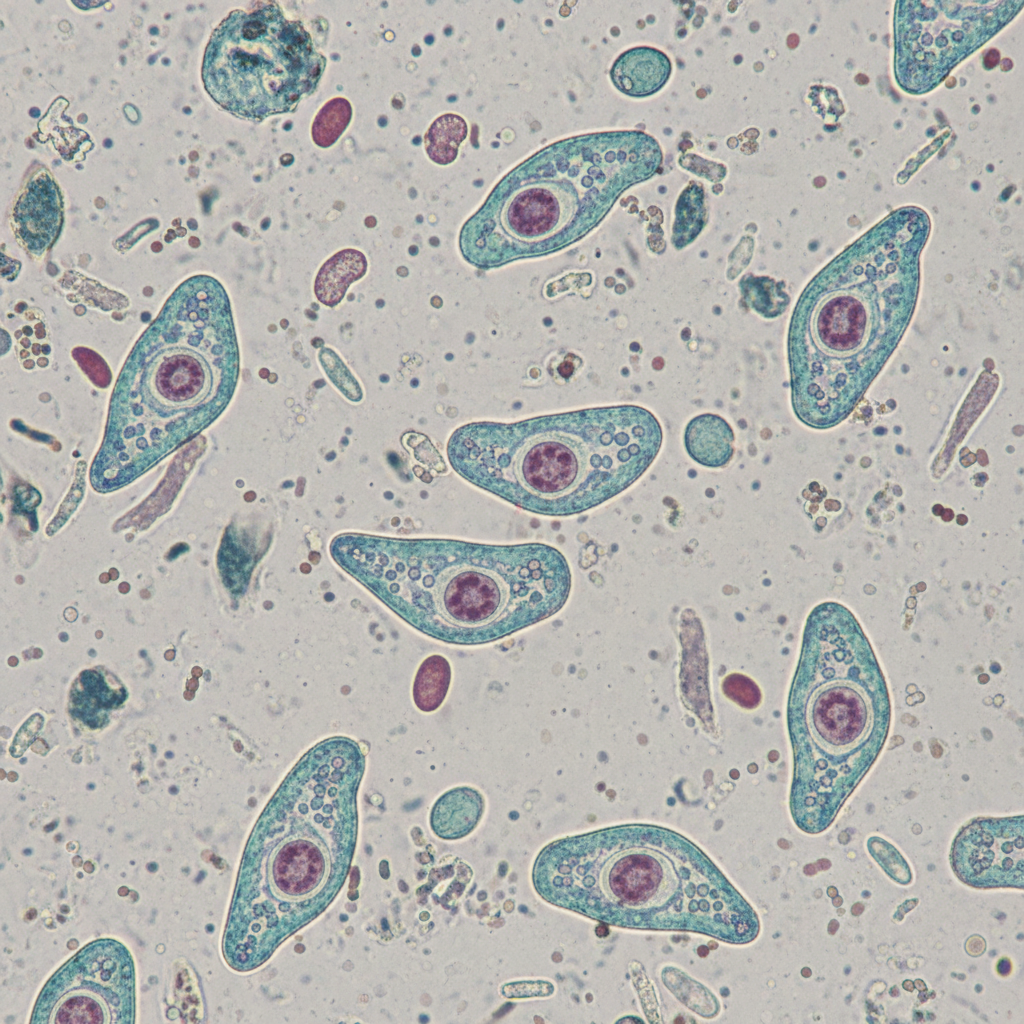
A 31-year-old man comes to the physician because of a 2-day history of abdominal pain and diarrhea. He reports that his stools are streaked with blood and mucus. He returned from a vacation in the Philippines 3 weeks ago. His vital signs are within normal limits. Abdominal examination shows hyperactive bowel sounds. A photomicrograph of a trichrome-stained wet mount of a stool specimen is shown. Which of the following organisms is the most likely cause of this patient's symptoms?

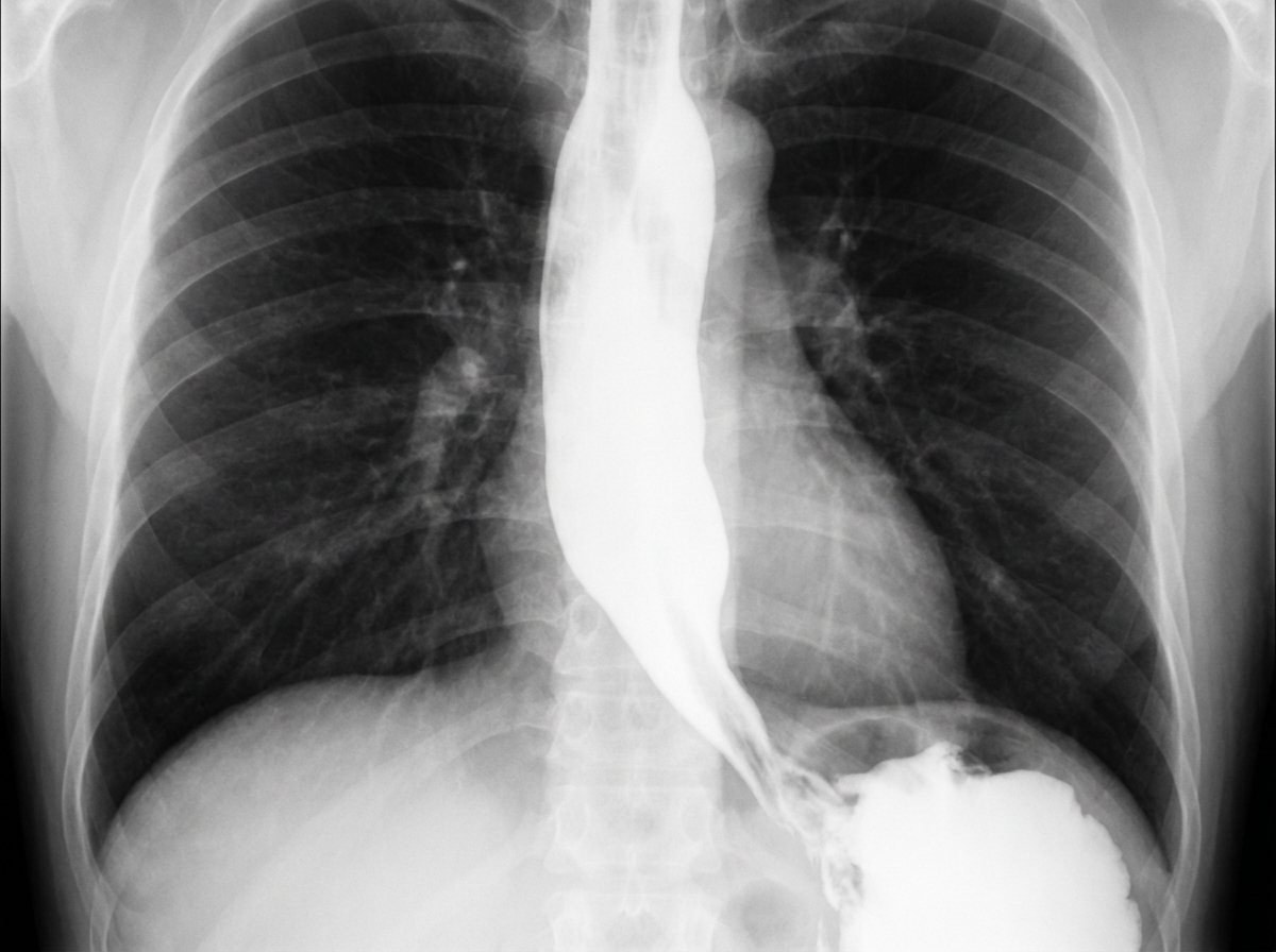
A 62-year-old man is referred to a gastroenterologist because of difficulty swallowing for the past 5 months. He has difficulty swallowing both solid and liquid foods, but there is no associated pain. He denies any shortness of breath or swelling in his legs. He immigrated from South America 10 years ago. He is a non-smoker and does not drink alcohol. His physical examination is unremarkable. A barium swallow study was ordered and the result is given below. Esophageal manometry confirms the diagnosis. What is the most likely underlying cause of this patient’s condition?

A 48-year-old man presents to the clinic with several weeks of watery diarrhea and right upper quadrant pain with fever. He also endorses malaise, nausea, and anorexia. He is HIV-positive and is currently on antiretroviral therapy. He admits to not being compliant with his current medications. His temperature is 37°C (98.6°F), respiratory rate is 15/min, pulse is 70/min, and blood pressure is 100/84 mm Hg. A physical examination is performed which is within normal limits. His blood tests results are given below: Hb%: 11 gm/dL Total count (WBC): 3,400 /mm3 Differential count: Neutrophils: 70% Lymphocytes: 25% Monocytes: 5% CD4+ cell count: 88/mm3 Stool microscopy results are pending. What is the most likely diagnosis?
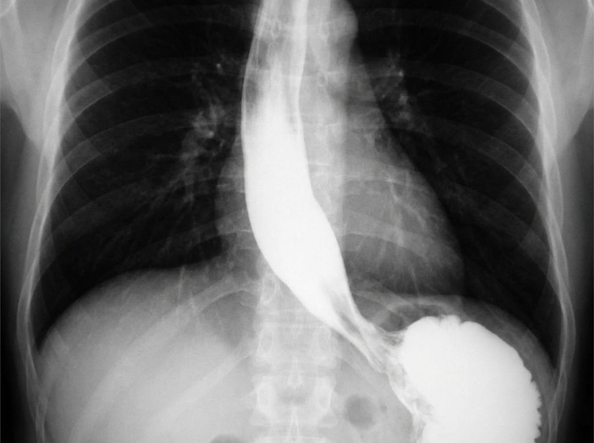
A 48-year-old man from Argentina presents to your office complaining of difficulty swallowing for the past few months. He is accompanied by his wife who adds that his breath has started to smell horrible. The patient says that he feels uncomfortable no matter what he eats or drinks. He also has lost 5 kg (11 lb) in the last 2 months. The patient is afebrile, and his vital signs are within normal limits. Physical exam is unremarkable. A barium swallow study along with esophageal manometry is performed and the results are shown in the image below. Manometry shows very high pressure at the lower esophageal sphincter. Which of the following is the most likely etiology of this patient’s symptoms?

A 36-year-old man is brought to the emergency department for right upper quadrant abdominal pain that began 3 days ago. The pain is nonradiating and has no alleviating or exacerbating factors. He denies any nausea or vomiting. He immigrated from Mexico 6 months ago and currently works at a pet shop. He has been healthy except for 1 week of bloody diarrhea 5 months ago. He is 182 cm (5 ft 11 in) tall and weighs 120 kg (264 lb); BMI is 36 kg/m2. His temperature is 101.8°F (38.8°C), pulse is 85/min, respirations are 14/min, and blood pressure is 120/75 mm Hg. Lungs are clear to auscultation. He has tenderness to palpation in the right upper quadrant. Laboratory studies show: Hemoglobin 11.7 g/dL3 Leukocyte Count 14,000/mm Segmented neutrophils 74% Eosinophils 2% Lymphocytes 17% Monocytes 7% Platelet count 140,000/mm3 Serum Na+ 139 mEq/L Cl- 101 mEq/L K+ 4.4 mEq/L HCO3- 25 mEq/L Urea nitrogen 8 mg/dL Creatinine 1.6 mg/dL Total bilirubin 0.4 mg/dL AST 76 U/L ALT 80 U/L Alkaline phosphatase 103 U/L Ultrasonography of the abdomen shows a 4-cm round, hypoechoic lesion in the right lobe of the liver with low-level internal echoes. Which of the following is the most likely diagnosis?
A 26-year-old woman comes to the physician because of several days of fever, abdominal cramps, and diarrhea. She drank water from a stream 1 week ago while she was hiking in the woods. Abdominal examination shows increased bowel sounds. Stool analysis for ova and parasites shows flagellated multinucleated trophozoites. Further evaluation shows the presence of antibodies directed against the pathogen. Secretion of these antibodies most likely requires binding of which of the following?
A 30-year-old Caucasian male is brought to the emergency room for recurrent diarrhea. He has had multiple upper respiratory infections since birth and does not take any medications at home. It is determined that Giardia lamblia is responsible for the recurrent diarrhea. The physician performs a serum analysis and finds normal levels of mature B lymphocytes. What other finding on serum analysis predisposes the patient to recurrent diarrheal infections?
Explanation: ***IgE*** - **IgE** antibodies are crucial in the immune response against helminthic parasites, including *N. americanus*, by sensitizing **mast cells** and **eosinophils**. - When **IgE** binds to the surface of parasites, the **Fc receptor** on eosinophils recognizes the Fc portion of IgE, leading to the release of cytotoxic granules that destroy the parasite (antibody-dependent cellular cytotoxicity). *IgA* - **IgA** is primarily found in **mucosal secretions** and plays a role in defending against pathogens at mucosal surfaces, but it is not the primary isotype involved in eosinophil-mediated **ADCC** against helminths. - While IgA can bind to some immune cells, its main function is to **neutralize toxins** and prevent microbial adhesion at mucosal sites. *IgG* - **IgG** is the most abundant antibody in serum and is involved in various immune functions, including **opsonization**, **neutralization**, and **complement activation**. - Although IgG can mediate ADCC by **NK cells** and **macrophages**, it is not the primary isotype for eosinophil-mediated killing of helminths, which is dominated by IgE. *IgM* - **IgM** is typically the first antibody produced during a primary immune response and is very effective at **activating the complement system**. - Its large pentameric structure also limits its diffusion into tissues, and it does not play a direct role in eosinophil-mediated ADCC against helminthic parasites. *IgD* - **IgD** primarily functions as a **B cell receptor** on naive B lymphocytes, signaling for their activation and differentiation. - It is present in very low concentrations in serum and its role in host defense against parasites or in ADCC is negligible.
Explanation: ***Giardiasis*** - The patient's history of drinking untreated stream water after a camping trip, followed by **greasy, foul-smelling diarrhea**, flatulence, nausea, and weight loss, is highly characteristic of **giardiasis**. - The stool microscopy finding of **motile protozoa** without blood cells or pus cells confirms the presence of *Giardia lamblia* infection, which is a common cause of such symptoms in campers. *Traveler’s diarrhea due to ETEC* - **ETEC (Enterotoxigenic *Escherichia coli*)** typically causes watery diarrhea, often without the prominent greasy, foul-smelling, and chronic nature seen in this case. - While ETEC is a common cause of traveler's diarrhea, the **microscopic finding of motile protozoa** in the stool definitively rules out a bacterial cause like ETEC. *Irritable bowel syndrome* - **Irritable bowel syndrome (IBS)** is a chronic functional disorder of the bowel, characterized by abdominal pain, bloating, and altered bowel habits (diarrhea, constipation, or both), but it does not have an acute onset directly linked to water consumption or involve **motile protozoa** in the stool. - IBS is a diagnosis of exclusion and would not be considered in the presence of a clear infectious etiology identified by stool examination. *C. difficile colitis* - **Clostridioides difficile colitis** typically presents with watery to bloody diarrhea, abdominal pain, and fever, usually following **antibiotic use** or in hospitalized patients, none of which are reported here. - The stool microscopy would show evidence of *C. difficile* toxins, not **motile protozoa**, and would often reveal pus cells or inflammatory markers. *Traveler’s diarrhea due to Norovirus* - **Norovirus** typically causes acute onset of vomiting, watery diarrhea, and abdominal cramps, often resolving within a few days, but it is a **viral infection**. - The detection of **motile protozoa** on stool microscopy rules out a viral etiology like Norovirus.
Explanation: ***Cryptosporidium parvum*** - This patient's **immunosuppressed state** (HIV with CD4 count of 85/mm3), severe **watery diarrhea**, and history of drinking **spring water** are highly suggestive of cryptosporidiosis. - The finding of **oocysts** on modified acid-fast stain is the definitive diagnostic test for *Cryptosporidium*. *Entamoeba histolytica* - While *Entamoeba histolytica* can cause diarrhea, it typically presents with **bloody stools (dysentery)** and may cause **liver abscesses**, which are not described here. - Diagnosis involves identifying trophozoites or cysts in stool, not acid-fast oocysts. *Cytomegalovirus* - **CMV colitis** can cause diarrhea in immunocompromised patients, but it is typically diagnosed by **biopsy showing inclusions** and is not characterized by acid-fast oocysts in stool. - CMV often presents with systemic symptoms, and while abdominal pain is common, the specific stool findings point elsewhere. *Giardia lamblia* - *Giardia lamblia* causes **greasy, foul-smelling stools** and **malabsorption**, often without fever, and is diagnosed by finding **cysts or trophozoites** in stool, which are not acid-fast oocysts. - While acquired from contaminated water, the clinical picture and diagnostic test differ markedly. *Rotavirus* - **Rotavirus** is a common cause of severe watery diarrhea, especially in **infants and young children**, but it is less common in adults without specific risk factors and is not diagnosed by acid-fast oocysts. - It does not specifically target immunocompromised individuals like *Cryptosporidium* does in this context.
Explanation: ***Entamoeba histolytica*** - The patient's symptoms of **bloody, mucoid diarrhea** (dysentery) after travel to the **Philippines** are highly suggestive of **amebic dysentery** caused by *Entamoeba histolytica*. - A **trichrome-stained wet mount** of stool revealing trophozoites or cysts of *Entamoeba histolytica* in this clinical context would confirm the diagnosis. *Campylobacter jejuni* - While *Campylobacter jejuni* can cause **bloody diarrhea** and is a common cause of **traveler's diarrhea**, it is a **bacterial infection**, and the question implies a parasitic etiology with the mention of a trichrome-stained wet mount. - Infection is typically associated with consumption of undercooked poultry or contaminated water. *Giardia lamblia* - *Giardia lamblia* causes **giardiasis**, which typically presents with **non-bloody, watery diarrhea**, **steatorrhea**, abdominal cramps, and bloating. - It does not usually cause **dysentery** or bloody stools. *Cryptosporidium parvum* - *Cryptosporidium parvum* causes **cryptosporidiosis**, characterized by **profuse, watery diarrhea** and abdominal cramps. - While it can be severe in immunocompromised individuals, it typically does not cause **bloody or mucoid stools** (dysentery). *Shigella dysenteriae* - *Shigella dysenteriae* causes **shigellosis**, a severe form of dysentery with **bloody, mucoid stools**, fever, and tenesmus. - While it fits the clinical picture of dysentery, the diagnostic method mentioned (trichrome-stained wet mount) is primarily used for identifying **parasitic organisms**, not bacteria like *Shigella*.
Explanation: ***Chagas disease*** - The patient's history of living in **South America** and presenting with **dysphagia for both solids and liquids** (suggesting a motility disorder), along with the **barium swallow image showing esophageal dilation and a 'bird's beak' appearance** at the gastroesophageal junction, are highly characteristic of achalasia caused by Chagas disease. - Chagas disease, caused by *Trypanosoma cruzi*, leads to the destruction of **myenteric plexus neurons** in the esophagus, resulting in achalasia (failure of the lower esophageal sphincter to relax) and megaesophagus. *Pharyngoesophageal diverticulum* - This typically presents as **Zenker's diverticulum**, causing **difficulty initiating a swallow**, regurgitation of undigested food, and sometimes halitosis, which is different from the described dysphagia for both solids and liquids. - A Zenker's diverticulum would appear as a **pouch-like protrusion** in the posterior pharynx, not the diffuse esophageal dilation seen in the image. *Esophageal rupture* - Esophageal rupture (Boerhaave syndrome) is an acute, life-threatening condition associated with **severe chest pain, vomiting, and crepitus**, not a chronic, progressive dysphagia without pain. - Imaging would reveal **extravasation of contrast** into the mediastinum or pleural space, not the smooth dilation and distal narrowing observed. *Gastroesophageal reflux disease* - While chronic GERD can lead to **strictures** and dysphagia, it typically causes **heartburn**, regurgitation, and sometimes odynophagia, and the dysphagia is usually progressive for solids first. - The barium swallow would show reflux or a stricture, not the **classic achalasia findings** of a dilated esophagus tapering to a narrow distal segment. *Squamous cell carcinoma of the esophagus* - Squamous cell carcinoma usually presents with **progressive dysphagia, initially for solids**, and is often associated with weight loss, smoking, and alcohol use, none of which are present in this patient. - A tumor would typically appear as an **irregular, focal narrowing or mass** on barium swallow, not the smooth, diffuse dilation seen in this image.
Explanation: ***Cryptosporidiosis*** - This patient's **HIV-positive status** with a **CD4+ count of 88/mm3** indicates severe immunosuppression, making them highly susceptible to opportunistic infections. The combination of **watery diarrhea**, **right upper quadrant pain with fever** (suggestive of biliary involvement), and general malaise is characteristic of cryptosporidiosis in immunocompromised individuals. - **Cryptosporidium** infection often causes **chronic, severe watery diarrhea** in patients with AIDS, and can also lead to **cholangitis or cholecystitis**, explaining the right upper quadrant pain and fever. *C. difficile colitis* - While *C. difficile* can cause watery diarrhea, especially in hospitalized patients or those on antibiotics, the **right upper quadrant pain with fever** and the patient's profound **immunocompromise (CD4+ <200)** makes opportunistic infections like *Cryptosporidium* more likely. - *C. difficile* typically presents with **colitis**, which may include abdominal pain but less commonly high-grade fever or specific right upper quadrant pain indicative of biliary involvement. *Traveler’s diarrhea due to ETEC* - **ETEC (Enterotoxigenic *E. coli*)** is a common cause of traveler's diarrhea, usually self-limiting and associated with recent travel, which is not mentioned here. - While it causes watery diarrhea, it typically does not present with **fever or right upper quadrant pain** suggestive of biliary disease, especially not in a patient with severe immunosuppression where opportunistic pathogens are more expected. *Norovirus infection* - **Norovirus** causes acute gastroenteritis with **vomiting and watery diarrhea**, sometimes low-grade fever, but it is typically a self-limiting illness lasting 1-3 days. - It does not explain the **right upper quadrant pain with fever** suggestive of biliary involvement, nor the prolonged "several weeks" duration of symptoms. *Irritable bowel syndrome* - **Irritable bowel syndrome (IBS)** is a functional gastrointestinal disorder characterized by chronic abdominal pain and altered bowel habits (diarrhea, constipation, or both), but it does not cause **fever, laboratory abnormalities (low CD4 count), or acute onset of symptoms** in an immunocompromised patient. - IBS is a diagnosis of exclusion and does not account for the systemic symptoms like **fever, malaise**, or the specific right upper quadrant pain.
Explanation: ***Trypanosoma cruzi infection*** - The patient's origin from **Argentina** (an endemic area for **Chagas disease**), coupled with symptoms of **dysphagia**, **weight loss**, and **halitosis**, strongly suggests **achalasia** secondary to *Trypanosoma cruzi* infection. - **Esophageal manometry** showing **very high pressure at the lower esophageal sphincter** and **absent or diminished peristalsis** is characteristic of achalasia, which in this context points to **Chagasic achalasia**. *Food allergy* - **Food allergies** typically present with acute symptoms such as **hives, angioedema, gastrointestinal upset**, or **anaphylaxis**, rather than chronic dysphagia and weight loss. - While eosinophilic esophagitis (a type of food allergy) can cause dysphagia, it usually presents with **esophageal strictures** or **rings** and is not associated with the distinct manometric findings of achalasia. *Outpouching of the mucosa and submucosa* - An **outpouching of the mucosa and submucosa** refers to a **Zenker's diverticulum**, which can cause dysphagia, halitosis, and regurgitation. - However, Zenker's diverticulum is typically located in the **upper esophagus** and would not cause increased lower esophageal sphincter pressure on manometry. *Malignant proliferation of squamous cells* - A **malignant proliferation of squamous cells** refers to **esophageal squamous cell carcinoma**, which can cause progressive dysphagia and weight loss. - While possible in an older patient, the characteristic manometric findings (high LES pressure) are more indicative of achalasia rather than a primary obstructive tumor. *Pyloric stenosis* - **Pyloric stenosis** is an obstruction at the gastric outlet, causing **projectile vomiting** and **weight loss**, primarily seen in infants. - In adults, it is usually due to peptic ulcer disease or malignancy and would not cause esophageal symptoms or increased lower esophageal sphincter pressure.
Explanation: ***Amebiasis*** - The patient's history of recent immigration from Mexico, prior **bloody diarrhea**, and current presentation with **right upper quadrant pain**, fever, leukocytosis, and an **abscess-like lesion in the liver** are highly suggestive of an **amoebic liver abscess**. - **Entamoeba histolytica**, often acquired through contaminated food or water, can cause colitis leading to bloody diarrhea and subsequently spread hematogenously to the liver. *Hepatic hydatid cyst* - **Hydatid cysts** (Echinococcosis) are typically slow-growing and often asymptomatic for many years before causing symptoms. - While they can be found in the liver, the presentation with **acute fever** and **leukocytosis** is less common than with an abscess. *Liver hemangioma* - A **liver hemangioma** is a benign vascular tumor, usually asymptomatic and discovered incidentally. - It does not cause fever, elevated white blood cell count, or abdominal pain in this acute manner. *Pyogenic liver abscess* - While a **pyogenic liver abscess** can present with similar symptoms (fever, RUQ pain, leukocytosis) and imaging findings, the history of **bloody diarrhea** and immigration from an endemic area points more specifically to amoebic etiology. - Pyogenic abscesses are often associated with other underlying conditions such as biliary tract disease or diverticulitis. *Hepatocellular carcinoma* - **Hepatocellular carcinoma (HCC)** typically occurs in patients with underlying chronic liver disease (e.g., cirrhosis, hepatitis B or C). - While it can present with abdominal pain and a liver mass, an acute febrile illness and a history of bloody diarrhea are not typical features of HCC.
Explanation: ***CD40 to CD40 ligand*** - The interaction between **CD40 on B cells** and **CD40 ligand (CD40L) on activated T helper cells** is crucial for **T cell-dependent B cell activation** and antibody class switching. - This binding leads to the maturation of the immune response, including the secretion of **high-affinity antibodies** like IgA, which is especially important for mucosal immunity against pathogens like *Giardia lamblia* (the likely cause of the patient's symptoms). *CD28 to B7 protein* - The binding of **CD28 on T cells** to **B7 protein (CD80/86) on antigen-presenting cells (APCs)** provides the **second co-stimulatory signal** required for T cell activation. - While essential for T cell activation, this interaction primarily supports T cell proliferation and differentiation, rather than directly mediating antibody secretion by B cells. *CD80/86 to CTLA-4* - **CTLA-4 (cytotoxic T-lymphocyte-associated protein 4)** is a receptor on T cells that binds to **CD80/86 (B7)** on APCs with higher affinity than CD28. - This interaction provides an **inhibitory signal** that downregulates T cell activation, serving as a negative feedback mechanism, and does not promote antibody secretion. *gp120 to CD4* - The **gp120 glycoprotein** on the surface of **HIV** binds to the **CD4 receptor** on T helper cells, initiating the entry of the virus into the cell. - This interaction is specific to HIV infection and is not involved in the normal process of antibody secretion in response to other pathogens. *CD8 to MHC I* - **CD8** is a co-receptor expressed on **cytotoxic T lymphocytes (CTLs)** that binds to **MHC class I molecules** on target cells. - This interaction is essential for the recognition of virally infected or cancerous cells by CTLs, leading to their destruction, but it is not directly involved in antibody production.
Explanation: ***Deficiency in IgA*** - **Selective IgA deficiency** is the most common primary immunodeficiency and typically presents with recurrent sinopulmonary and gastrointestinal infections. - **IgA** is crucial for mucosal immunity, and its deficiency renders the patient susceptible to infections like *Giardia lamblia*, which colonizes the gut. *Deficiency in IgG* - While IgG deficiency can lead to recurrent infections, it typically involves a broader range of pathogens and often includes more severe bacterial infections. - The presence of **normal mature B lymphocytes** in this patient suggests a defect in immunoglobulin class switching or secretion rather than a complete absence of antibody-producing cells. *Deficiency in neutrophils* - **Neutrophil deficiency** (neutropenia) primarily leads to recurrent bacterial and fungal infections, often presenting with skin abscesses and systemic infections. - It does not specifically predispose an individual to protozoal infections like *Giardia lamblia* or recurrent upper respiratory tract infections in this manner. *Deficiency in NK cells* - **Natural killer (NK) cells** are important for antiviral and antitumor immunity. - A deficiency in NK cells would typically manifest as increased susceptibility to viral infections (herpesviruses) and certain cancers, not *Giardia lamblia* or recurrent upper respiratory infections, which are often bacterial or viral in origin. *Deficiency in CD8+ T cells* - **CD8+ T cells** are crucial for killing virally infected cells and some tumor cells. - A deficiency would lead to increased susceptibility to severe viral infections and certain intracellular bacteria, not typically chronic *Giardia* infection or recurrent bacterial upper respiratory infections.
Explanation: ***Giardia lamblia*** - The patient's symptoms of **watery, non-bloody, foul-smelling, greasy stools** (steatorrhea) with abdominal cramping after a camping trip are classic for giardiasis. The image shows a **trophozoite of Giardia lamblia**, characterized by its pear shape, multiple flagella, and two nuclei, often described as having an "old man's face" appearance. - The **epidemiological context** (camping trip, son with similar symptoms) suggests exposure to contaminated water, a common source of *Giardia* infection. *Yersinia enterocolitica* - This pathogen typically causes **bloody diarrhea**, fever, and sometimes abdominal pain that can mimic appendicitis (*pseudoappendicitis*), which are not present in this case. - It's mainly associated with consuming **undercooked pork** or contaminated milk products, not typically recreational water exposure. *Bacillus cereus* - This bacterium causes **food poisoning** with either an emetic form (vomiting) due to preformed toxins in **fried rice** or a diarrheal form (watery diarrhea) associated with meat and vegetable dishes. - The incubation periods are usually short (1-6 hours for emetic, 6-15 hours for diarrheal), which does not fit the 2-week duration described. *Clostridium difficile* - *C. difficile* infection is characterized by **watery diarrhea** and **abdominal cramps**, often following **antibiotic use** or in healthcare settings, which are not mentioned here. - While it can cause severe diarrhea, the stool is usually not described as greasy, and the image does not show *C. difficile* organisms or their toxins. *Campylobacter jejuni* - This bacterium is a common cause of **bacterial gastroenteritis**, typically presenting with **bloody diarrhea**, fever, and abdominal pain. - It is often acquired from contaminated **poultry** or unpasteurized milk, and is associated with complications like **Guillain-Barré syndrome**, none of which are suggested by the clinical picture or image.
Explanation: ***Reactivation of dormant liver stage*** - The patient's history of **recurrent malaria-like symptoms** after a trip to Indonesia and previous successful treatment with chloroquine strongly suggests malaria caused by *Plasmodium vivax* or *P. ovale*, which are known for forming **dormant hypnozoites in the liver**. - **Chloroquine** only targets the asexual erythrocytic stages and does not eliminate these dormant liver stages, leading to relapses when they reactivate and release merozoites into the bloodstream. *Decline in circulating antibodies* - While a decline in antibodies can increase susceptibility to reinfection, it is less likely to explain the specific pattern of **relapse malaria** seen with *P. vivax* or *P. ovale*. - The primary mechanism for recurrence in these species is the **activation of hypnozoites**, not simply a waning immune response to new exposure. *Dissemination within macrophages* - This mechanism is characteristic of infections that can persist intracellularly within macrophages, such as certain bacterial or parasitic infections like **leishmaniasis**. - It is not a typical mechanism for the recurrence of *Plasmodium* species responsible for malaria. *Natural drug resistance* - Although drug resistance to chloroquine exists, especially in *P. falciparum* and increasingly *P. vivax*, the term "natural" implies an inherent resistance in the organism from the outset. - The patient initially responded to chloroquine, indicating it was effective against the erythrocytic stages, making **innate resistance** less probable as the sole cause of relapse. *Reinfection by *Anopheles* mosquito* - While reinfection is possible if the patient returned to an endemic area and was bitten again, the presentation of a **relapse 9 months later** after initial treatment is a classic pattern for *P. vivax* or *P. ovale*, which can remain dormant and reactivate without new exposure. - The symptoms occurred without a recent travel history back to an endemic region, making a new infection less likely than a relapse from dormant hypnozoites.
Explanation: ***Metronidazole and iodoquinol*** - The patient's symptoms (bloody diarrhea, abdominal pain, fever, liver cyst) and risk factors (sexual activity with men and women) are highly suggestive of an **amoebic liver abscess** caused by *Entamoeba histolytica*. - **Metronidazole** is the drug of choice for invasive amoebiasis (including liver abscess), while **iodoquinol** (or paromomycin) treats the intestinal luminal cysts to prevent recurrence and transmission. *Supportive therapy* - While supportive care is important for managing symptoms like fever and dehydration, it does not address the underlying **amoebic infection** or the liver abscess. - Delaying specific antimicrobial therapy can lead to worsening of the abscess, potential rupture, and increased morbidity. *Sulfadiazine and pyrimethamine* - This combination is the standard treatment for **toxoplasmosis**, a protozoal infection that typically affects immunocompromised individuals and can cause encephalitis or disseminated disease. - It is not effective against *Entamoeba histolytica* and would not resolve an amoebic liver abscess. *Nifurtimox* - **Nifurtimox** is an antiparasitic medication specifically used to treat **Chagas disease**, caused by *Trypanosoma cruzi*. - Chagas disease presents with different clinical manifestations and is transmitted by blood-sucking triatomine bugs, which does not fit the patient's presentation. *Amphotericin* - **Amphotericin B** is a broad-spectrum **antifungal agent** used to treat severe systemic fungal infections. - It has no activity against *Entamoeba histolytica* or other protozoal infections causing similar symptoms.
Explanation: ***Nematode*** - The patient's symptoms (fatigue, pallor) and lab results (**microcytic anemia** with **low hemoglobin**, **low MCV**, **low iron**, and **low ferritin**) are highly suggestive of **iron deficiency anemia**. The history of backpacking worldwide increases the suspicion of **hookworm infection**, which is a nematode that causes chronic gastrointestinal blood loss leading to iron deficiency. - **Hookworms** (e.g., *Ancylostoma duodenale*, *Necator americanus*) attach to the intestinal wall, causing persistent blood loss as they feed, which depletes iron stores over time. *Mosquito-borne protozoa* - **Mosquito-borne protozoa** primarily refers to *Plasmodium* species which cause malaria. While malaria can cause anemia, it typically presents with **hemolytic anemia** (elevated LDH, jaundice), intermittent fevers, and splenomegaly, not the profound iron deficiency seen here. - The anemia in malaria is usually **normocytic or macrocytic** due to increased erythropoiesis, and profound iron deficiency is not the primary mechanism. *Double-stranded virus* - **Double-stranded viruses** (e.g., adenoviruses, herpesviruses, some papillomaviruses) typically cause acute infections, and while some can lead to anemia through bone marrow suppression or chronic inflammation, they do not directly cause **iron deficiency anemia** with this specific lab profile. - The clinical picture of chronic fatigue and iron depletion after travel is not characteristic of common viral infections caused by double-stranded viruses. *Single-stranded virus* - **Single-stranded viruses** (e.g., influenza, measles, HIV, dengue, enteroviruses) cause a wide range of illnesses. While some can lead to anemia, often through **bone marrow suppression** or chronic inflammation, they are not typically associated with the profound **iron deficiency** and microcytic anemia seen in this patient. - Travel history can be relevant for some single-stranded viral infections (e.g., HIV, dengue), but the specific laboratory findings point away from a primary viral etiology for the anemia. *Tick-borne protozoa* - **Tick-borne protozoa** most commonly refers to *Babesia* species, which cause babesiosis. This disease primarily leads to **hemolytic anemia** (intravascular hemolysis, elevated LDH), fever, chills, and fatigue, which is distinct from the **iron deficiency anemia** presented in the case. - **Babesiosis** would typically present with signs of hemolysis, such as elevated LDH and sometimes hemoglobinuria, which are absent in this patient.
Explanation: **Plasmodium falciparum** - The image shows **multiple ring forms** and **applique forms** within red blood cells, which are characteristic of *Plasmodium falciparum* malaria. The clinical presentation of **headache, muscle pain, recurrent fever spikes without a noticeable rhythm, jaundice, splenomegaly, and anemia (Hb 10 g/dL)** in a traveler returning from India and Africa is highly consistent with malaria, especially given the chloroquine chemoprophylaxis which is often ineffective against chloroquine-resistant strains of *P. falciparum*. - *P. falciparum* can cause severe disease, including **anemia** due to red blood cell destruction and **jaundice** due to hemolysis and liver involvement, and is notorious for its **irregular fever patterns** early in the infection cycle. *Chikungunya virus* - Chikungunya typically presents with **high fever, severe polyarthralgia**, and rash, but does not cause the parasitemia or specific red blood cell morphology seen in the image. - While present in endemic regions like India and Africa, it does not lead to **anemia, splenomegaly, or jaundice** to the extent seen in this patient, nor does it appear on a blood smear as intracellular parasites. *Trypanosoma cruzi* - *Trypanosoma cruzi* causes **Chagas disease**, which is endemic to **Central and South America**, not India or Africa. - While it can be found in blood smears during the acute phase (trypomastigotes), its morphology differs significantly from the ring forms seen, and the overall clinical picture of **fever, jaundice, and marked splenomegaly with characteristic RBC parasites** does not fit Chagas disease. *Leishmania donovani* - *Leishmania donovani* causes **visceral leishmaniasis (kala-azar)**, characterized by **prolonged fever, splenomegaly, hepatomegaly, pancytopenia**, and weight loss. - While present in India and Africa, the parasites (**amastigotes**) are typically found within **macrophages** in bone marrow, spleen, or liver aspirates, not as ring forms within red blood cells on a peripheral blood smear. *Trypanosoma brucei* - *Trypanosoma brucei* causes **African sleeping sickness**, which involves **fever, headache, joint pain, neurological symptoms**, and lymphadenopathy (Winterbottom's sign). - The parasites (trypomastigotes) are observed extracellularly in the blood, lymph, or CSF, and have a distinct **elongated, flagellated morphology** that is completely different from the intracellular ring forms seen in the provided image.
Explanation: ***Protozoa*** - The patient's history of **Pneumocystis jirovecii pneumonia** (PJP) suggests an **immunocompromised state**, likely due to HIV/AIDS. - In such patients, **ring-enhancing brain lesions** are highly characteristic of **cerebral toxoplasmosis**, an opportunistic infection caused by the protozoan *Toxoplasma gondii*. *Virus* - While viruses like **CMV** or **JC virus** (causing PML) can affect the brain in immunocompromised patients, they typically present with different imaging features (e.g., non-enhancing lesions in PML) and are less likely to cause multiple ring-enhancing lesions. - Though HIV can cause **HIV encephalopathy**, it typically involves **diffuse atrophy** and **white matter changes**, rather than distinct ring-enhancing lesions. *Neoplasm* - **Primary central nervous system lymphoma (PCNSL)** can present with ring-enhancing lesions, especially in HIV-positive individuals. - However, given the association with PJP, **infectious etiologies** like toxoplasmosis are generally more common as the initial diagnosis for multiple ring-enhancing lesions in this patient population. *Bacteria* - **Bacterial brain abscesses** can cause ring-enhancing lesions but are less common in disseminated opportunistic infections in HIV/AIDS compared to protozoal or fungal infections. - They also typically present with a more **acute inflammatory picture** and may be preceded by a source of bacterial infection (e.g., endocarditis, sinusitis) not mentioned here. *Prion* - **Prion diseases** (e.g., Creutzfeldt-Jakob disease) cause rapidly progressive dementia and characteristic EEG and MRI findings (e.g., cortical ribboning, basal ganglia hyperintensity) that do not typically include multiple ring-enhancing lesions. - They are also not associated with the immunocompromised state indicated by PJP.
Explanation: ***Leishmania braziliensis*** - The patient's history of travel to **Guatemala**, a region endemic for **cutaneous leishmaniasis**, along with the characteristic skin lesion (**solitary, round, pink plaque with central ulceration**) and **axillary lymphadenopathy**, is highly suggestive of infection with *Leishmania braziliensis*. - The microscopic image would typically show **amastigotes** within **macrophages**, which are pathognomonic for leishmaniasis. *Treponema pallidum* - This bacterium causes **syphilis**, which can present with a **chancre** (painless ulcer) in its primary stage. - However, the chancre is typically firm, solitary, and non-itchy, and the patient's travel history and specific lesion morphology are more consistent with leishmaniasis. *Borrelia burgdorferi* - This spirochete causes **Lyme disease**, characterized by **erythema migrans** (a migrating rash with central clearing) in its early stage. - The rash seen in this case does not resemble erythema migrans, and regional lymphadenopathy is less prominent compared to leishmaniasis. *Trypanosoma brucei* - This protozoan causes **African sleeping sickness**, which is endemic to **sub-Saharan Africa**, not Central America. - Initial symptoms may include a **trypanosomal chancre** at the bite site, followed by systemic symptoms and neurological involvement, which are not described here. *Ancylostoma duodenale* - This is a type of **hookworm** that causes parasitic infection, primarily affecting the **gastrointestinal tract**, leading to **anemia**. - Skin manifestations are typically **pruritic larvae currens** (creeping eruption) due to larval migration, which is different from the described ulcerated plaque.
Malaria parasites and life cycle
Practice Questions
Intestinal protozoa (Giardia, Entamoeba)
Practice Questions
Toxoplasma gondii
Practice Questions
Trypanosomes
Practice Questions
Leishmania species
Practice Questions
Nematodes (roundworms)
Practice Questions
Cestodes (tapeworms)
Practice Questions
Trematodes (flukes)
Practice Questions
Blood and tissue parasites
Practice Questions
Antiparasitic medications
Practice Questions
Laboratory diagnosis of parasites
Practice Questions
Global health impact of parasitic infections
Practice Questions
Ectoparasites and vector-borne diseases
Practice Questions
Get full access to all questions, explanations, and performance tracking.
Start For Free