
Liver disease — MCQs

On this page
A 48-year-old man presents to his primary care physician with a 6-month history of increasing joint pain and stiffness. He says that the pain is primarily located in his knees and occurs in sharp bursts that are accompanied by redness and warmth. His past medical history is significant for diabetes though he is not currently taking any medications. He also suffers from occasional diarrhea with fatty stools. Physical exam reveals mild swelling and redness in his knees bilaterally. Furthermore, he is found to be very tan despite the fact that he says he stays out of the sun. He notes that he has always been significantly more tan than anyone else in his family. This patient is most likely predisposed to which of the following diseases?
A 67-year-old man presents with fatigue, progressive abdominal distention and yellow skin coloration for the past 2 weeks. He denies fever, chills, or other symptoms. Past medical history is unremarkable. He reports heavy alcohol consumption for the past several years but says he quit recently. On physical examination, the patient appears jaundiced and is ill-appearing. There is shifting dullness present on abdominal percussion with a positive fluid wave. Sclera are icteric. Bilateral gynecomastia is present. Laboratory findings are significant for the following: Hgb 13 g/dL Leukocyte count 4,500/mm3 Platelets 86,000/mm3 Aspartate transaminase (AST) 108 U/L Alanine transaminase (ALT) 55 U/L GGT 185 U/L Urea 23 mg/dL Iron 120 μg/dL Ferritin 180 μg/dL Transferrin saturation 40% Which of the following is the most likely diagnosis in this patient?
A 54-year-old man with alcoholism comes to the emergency department because of vomiting blood for 6 hours. He has had 3–4 episodes in which he has vomited dark red blood during this period. He has had no epigastric pain or tarry stools. On arrival, his temperature is 37.3°C (99.1°F), pulse is 134/min, and blood pressure is 80/50 mm Hg. He is resuscitated with 0.9% saline and undergoes an emergency upper endoscopy, which shows actively bleeding varices. Band ligation of the varices is done and hemostasis is achieved. He is diagnosed with Child class B cirrhosis. He is concerned about the possibility of recurrence of such an episode. He is asked to abstain from alcohol, to which he readily agrees. In addition to non-selective beta-blocker therapy, which of the following is the most appropriate recommendation to prevent future morbidity and mortality from this condition?
A 56-year-old woman with a history of alcoholic cirrhosis and recurrent esophageal varices who recently underwent transjugular intrahepatic portosystemic shunt (TIPS) placement is brought to the emergency room by her daughter due to confusion and agitation. Starting this morning, the patient has appeared sleepy, difficult to arouse, and slow to respond to questions. Her temperature is 97.6°F (36.4°C), blood pressure is 122/81 mmHg, pulse is 130/min, respirations are 22/min, and oxygen saturation is 98% on room air. She repeatedly falls asleep and is combative during the exam. Laboratory values are notable for a potassium of 3.0 mEq/L. The patient is given normal saline with potassium. Which of the following is the most appropriate treatment for this patient?
A 45-year-old homeless man is brought to the emergency department by the police. He was found intoxicated and passed out in a library. The patient has a past medical history of IV drug abuse, diabetes, alcohol abuse, and malnutrition. The patient has been hospitalized previously for multiple episodes of pancreatitis and sepsis. Currently, the patient is minimally responsive and only withdraws his extremities in response to painful stimuli. His temperature is 99.5°F (37.5°C), blood pressure is 90/48 mmHg, pulse is 150/min, respirations are 17/min, and oxygen saturation is 95% on room air. Physical exam is notable for tachycardia, a diastolic murmur at the left lower sternal border, and bilateral crackles on pulmonary exam. The patient is started on IV fluids, vancomycin, and piperacillin-tazobactam. Laboratory values are ordered as seen below. Hemoglobin: 9 g/dL Hematocrit: 30% Leukocyte count: 11,500/mm^3 with normal differential Platelet count: 297,000/mm^3 Serum: Na+: 139 mEq/L Cl-: 100 mEq/L K+: 4.0 mEq/L HCO3-: 28 mEq/L BUN: 33 mg/dL Glucose: 60 mg/dL Creatinine: 1.7 mg/dL Ca2+: 9.7 mg/dL PT: 20 seconds aPTT: 60 seconds AST: 1,010 U/L ALT: 950 U/L The patient is admitted to the medical floor. Five days later, the patient's neurological status has improved. His temperature is 99.5°F (37.5°C), blood pressure is 130/90 mmHg, pulse is 90/min, respirations are 11/min, and oxygen saturation is 99% on room air. Laboratory values are repeated as seen below. Hemoglobin: 10 g/dL Hematocrit: 32% Leukocyte count: 9,500/mm^3 with normal differential Platelet count: 199,000/mm^3 Serum: Na+: 140 mEq/L Cl-: 102 mEq/L K+: 4.3 mEq/L HCO3-: 24 mEq/L BUN: 31 mg/dL Glucose: 100 mg/dL Creatinine: 1.6 mg/dL Ca2+: 9.0 mg/dL PT: 40 seconds aPTT: 90 seconds AST: 150 U/L ALT: 90 U/L Which of the following is the best description of this patient’s current status?
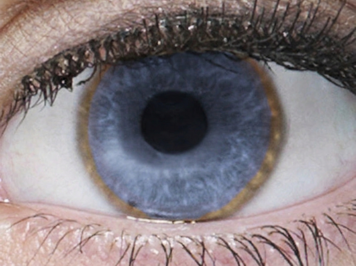
A 17-year-old man presents to his primary care physician with a bilateral tremor of the hands. He is a senior in high school and during the year, his grades have plummeted to the point that he is failing. He says his memory is now poor, and he has trouble focusing on tasks. His behavior has changed in the past 6 months, in that he has frequent episodes of depression, separated by episodes of bizarre behavior, including excessive alcohol drinking and shoplifting. His parents have started to suspect that he is using street drugs, which he denies. His handwriting has become very sloppy. His parents have noted slight slurring of his speech. Family history is irrelevant. Physical examination reveals upper extremity tremors, mild dystonia of the upper extremities, and mild incoordination involving his hands. The patient’s eye is shown. Which of the following best represents the etiology of this patient illness?
A 48-year-old man with a history of diabetes mellitus presents to his primary care physician with lethargy, joint pain, and impotence. Lab evaluation is notable for a ferritin of 1400 ug/L (nl <300 ug/L), increased total iron, increased transferrin saturation, and decreased total iron binding capacity. All of the following are true regarding this patient's condition EXCEPT:
A 71-year-old woman comes to the physician because of an 8-month history of fatigue. Laboratory studies show a hemoglobin concentration of 13.3 g/dL, a serum creatinine concentration of 0.9 mg/dL, and a serum alkaline phosphatase concentration of 100 U/L. Laboratory evaluation of which of the following parameters would be most helpful in determining the cause of this patient's symptoms?
A 47-year-old woman comes to the physician because of a 3-week history of generalized fatigue, mild fever, abdominal pain, and nausea. She attended the state fair over a month ago, where she tried a number of regional foods, and wonders if it might have been caused by something she ate. She has also noticed darkening of her urine, which she attributes to not drinking enough water recently. She has type 2 diabetes mellitus. She drinks 1–2 beers daily. She works as nursing assistant in a rehabilitation facility. Current medications include glyburide, sitagliptin, and a multivitamin. She appears tired. Her temperature is 38.1°C (100.6°F), pulse is 99/min, and blood pressure is 110/74 mm Hg. Examination shows mild scleral icterus. The liver is palpated 2–3 cm below the right costal margin and is tender. Laboratory studies show: Hemoglobin 10.6 g/dL Leukocyte count 11600/mm3 Platelet count 221,000/mm3 Serum Urea nitrogen 26 mg/dL Glucose 122 mg/dL Creatinine 1.3 mg/dL Bilirubin 3.6 mg/dL Total 3.6 mg/dL Direct 2.4 mg/dL Alkaline phosphatase 72 U/L AST 488 U/L ALT 798 U/L Hepatitis A IgG antibody (HAV-IgG) positive Hepatitis B surface antigen (HBsAg) positive Hepatitis B core IgG antibody (anti-HBc) positive Hepatitis B envelope antigen (HBeAg) positive Hepatitis C antibody (anti-HCV) negative Which of the following is the most likely diagnosis?
A 32-year-old woman comes to the emergency department for a 2-week history of right upper quadrant abdominal pain. She has also been feeling tired and nauseous for the past 5 weeks. She has a history of depression and suicidal ideation. She is a social worker for an international charity foundation. She used intravenous illicit drugs in the past but quit 4 months ago. Her only medication is sertraline. Her temperature is 37.8°C (100.0°F), pulse is 100/min, and blood pressure is 128/76 mm Hg. She is alert and oriented. Scleral icterus is present. Abdominal examination shows tenderness to palpation in the right upper quadrant. The liver edge is palpated 3 cm below the right costal margin. There is no rebound tenderness or guarding. The abdomen is non-distended and the fluid wave test is negative. She is able to extend her arms with wrists in full extension and hold them steady without flapping. Laboratory studies show: Hemoglobin 13.8 g/dL Leukocytes 13,700/mm3 Platelets 165,000/mm3 Prothrombin time 14 seconds Partial thromboplastin time 35 seconds Serum: Total bilirubin 4.8 mg/dL Direct bilirubin 1.3 mg/dL Aspartate aminotransferase 1852 U/L Alanine aminotransferase 2497 U/L Urea nitrogen 21 mg/dL Creatinine 1.2 mg/dL Hepatitis A IgM antibody Negative Hepatitis B surface antigen Negative Hepatitis B surface antibody Negative Hepatitis B core IgM antibody Positive Hepatitis C antibody Positive Hepatitis C RNA Negative Urine beta-hCG Negative Which of the following is the most appropriate next step in management?
Practice by Chapter
Viral hepatitis (A, B, C, D, E)
Practice Questions
Alcoholic liver disease
Practice Questions
Non-alcoholic fatty liver disease
Practice Questions
Drug-induced liver injury
Practice Questions
Autoimmune hepatitis
Practice Questions
Cirrhosis management
Practice Questions
Portal hypertension complications
Practice Questions
Ascites diagnosis and management
Practice Questions
Hepatic encephalopathy
Practice Questions
Spontaneous bacterial peritonitis
Practice Questions
Hepatorenal syndrome
Practice Questions
Hepatocellular carcinoma
Practice Questions
Liver transplantation
Practice Questions
Want unlimited practice?
Get full access to all questions, explanations, and performance tracking.
Scan to download app