
Gastroenterology — MCQs

On this page
A previously healthy 41-year-old woman comes to the physician for the evaluation of recurrent episodes of palpitations and sweating over the past month. Her symptoms typically start after swimming practice and improve after drinking ice tea and eating some candy. She has also had a 5-kg (11-lb) weight gain over the past 3 months. She works as a nurse. Physical examination shows no abnormalities. Fasting serum studies show: Glucose 38 mg/dL Insulin 260 μU/mL (N=11–240) Proinsulin 65 μU/mL (N <20% of total insulin) C-peptide 5.0 ng/mL (N=0.8–3.1) Insulin secretagogues absent Which of the following is the most likely cause of her symptoms?
A 32-year-old female comes to the physician because of recurrent episodes of abdominal pain, bloating, and loose stools lasting several days to a couple weeks. She has had these episodes since she was 24 years old but they have worsened over the last 6 weeks. The site of the abdominal pain and the intensity of pain vary. She has around 3–4 bowel movements per day during these episodes. Menses are regular at 31 day intervals with moderate flow; she has moderate pain in her lower abdomen during menstruation. She moved from a different city 2 months ago to start a new demanding job. Her mother has been suffering from depression for 10 years. She does not smoke or drink alcohol. Her own medications include multivitamins and occasionally naproxen for pain. Temperature is 37.4°C (99.3°F), pulse is 88/min, and blood pressure is 110/82 mm Hg. Abdominal examination shows no abnormalities. Laboratory studies show: Hemoglobin 14.1 g/dL Leukocyte count 8100/mm3 Erythrocyte sedimentation rate 15 mm/h Serum Glucose 96 mg/dL Creatinine 1.1 mg/dL IgA anti-tissue transglutaminase antibody negative Urinalysis shows no abnormalities. Further evaluation is most likely to show which of the following in this patient?
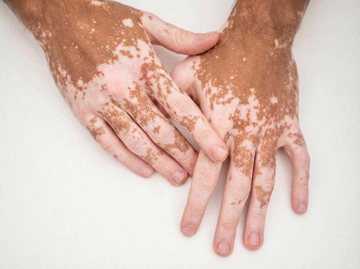
A 14-year-old boy comes to the physician because of multiple patches on his trunk and thighs that are lighter than the rest of his skin. He also has similar depigmented lesions on his hands and feet and around the mouth. The patches have gradually increased in size over the past 2 years and are not associated with itchiness, redness, numbness, or pain. His family emigrated from Indonesia 8 years ago. An image of the skin lesions is shown. What is the most likely cause of this patient's skin findings?
For which of the following patients would you recommend prophylaxis against mycobacterium avium-intracellulare?
A 65-year-old man presents to his primary care physician for a yearly checkup. He states he feels he has been in good health other than minor fatigue, which he attributes to aging. The patient has a past medical history of hypertension and is currently taking chlorthalidone. He drinks 1 glass of red wine every night. He has lost 5 pounds since his last appointment 4 months ago. His temperature is 99.2°F (37.3°C), blood pressure is 147/98 mmHg, pulse is 80/min, respirations are 14/min, and oxygen saturation is 99% on room air. Physical exam reveals an obese man in no acute distress. Laboratory values are ordered as seen below. Hemoglobin: 9 g/dL Hematocrit: 27% Mean corpuscular volume: 72 µm^3 Leukocyte count: 6,500/mm^3 with normal differential Platelet count: 193,000/mm^3 Serum: Na+: 139 mEq/L Cl-: 101 mEq/L K+: 4.3 mEq/L HCO3-: 25 mEq/L BUN: 20 mg/dL Glucose: 99 mg/dL Creatinine: 1.1 mg/dL Ca2+: 9.0 mg/dL AST: 32 U/L ALT: 20 U/L 25-OH vitamin D: 15 ng/mL Which of the following is the best next step in management?
A 32-year-old woman comes to the physician because of flank pain, myalgia, and reddish discoloration of her urine for the past 2 days. One week ago, she had a fever and a sore throat and was prescribed antibiotics. She is otherwise healthy and has no history of serious illness. Her temperature is 37.9°C (100.2°F), pulse is 70/min, and blood pressure is 128/75 mm Hg. Physical examination shows a soft abdomen and no costovertebral angle tenderness. Examination of the mouth and pharynx shows no abnormalities. There is a faint maculopapular rash over the trunk and extremities. Serum creatinine is 2.4 mg/dL. Urinalysis shows: Protein 2+ Blood 2+ RBC 20–30/hpf WBC 12/hpf Bacteria none Which of the following is the most likely diagnosis?
A 41-year-old woman comes to the primary care physician’s office with a 7-day history of headaches, sore throat, diarrhea, fatigue, and low-grade fevers. The patient denies any significant past medical history, recent travel, or recent sick contacts. On review of systems, the patient endorses performing sex acts in exchange for money and recreational drugs over the last several months. You suspect primary HIV infection, but the patient refuses further evaluation. At a follow-up appointment 1 week later, she reports that she had been previously tested for HIV, and it was negative. Physical examination does not reveal any external abnormalities of her genitalia. Her heart and lung sounds are normal on auscultation. Her vital signs show a blood pressure of 123/82 mm Hg, heart rate of 82/min, and a respiratory rate of 16/min. Of the following options, which is the next best step in patient management?
A 46-year-old man presents to the clinic with a 2-week history of fever, fatigue, and coughing up blood. On questioning, he notes that he has also experienced some weight loss over the past 4 months and a change in the color of his urine, with intermittent passage of dark-colored urine during that time. The man does not have a prior history of cough or hemoptysis and has not been in contact with anyone with a chronic cough. The cough was originally productive of rust-colored sputum, but it has now progressed to the coughing up of blood and sputum at least twice daily. Sputum production is approximately 2 spoonfuls per coughing episode. Vital signs include: temperature 36.7°C (98.0°F), respiratory rate 42/min, and pulse 88/min. Physical examination reveals an anxious but tired-looking man with mild respiratory distress and mild pallor. Laboratory and antibody tests are ordered and the findings include the following: Laboratory test Hematocrit 34% Hepatitis antibody test negative Hepatitis C antibody test negative 24-hour urinary protein 2 g Urine microscopy more than 5 RBC under high power microscopy Antibody test C-ANCA negative Anti MPO/P-ANCA positive Serum urea 140 mg/dL Serum creatinine 2.8 mg/dL Renal biopsy shows glomerulonephritis with crescent formation. Which of the following is the most likely diagnosis in this patient?
A 43-year-old HIV positive male presents with signs and symptoms concerning for a fungal infection. He is currently not on antiretrovirals and his CD4 count is 98. Which of the following candidal infections could be seen in this patient but would be very rare in an immunocompetent host?
A 40-year-old woman comes to the physician for right lower abdominal pain for 6 months. She has multiple non-bloody, watery bowel movements daily and experiences abdominal cramping. Sometimes, she feels sudden palpitations, is short of breath, and her face becomes red. She has lost 7 kg over the past 3 months. She went on a 3-week hiking trip to Cambodia 6 months ago. She has smoked a pack of cigarettes daily for 15 years. Her temperature is 37˚C (98.6°F), her pulse is 72/min and her blood pressure is 125/70 mm Hg. On physical examination, tiny blood vessels are noted on her face and arms. Lung auscultation shows bilateral wheezing. The abdomen is soft and nondistended. There is localized tenderness to the right lower quadrant, but no rebound tenderness or guarding. Laboratory studies show: Leukocyte count 4,600 /mm3 Segmented neutrophils 61 % Eosinophils 2 % Platelet count 254,000 /mm3 Hemoglobin 13.1 g/dL Serum Aspartate aminotransferase (AST) 110 IU/L Alanine aminotransferase (ALT) 128 IU/L C-reactive protein 8 mg/dL (N = 0–10) Which of the following is the most likely diagnosis?
Practice by Chapter
GERD and esophageal disorders
Practice Questions
Peptic ulcer disease
Practice Questions
Helicobacter pylori infection
Practice Questions
Celiac disease
Practice Questions
Irritable bowel syndrome
Practice Questions
Diverticular disease
Practice Questions
GI bleeding (upper and lower)
Practice Questions
Small intestinal bacterial overgrowth
Practice Questions
Malabsorption syndromes
Practice Questions
Colorectal cancer screening
Practice Questions
Functional GI disorders
Practice Questions
Anorectal disorders
Practice Questions
GI motility disorders
Practice Questions
Want unlimited practice?
Get full access to all questions, explanations, and performance tracking.
Scan to download app