
Gastroenterology — MCQs

On this page
A 59-year-old man presents to the emergency department with a 6 day history of persistent fevers. In addition, he has noticed that he feels weak and sometimes short of breath. His past medical history is significant for congenital heart disease though he doesn't remember the specific details. He has been unemployed for the last 3 years and has been occasionally homeless. Physical exam reveals nailbed splinter hemorrhages and painful nodes on his fingers and toes. Blood cultures taken 12 hours apart grow out Streptococcus gallolyticus. Which of the following is most likely associated with this patient's disease?
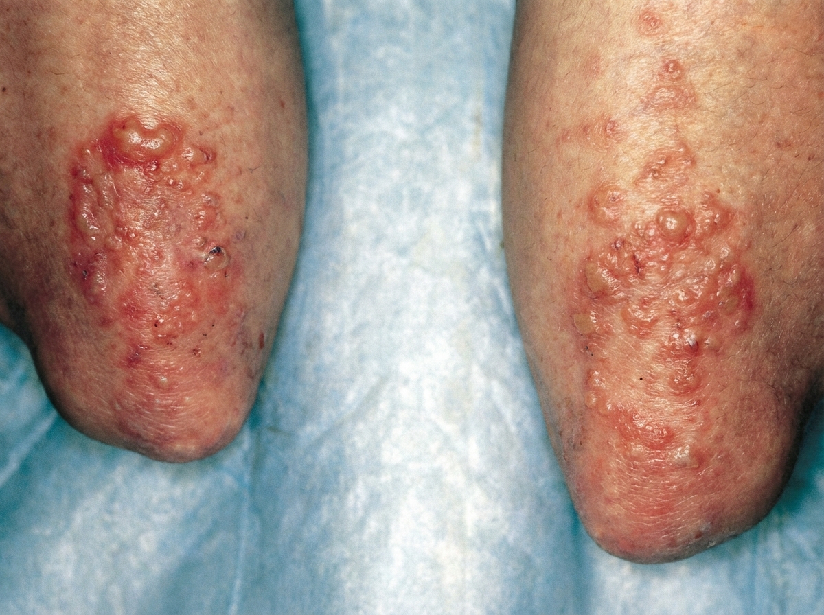
A 23-year-old woman comes to the physician because of a 2-month history of diarrhea, flatulence, and fatigue. She reports having 3–5 episodes of loose stools daily that have an oily appearance. The symptoms are worse after eating. She also complains of an itchy rash on her elbows and knees. A photograph of the rash is shown. Further evaluation of this patient is most likely to show which of the following findings?
A 28-year-old male comes to the physician for worsening back pain. The pain began 10 months ago, is worse in the morning, and improves with activity. He has also had bilateral hip pain and difficulty bending forward during exercise for the past 3 months. He has celiac disease and eats a gluten-free diet. Examination shows a limited range of spinal flexion. Flexion, abduction, and external rotation of both hips produces pain. Further evaluation of this patient is most likely to show which of the following laboratory findings?
A 13-year-old girl is admitted to the hospital due to muscle weakness, pain, and arthralgia in her wrist joints. The patient says, "I am having trouble walking home after school, especially climbing steep hills." She also complains of malaise. On physical examination, a heliotrope rash is observed around her eyes, and multiple hyperkeratotic, flat, red papules with central atrophy are present on the back of the metacarpophalangeal and interphalangeal joints. Deposits of calcium are also noted on the pads of her fingers. Her serum creatine kinase levels are elevated. Which of the following antibodies is most likely to be found in this patient?
A 38-year-old woman presents to the primary care physician with a complaint of painless hematuria over the last 5 days. History reveals that she has a 20 pack-year smoking history, and her last menses was 10 days ago. Her blood pressure is 130/80 mm Hg, heart rate is 86/min, respiratory rate is 19/min, and temperature is 36.6°C (98.0°F). Physical examination is within normal limits. Laboratory studies show: Creatinine 0.9 mg/dL Blood urea nitrogen 15 mg/dL Prothrombin time 12.0 sec Partial thromboplastin time 28.1 sec Platelet count 250,000/mm3 Urine microscopy reveals 15 RBC/HPF and no leukocytes, casts, or bacteria. Which of the following is the best next step for this patient?
A 27-year-old man comes to the physician because of worsening abdominal pain over the last several months. He has also had recent feelings of sadness and a lack of motivation at work, where he is employed as a computer programmer. He denies suicidal thoughts. He has a history of multiple kidney stones. He has a family history of thyroid cancer in his father and uncle, who both underwent thyroidectomy before age 30. His temperature is 37°C (98°F), blood pressure is 138/86 mm Hg, and pulse is 87/min. Physical examination shows diffuse tenderness over the abdomen and obesity but is otherwise unremarkable. Serum studies show: Na+ 141 mEq/L K+ 3.6 mEq/L Glucose 144 mg/dL Ca2+ 12.1 mg/dL Albumin 4.1 g/dL PTH 226 pg/mL (normal range 12–88 pg/mL) Results of a RET gene test return abnormal. The physician refers him to an endocrine surgeon. Which of the following is the most appropriate next step in diagnosis?
A 43-year-old woman comes to the physician because of worsening heartburn and abdominal pain for the past 4 months. During this period she has also had multiple episodes of greasy diarrhea. Six months ago, she had similar symptoms and was diagnosed with a duodenal ulcer. Her mother died of complications from uncontrolled hypoglycemia and had primary hyperparathyroidism. The patient does not drink alcohol or smoke cigarettes. Her only medications are pantoprazole and ranitidine. Her epigastric region is tender when palpated. An esophagogastroduodenoscopy shows a friable ulcer in the distal duodenum. Further evaluation is most likely to show which of the following?
A 58-year-old man presents to the Emergency Department after 3 hours of intense suprapubic pain associated with inability to urinate for the past day or two. His medical history is relevant for benign prostatic hyperplasia (BPH) that has been under treatment with prazosin and tadalafil. Upon admission, he is found to have a blood pressure of 180/100 mm Hg, a pulse of 80/min, a respiratory rate of 23/min, and a temperature of 36.5°C (97.7°F). He weighs 84 kg (185.1 lb) and is 175 cm (5 ft 7 in) tall. Physical exam, he has suprapubic tenderness. A bladder scan reveals 700 ml of urine. A Foley catheter is inserted and the urine is drained. Initial laboratory tests and their follow up 8 hours after admission are shown below. Admission 8 hours after admission Serum potassium 4.2 mmol/L Serum potassium 4.0 mmol/L Serum sodium 140 mmol/L Serum sodium 142 mmol/L Serum chloride 102 mmol/L Serum chloride 110 mmol/L Serum creatinine 1.4 mg/dL Serum creatinine 1.6 mg/dL Serum blood urea nitrogen 64 mg/dL Serum blood urea nitrogen 62 mg/dL Urine output 250 mL Urine output 260 mL A senior attending suggests a consultation with Nephrology. Which of the following best justifies this suggestion?
A 49-year-old man comes to the physician because of tender, red nodules that appeared on his chest 3 days ago. Three weeks ago, he had similar symptoms in his right lower limb and another episode in his left foot; both episodes resolved spontaneously. He also has diarrhea and has had a poor appetite for 1 month. He has a history of dry cough and joint pain, for which he takes albuterol and aspirin as needed. He has smoked 2 packs of cigarettes daily for 15 years. He does not drink alcohol. Physical examination shows a linear, erythematous lesion on the right anterior chest wall, through which a cord-like structure can be palpated. The lungs are clear to auscultation. The abdomen is soft, nontender, and non-distended. Examination of the legs is normal. An ultrasound of the legs shows no abnormalities. Which of the following is the most appropriate next step in diagnosis of the underlying condition?
A 21-year-old man presents to the emergency room with abdominal pain and nausea for the past 5 hours. The pain is diffusely spread and of moderate intensity. The patient also says he has not felt like eating since yesterday. He has no past medical history and is not on any medications. He regularly drinks 2–4 beers per day but does not smoke or use illicit substances. Vitals show a pulse of 120/min, a respiratory rate of 26/min, a blood pressure of 110/60 mm Hg, and a temperature of 37.8°C (100.0°F). Examination reveals a soft, diffusely tender abdomen with no guarding. Bowel sounds are present. His mucous membranes are slightly dry and there is a fruity smell to his breath. Laboratory tests show: Laboratory test pH 7.31 Serum glucose (random) 450 mg/dL Serum electrolytes Sodium 149 mEq/L Potassium 5 mEq/L Chloride 99 mEq/L Bicarbonate 16 mEq/L Serum creatinine 1.0 mg/dL Blood urea nitrogen 15 mg/dL Urinalysis Proteins Negative Glucose Positive Ketones Positive Leucocytes Negative Nitrites Negative Red blood cells (RBC) Negative Casts Negative Which of the following explains this patient's presentation?
Practice by Chapter
GERD and esophageal disorders
Practice Questions
Peptic ulcer disease
Practice Questions
Helicobacter pylori infection
Practice Questions
Celiac disease
Practice Questions
Irritable bowel syndrome
Practice Questions
Diverticular disease
Practice Questions
GI bleeding (upper and lower)
Practice Questions
Small intestinal bacterial overgrowth
Practice Questions
Malabsorption syndromes
Practice Questions
Colorectal cancer screening
Practice Questions
Functional GI disorders
Practice Questions
Anorectal disorders
Practice Questions
GI motility disorders
Practice Questions
Want unlimited practice?
Get full access to all questions, explanations, and performance tracking.
Scan to download app