
Gastroenterology — MCQs

On this page
A 35-year-old woman is presenting for a general wellness checkup. She is generally healthy and has no complaints. The patient does not smoke, drinks 1 alcoholic drink per day, and exercises 1 day per week. She recently had silicone breast implants placed 1 month ago. Her family history is notable for a heart attack in her mother and father at the age of 71 and 55 respectively. Her father had colon cancer at the age of 70. Her temperature is 99.0°F (37.2°C), blood pressure is 121/81 mmHg, pulse is 77/min, respirations are 14/min, and oxygen saturation is 98% on room air. Physical exam is unremarkable. Which of the following is the most appropriate initial step in management?
A 45-year-old woman comes to see you for a second opinion regarding an upcoming surgery for pancreatic insulinoma. While taking a surgical history, she tells you she previously had a pituitary tumor resected. For which additional neoplasms might you consider testing her?
A 19-year-old woman presents to the physician for a routine health maintenance examination. She has a past medical history of gastroesophageal reflux disease. She recently moved to a new city to begin her undergraduate studies. Her father was diagnosed with colon cancer at age 46. Her father's brother died because of small bowel cancer. Her paternal grandfather died because of stomach cancer. She takes a vitamin supplement. Current medications include esomeprazole and a multivitamin. She smoked 1 pack of cigarettes daily for 3 years but quit 2 years ago. She drinks 1–2 alcoholic beverages on the weekends. She appears healthy. Vital signs are within normal limits. Physical examination shows no abnormalities. Colonoscopy is unremarkable. Germline testing via DNA sequencing in this patient shows mutations in DNA repair genes MLH1 and MSH2. Which of the following will this patient most likely require at some point in her life?
A 31-year-old man presents with jaundice, scleral icterus, dark urine, and pruritus. He also says that he has been experiencing abdominal pain shortly after eating. He says that symptoms started a week ago and have not improved. The patient denies any associated fever or recent weight-loss. He is afebrile and vital signs are within normal limits. On physical examination, the patient’s skin appears yellowish. Scleral icterus is present. Remainder of physical examination is unremarkable. Laboratory findings are significant for: Conjugated bilirubin 5.1 mg/dL Total bilirubin 6.0 mg/dL AST 24 U/L ALT 22 U/L Alkaline phosphatase 662 U/L A contrast CT of the abdomen is unremarkable. An ultrasound of the right upper quadrant reveals a normal gallbladder, but the common bile duct is not visible. Which of the following is the next best step in the management of this patient?
A 54-year-old man comes to the physician because of dysphagia and hoarseness of voice for the past 3 months. Initially, he had difficulty swallowing solid food but now has difficulty swallowing porridge and liquids as well. He has recently been choking on his oral secretions. During this period, he has had an 8.2-kg (18-lb) weight loss. He has noticed increasing weakness of both arms over the past year. He appears ill. His temperature is 36.8°C (98.2°F), pulse is 74/min, respirations are 14/min, and blood pressure is 114/74 mmHg. Examination shows tongue atrophy and pooled oral secretions. There is diffuse muscle atrophy with occasional twitching. He is unable to lift his arms above the chest level. Deep tendon reflexes are 3+ in all extremities. Sensation to pinprick, light touch, and vibration is intact. Laboratory studies show: Hemoglobin 16.1 g/dL Leukocyte count 10,900/mm3 Erythrocyte sedimentation rate 20 mm/h Serum Na+ 133 mEq/L K+ 4.2 mEq/L Cl- 101 mEq/L Urea nitrogen 12 mg/dL Creatinine 1.1 mg/dL Creatine kinase 320 U/L Albumin 4.3 mg/dL Lactate dehydrogenase 307 U/L An esophagogastroduodenoscopy shows no abnormalities. Which of the following is the most likely cause of this patient's symptoms?
A 28-year-old graduate student visits the university health clinic for 3-weeks of epigastric pain that worsens with meals, associated with retrosternal pain, early satiety, and bloating. She denies vomiting blood or blood in her stool. She has been consuming large volumes of caffeinated-drinks and fast-food for a month, as she has been studying for her tests. Her family and personal history are unremarkable with no history of gastrointestinal cancer. Her vital signs are within normal limits. Physical examination is only positive for a mild epigastric tenderness. Which of the following is the most appropriate approach in this case?
A 60-year-old patient presents to the urgent care clinic with complaints of pain and abdominal distention for the past several weeks. The pain began with a change in bowel habits 3 months ago, and he gradually defecated less until he became completely constipated, which led to increasing pain and distention. He also mentions that he has lost weight during this period, even though he has not changed his diet. When asked about his family history, the patient reveals that his brother was diagnosed with colorectal cancer at 65 years of age. An abdominal radiograph and CT scan were done which confirmed the diagnosis of obstruction. Which of the following locations in the digestive tract are most likely involved in this patient’s disease process?
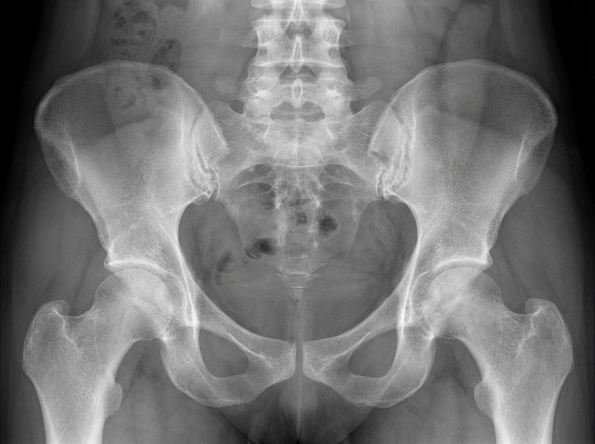
A 32-year-old woman comes to her physician because of increasing back pain for the past 10 months. The pain is worse in the morning when she wakes up and improves with activity. She used to practice yoga, but stopped 5 months ago as bending forward became increasingly difficult. She has also had bilateral hip pain for the past 4 months. She has not had any change in urination. She has celiac disease and eats a gluten-free diet. Her temperature is 37.1°C (98.8°F), pulse is 65/min, respirations are 13/min, and blood pressure is 116/72 mmHg. Examination shows the range of spinal flexion is limited. Flexion, abduction, and external rotation of bilateral hips produces pain. An x-ray of her pelvis is shown. Further evaluation of this patient is likely to show which of the following?
A 25-year-old woman presents to a physician for a new patient physical exam. Aside from occasional shin splints, she has a relatively unremarkable medical history. She takes oral contraceptive pills as scheduled and a multivitamin daily. She reports no known drug allergies. All of her age appropriate immunizations are up to date. Her periods have been regular, occurring once every 28 to 30 days with normal flow. She is sexually active with two partners, who use condoms routinely. She works as a cashier at the local grocery store. Her mother has diabetes and coronary artery disease, and her father passed away at age 45 after being diagnosed with colon cancer at age 40. Her grand-aunt underwent bilateral mastectomies after being diagnosed with breast cancer at age 60. Her physical exam is unremarkable. Which of the following is the best recommendation for this patient?
A 38-year-old man comes to the physician because of progressive pain and swelling of his left knee for the past 2 days. He has been taking ibuprofen for the past 2 days without improvement. Four days ago, he scraped his left knee while playing baseball. He has a 2-month history of progressive pain and stiffness in his back. The pain starts after waking up and lasts for 20 minutes. He has type 2 diabetes mellitus. His older sister has rheumatoid arthritis. He is 170 cm (5 ft 7 in) tall and weighs 91 kg (201 lb); BMI is 31.5 kg/m2. Temperature is 39°C (102.2°F), pulse is 90/min, and blood pressure is 135/85 mm Hg. Examination shows an erythematous, tender, and swollen left knee; range of motion is limited. There are abrasions over the lateral aspect of the left knee. The remainder of the examination shows no abnormalities. Laboratory studies show a leukocyte count of 13,500/mm3 and an erythrocyte sedimentation rate of 70 mm/h. Which of the following is the most appropriate next step in management?
Practice by Chapter
GERD and esophageal disorders
Practice Questions
Peptic ulcer disease
Practice Questions
Helicobacter pylori infection
Practice Questions
Celiac disease
Practice Questions
Irritable bowel syndrome
Practice Questions
Diverticular disease
Practice Questions
GI bleeding (upper and lower)
Practice Questions
Small intestinal bacterial overgrowth
Practice Questions
Malabsorption syndromes
Practice Questions
Colorectal cancer screening
Practice Questions
Functional GI disorders
Practice Questions
Anorectal disorders
Practice Questions
GI motility disorders
Practice Questions
Want unlimited practice?
Get full access to all questions, explanations, and performance tracking.
Scan to download app