
Gastroenterology — MCQs

On this page
A 39-year-old man comes to the emergency department because of fever, urinary frequency, and lower back pain for the last 3 days. During this period, he has also had pain with the 3 times he has defecated. He is sexually active with one female partner and does not use condoms. His father died of colon cancer at the age of 67 years. The patient has smoked one pack of cigarettes daily for 14 years and drinks alcohol occasionally. His temperature is 39.1°C (102.3°F), pulse is 114/min, and blood pressure is 140/90 mm Hg. Physical examination shows mild suprapubic pain on deep palpation and a swollen, tender prostate. The remainder of the examination shows no abnormalities. His hemoglobin concentration is 15.4 g/dL, leukocyte count is 18,400/mm3, and platelet count is 260,000/mm3. Which of the following is the most appropriate next step in the management of this patient's condition?
A 54-year-old man comes to the physician because of diarrhea that has become progressively worse over the past 4 months. He currently has 4–6 episodes of foul-smelling stools per day. Over the past 3 months, he has had fatigue and a 5-kg (11-lb) weight loss. He returned from Bangladesh 6 months ago after a year-long business assignment. He has osteoarthritis and hypertension. Current medications include amlodipine and naproxen. He appears pale and malnourished. His temperature is 37.3°C (99.1°F), pulse is 76/min, and blood pressure is 140/86 mm Hg. Examination shows pale conjunctivae and dry mucous membranes. Angular stomatitis and glossitis are present. The abdomen is distended but soft and nontender. Rectal examination shows no abnormalities. Laboratory studies show: Hemoglobin 8.9 g/dL Leukocyte count 4100/mm3 Platelet count 160,000/mm3 Mean corpuscular volume 110 μm3 Serum Na+ 133 mEq/L Cl- 98 mEq/l K+ 3.3 mEq/L Creatinine 1.1 mg/dL IgA 250 mg/dL Anti-tissue transglutaminase, IgA negative Stool culture and studies for ova and parasites are negative. Test of the stool for occult blood is negative. Fecal fat content is 22 g/day (N < 7). Fecal lactoferrin is negative and elastase is within normal limits. Which of the following is the most appropriate next step in diagnosis?
A 70-year-old man comes to the physician because of episodes of watery stools for the past 6 weeks. During this period, he has also had recurrent episodes of reddening of the face, neck, and chest that last up to 30 minutes, especially following alcohol consumption. He has hypertension. He smoked one pack of cigarettes daily for 20 years but quit 8 years ago. He drinks two glasses of wine daily. Current medications include enalapril. He appears pale. He is 185 cm (6 ft 1 in) tall and weighs 67 kg (147.7 lb); BMI is 19.6 kg/m2. His temperature is 36.7°C (98°F), pulse is 85/min, and blood pressure is 130/85 mm Hg. Scattered expiratory wheezing is heard throughout both lung fields. Cardiac examination shows no abnormalities. The abdomen is soft and mildly tender. The remainder of the physical examination shows no abnormalities. A complete blood count and serum concentrations of urea nitrogen and creatinine are within the reference range. Which of the following is the most likely diagnosis in this patient?
Please refer to the summary above to answer this question Which of the following is the most likely diagnosis? Patient information Age: 61 years Gender: F, self-identified Ethnicity: unspecified Site of care: emergency department History Reason for Visit/Chief Concern: "My belly really hurts." History of Present Illness: developed abdominal pain 12 hours ago pain constant; rated at 7/10 has nausea and has vomited twice has had two episodes of nonbloody diarrhea in the last 4 hours 12-month history of intermittent constipation reports no sick contacts or history of recent travel Past medical history: hypertension type 2 diabetes mellitus mild intermittent asthma allergic rhinitis Social history: diet consists mostly of high-fat foods does not smoke drinks 1–2 glasses of wine per week does not use illicit drugs Medications: lisinopril, metformin, albuterol inhaler, fexofenadine, psyllium husk fiber Allergies: no known drug allergies Physical Examination Temp Pulse Resp. BP O2 Sat Ht Wt BMI 38.4°C (101.1°F) 85/min 16/min 134/85 mm Hg – 163 cm (5 ft 4 in) 94 kg (207 lb) 35 kg/m2 Appearance: lying back in a hospital bed; appears uncomfortable Neck: no jugular venous distention Pulmonary: clear to auscultation; no wheezes, rales, or rhonchi Cardiac: regular rate and rhythm; normal S1 and S2; no murmurs, rubs, or gallops Abdominal: obese; soft; tender to palpation in the left lower quadrant; no guarding or rebound tenderness; normal bowel sounds Extremities: no edema; warm and well-perfused Skin: no rashes; dry Neurologic: alert and oriented; cranial nerves grossly intact; no focal neurologic deficits
A 28-year-old man comes to the physician because of a 3-month history of a recurrent pruritic rash on his face and scalp. He reports that he has been using a new shaving cream once a week for the past 5 months. A year ago, he was diagnosed with HIV and is currently receiving triple antiretroviral therapy. He drinks several six-packs of beer weekly. Vital signs are within normal limits. A photograph of the rash is shown. A similar rash is seen near the hairline of the scalp and greasy yellow scales are seen at the margins of the eyelids. Which of the following is the most likely diagnosis?

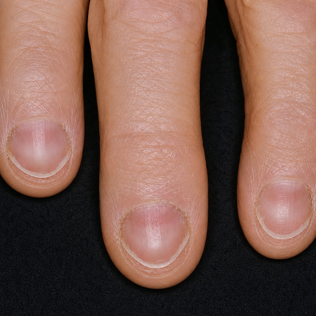
A 57-year-old man comes to the physician because of tiredness and dyspnea on exertion for several months. Recently, he has also noticed changes of his fingernails. A photograph of his nails is shown. Which of the following is the most likely underlying cause of these findings?
A 21-year-old Caucasian male presents to your office with wheezing and rhinitis. Laboratory results show peripheral eosinophilia and antibodies against neutrophil myeloperoxidase. What is the most likely diagnosis?
A 37-year-old African-American man presents to his primary care provider with a history of fatigue and nausea that started about 6 months ago. His symptoms have slowly gotten worse and now he has trouble climbing the stairs to his 3rd floor apartment without resting. Past medical history is significant for poorly controlled HIV and a remote history of heroin addiction. Today his temperature is 36.9°C (98.4°F), the blood pressure is 118/72 mm Hg, and the pulse is 75/min. Physical examination reveals morbid obesity and 1+ pitting edema of both lower extremities. Urine dipstick reveals 2+ proteinuria. Urinalysis shows no abnormal findings. Which of the following is the most likely etiology of this patient condition?
A 32-year-old man, otherwise healthy, presents with flank pain and severe nausea for the last 9 hours. He describes the pain as severe, intermittent, localized to the right flank, and radiates to the groin. His past medical history is significant for recurrent nephrolithiasis. The patient does not smoke and drinks alcohol socially. Today his temperature is 37.0°C (98.6°F), the pulse is 90/min, the respiratory rate is 25/min, and the oxygen saturation is 99% on room air. On physical examination, the patient is in pain and unable to lie still. The patient demonstrates severe costovertebral angle tenderness. The remainder of the exam is unremarkable. Non-contrast CT of the abdomen and pelvis reveals normal-sized kidneys with the presence of a single radiopaque stone lodged in the ureteropelvic junction and clusters of pyramidal medullary calcifications in both kidneys. Intravenous pyelography reveals multiple, small cysts measuring up to 0.3 cm in greatest dimension in medullary pyramids and papillae of both kidneys. Which of the following would you also most likely expect to see in this patient?
A 66-year-old woman comes to the emergency department because of a 1-day history of severe abdominal pain, nausea, and vomiting. She has also had profuse watery diarrhea with streaks of blood for the past 5 days. She had a urinary tract infection 3 weeks ago and was treated with a 14-day course of ciprofloxacin. She appears in severe distress. Her temperature is 39.3°C (102.7°F), pulse is 110/min, and blood pressure is 100/60 mm Hg. Examination shows a distended abdomen, tenderness in the lower quadrants, and hypoactive bowel sounds; rebound tenderness and abdominal rigidity are absent. Cardiopulmonary examination shows no abnormalities. Test of the stool for occult blood is positive. Laboratory studies show: Hemoglobin 10.2 g/dL Leukocyte count 28,000/mm3 Serum Na+ 133 mEq/L K+ 3.3 mEq/L Cl- 97 mEq/L Glucose 98 mg/dL Creatinine 1.3 mg/dL Two wide bore needles are inserted and intravenous fluids are administered. An abdominal x-ray of the patient would be most likely to show which of the following?
Practice by Chapter
GERD and esophageal disorders
Practice Questions
Peptic ulcer disease
Practice Questions
Helicobacter pylori infection
Practice Questions
Celiac disease
Practice Questions
Irritable bowel syndrome
Practice Questions
Diverticular disease
Practice Questions
GI bleeding (upper and lower)
Practice Questions
Small intestinal bacterial overgrowth
Practice Questions
Malabsorption syndromes
Practice Questions
Colorectal cancer screening
Practice Questions
Functional GI disorders
Practice Questions
Anorectal disorders
Practice Questions
GI motility disorders
Practice Questions
Want unlimited practice?
Get full access to all questions, explanations, and performance tracking.
Scan to download app