
Gastroenterology — MCQs

On this page
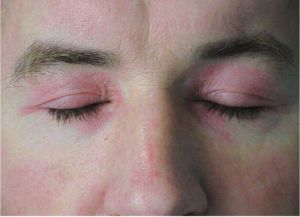
A 43-year-old woman comes to the physician because of a 3-week history of progressive weakness. She has had increased difficulty combing her hair and climbing stairs. She has hypertension. She has smoked a pack of cigarettes daily for 25 years. She does not drink alcohol. Her mother had coronary artery disease and systemic lupus erythematosus. Her current medications include chlorthalidone and vitamin supplements. Her temperature is 37.8°C (100.0°F), pulse is 71/min, and blood pressure is 132/84 mm Hg. Cardiopulmonary examination is unremarkable. A rash is shown that involves both her orbits. Skin examination shows diffuse erythema of the upper back, posterior neck, and shoulders. Which of the following antibodies are most likely to be present in this patient?
A 35-year old Caucasian woman visits a community clinic and is presenting with a long history of early satiety, diarrhea, fatigue, hair loss, and brittle nails. Her family history is insignificant. Her personal history is relevant for iron deficiency anemia and vitamin B12 deficiency, as seen in her lab reports a few months back. Her physical examination is unremarkable except for pale skin and mucous surfaces, and glossitis. She brings with herself an upper endoscopy report describing body and fundal atrophic gastritis. Which of the following tests would you expect to be positive in this patient?
A 52-year-old man presents to the emergency department with severe pain of the left first metatarsophalangeal joint. He says that the pain started 3 hours ago and describes it as sharp in character. The pain has been so severe that he has not been able to tolerate any movement of the joint. His past medical history is significant for hypertension for which he takes a thiazide diuretic. His diet consists primarily of red meat, and he drinks 5 bottles of beer per night. On physical exam, his left first metatarsophalangeal joint is swollen, erythematous, and warm to the touch. Which of the following characteristics would be seen with the most likely cause of this patient's symptoms?
A 46-year-old woman presents to your medical office complaining of ‘feeling tired’. The patient states that she has been having some trouble eating because her ‘tongue hurts’, but she has no other complaints. On examination, the patient has pale conjunctiva and skin and also appears tired. She has a smooth, red tongue that is tender to touch with a tongue depressor. The patient’s hands and feet feel cold. Fluoroscopic evaluation of the swallowing mechanism and esophagus is normal. Which of the following diagnoses is most likely?
A 53-year-old man comes to the emergency department for severe left knee pain for the past 8 hours. He describes it as an unbearable, burning pain that woke him up from his sleep. He has been unable to walk since. He has not had any trauma to the knee. Ten months ago, he had an episode of acute pain and swelling of the right great toe that subsided after treatment with indomethacin. He has hypertension, type 2 diabetes mellitus, psoriasis, and hyperlipidemia. Current medications include topical betamethasone, metformin, glipizide, losartan, and simvastatin. Two weeks ago, hydrochlorothiazide was added to his medication regimen to improve blood pressure control. He drinks 1–2 beers daily. He is 170 cm (5 ft 7 in) tall and weighs 110 kg (242 lb); BMI is 38.1 kg/m2. His temperature is 38.4°C (101.1°F). Examination shows multiple scaly plaques over his palms and soles. The left knee is erythematous, swollen, and tender; range of motion is limited by pain. Which of the following is the most appropriate next step in management?
A 22-year-old female college student comes to your clinic to establish care. She has no significant past medical history and her only complaint today is that she has had trouble maintaining a consistent weight. Her temperature is 98.6°F (37.0°C), blood pressure is 100/65 mmHg, pulse is 62/min, and respirations are 12/min. Her body mass index is 19.5. Her physical exam is significant for callused knuckles and dental enamel erosions. What laboratory abnormalities are likely to be found in this patient?
A 49-year-old man comes with odynophagia, abdominal pain, fatigue, headache, and fever for several weeks. The patient reports no chronic medical problems, no travel, and no recent sick exposures. Physical examination is significant only for an erythematous oral mucosa and cervical lymphadenopathy. His vital signs show a blood pressure of 121/72 mm Hg, heart rate of 82/min, and respiratory rate of 16/min. On a review of systems, the patient reports regular, unprotected sexual encounters with men and women. Of the following options, which disease must be excluded?
A 27-year old woman comes to the physician for a rash that began 5 days ago. The rash involves her abdomen, back, arms, and legs, including her hands and feet. Over the past month, she has also had mild fever, headache, and myalgias. She has no personal history of serious illness. She smokes 1 pack of cigarettes a day and binge drinks on the weekends. She uses occasional cocaine, but denies other illicit drug use. Vital signs are within normal limits. Physical examination shows a widespread, symmetric, reddish-brown papular rash involving the trunk, upper extremities, and palms. There is generalized, nontender lymphadenopathy. Skin examination further shows patchy areas of hair loss on her scalp and multiple flat, broad-based, wart-like papules around her genitalia and anus. Rapid plasma reagin and fluorescent treponemal antibody test are both positive. In addition to starting treatment, which of the following is the most appropriate next step in management?
A 70-year-old man presents with severe abdominal pain over the last 24 hours. He describes the pain as severe and associated with diarrhea, nausea, and vomiting. He says he has had a history of postprandial abdominal pain over the last several months. The patient denies any fever, chills, recent antibiotic use. Past medical history is significant for peripheral arterial disease and type 2 diabetes mellitus. The patient reports a 20 pack-year smoking history. His vital signs include blood pressure 90/60 mm Hg, pulse 100/min, respiratory rate 22/min, temperature 38.0°C (100.5°F), and oxygen saturation of 98% on room air. On physical examination, the patient is ill-appearing. His abdomen is severely tender to palpation and distended with no rebound or guarding. Pain is disproportionate to the exam findings. Rectal examination demonstrates bright red-colored stool. Abdominal X-ray is unremarkable. Stool culture was negative for C. difficile. A contrast-enhanced CT scan reveals segmental colitis involving the distal transverse colon. Which of the following is the most likely cause of this patient's symptoms?
A 66-year-old woman is brought to the emergency department because of fever, chills, night sweats, and progressive shortness of breath for 1 week. She also reports generalized fatigue and nausea. She has type 2 diabetes mellitus and hypothyroidism. Current medications include metformin, sitagliptin, and levothyroxine. She appears ill. Her temperature is 38.7°C (101.7°F), pulse is 104/min, and blood pressure is 160/90 mm Hg. Examination shows pale conjunctivae and small nontender hemorrhagic macules over her palms and soles. Crackles are heard at both lung bases. A grade 2/6 mid-diastolic murmur is heard best at the third left intercostal space and is accentuated by leaning forward. The spleen is palpated 1–2 cm below the left costal margin. Laboratory studies show: Hemoglobin 10.6 g/dL Leukocyte count 18,300/mm3 Erythrocyte sedimentation rate 48 mm/h Urine Protein 1+ Blood 2+ RBCs 20-30/hpf WBCs 0-2/hpf An echocardiography shows multiple vegetations on the aortic valve. Blood cultures grow S. gallolyticus. She is treated with ampicillin and gentamicin for 2 weeks and her symptoms resolve. A repeat echocardiography at 3 weeks shows mild aortic regurgitation with no vegetations. Which of the following is the most appropriate next step in management?
Practice by Chapter
GERD and esophageal disorders
Practice Questions
Peptic ulcer disease
Practice Questions
Helicobacter pylori infection
Practice Questions
Celiac disease
Practice Questions
Irritable bowel syndrome
Practice Questions
Diverticular disease
Practice Questions
GI bleeding (upper and lower)
Practice Questions
Small intestinal bacterial overgrowth
Practice Questions
Malabsorption syndromes
Practice Questions
Colorectal cancer screening
Practice Questions
Functional GI disorders
Practice Questions
Anorectal disorders
Practice Questions
GI motility disorders
Practice Questions
Want unlimited practice?
Get full access to all questions, explanations, and performance tracking.
Scan to download app