Expression of an mRNA encoding for a soluble form of the Fas protein prevents a cell from undergoing programmed cell death. However, after inclusion of a certain exon, this same Fas pre-mRNA eventually leads to the translation of a protein that is membrane bound, subsequently promoting the cell to undergo apoptosis. Which of the following best explains this finding?
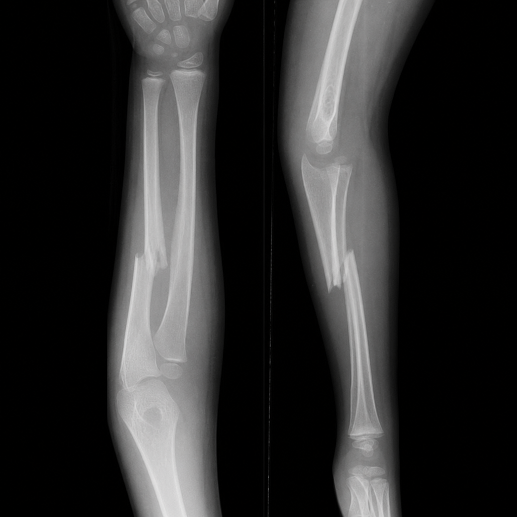
An investigator is studying the rate of wound healing by secondary intention. He performs a biopsy of a surgically debrided wound 1 day and 5 days after the initial surgical procedure. The second biopsy shows wound contraction, endothelial cell proliferation, and accumulation of macrophages. The cells responsible for wound contraction also secrete a protein that assembles in supercoiled triple helices. In which of the following structures does this protein type play an important structural role?
During protein translation, the triplet code of mRNA is read by a ribosome and assisted by elongation and translation factors until it reaches a stop codon (UAA, UAG, or UGA). When a stop codon is reached, a release factor binds, removing the peptide from the active ribosome and completing translation. What will happen if a mutation causes the recruitment of a release factor prior to the completion of a full peptide?
A researcher is trying to decipher how mRNA codons contain information about proteins. He first constructs a sequence of all cytosine nucleotides and sees that a string of prolines is synthesized. He knows from previous research that information is encoded in groups of 3 so generates the following sequences: ACCACCACC, CACCACCAC, and CCACCACCA. Surprisingly, he sees that new amino acids are produced with the first two sequences but that the third sequence is still a string of prolines. Which of the following biochemical principles explains why this phenomenon was observed?
A 3-year-old boy is diagnosed with an alpha-gal allergy, also known as mammalian meat allergy (MMA). This condition is mediated by a reaction to the carbohydrate, galactose-alpha-1,3-galactose. An experimental treatment has been developed to halt the N-linked oligosaccharide addition that occurs in the synthesis of this compound. Which of the following cellular structures is most likely targeted by this experimental drug?
A scientist wants to determine if a specific fragment is contained within genome X. She uses a restriction enzyme to digest the genome into smaller fragments to run on an agarose gel, with the goal of separating the resulting fragments. A nitrocellulose blotting paper is then used to transfer the fragments from the agarose gel. A radiolabeled probe containing a complementary sequence to the fragment she is searching for is incubated with the blotting paper. Which of the following is the RNA equivalent of this technique?
A 21-year-old man comes to the physician because of a 3-week history of yellow discoloration of his skin, right upper abdominal pain, and fatigue. Two years ago, he underwent right-sided pleurodesis for recurrent spontaneous pneumothorax. Pulmonary examination shows mild bibasilar crackles and expiratory wheezing. Laboratory studies show an elevation of serum transaminases. Histopathological examination of a tissue specimen obtained on liver biopsy shows PAS-positive globules within periportal hepatocytes. Genetic analysis shows substitution of lysine for glutamic acid at position 342 of a gene located on chromosome 14 that encodes for a protease inhibitor (Pi). This patient most likely has which of the following Pi genotypes?
A researcher is tracing the fate of C-peptide, a product of preproinsulin cleavage. Which of the following is a true statement regarding the fate of C-peptide?
A 21-year-old woman comes to the physician for an annual health maintenance examination. She has no particular health concerns. Laboratory studies show: Hemoglobin 11.2 g/dL Mean corpuscular volume 74 μm3 Mean corpuscular hemoglobin concentration 30% Hb/cell Red cell distribution width 14% (N=13–15) Genetic analysis shows a point mutation in intron 1 of a gene on the short arm of chromosome 11. A process involving which of the following components is most likely affected in this patient?
An investigator is studying the activity of N-terminal peptidase in eukaryotes. Sulfur-containing amino acids are radiolabeled and isolated using 35S. During translation of a non-mitochondrial human genome, some of the radiolabeled amino acids bind to the aminoacyl, peptidyl, and exit sites of a eukaryotic ribosome but others bind only to the peptidyl and exit sites. Only the radiolabeled amino acids that do not bind to the ribosomal aminoacyl-site can be excised by the N-terminal peptidase. Which of the following best describes the anticodon sequence of the transfer RNA charged by the amino acid target of the N-terminal peptidase?
Explanation: ***Alternative splicing*** - The scenario describes a single **pre-mRNA** producing two different protein forms (soluble vs. membrane-bound Fas) with distinct functions, depending on the inclusion or exclusion of a specific **exon**. This is the hallmark of alternative splicing. - **Alternative splicing** allows a single gene to encode multiple protein isoforms, leading to diverse cellular functions and regulation. *Histone deacetylation* - **Histone deacetylation** is a mechanism of gene regulation that typically represses gene expression by making DNA less accessible for transcription, not by altering the protein product of an already transcribed gene. - It affects whether a gene is turned "on" or "off," but doesn't explain how the same pre-mRNA produces different protein versions. *DNA missense mutation* - A **DNA missense mutation** would alter a single base pair in the DNA, potentially changing one amino acid in the resulting protein. - While it can lead to functional changes in a protein, it would not explain the complete inclusion or exclusion of an entire exon, which profoundly changes the protein's overall structure and membrane association in this manner. *Base excision repair* - **Base excision repair** is a DNA repair pathway that corrects small, non-bulky DNA lesions, such as damaged or modified bases. - This process is involved in maintaining genomic integrity and does not explain the differential processing of an mRNA transcript to produce two distinct protein isoforms. *Post-translational modifications* - **Post-translational modifications** (PTMs) occur after protein translation and involve chemical changes to the protein (e.g., phosphorylation, glycosylation). - While PTMs can alter protein function or localization, they do not explain how an entire exon's inclusion or exclusion leads to fundamentally different protein structures (soluble vs. membrane-bound).
Explanation: ***Reticular fibers*** - Wound contraction is mediated by **myofibroblasts**, which secrete collagen (primarily **Type I** and **Type III collagen**). - **Type III collagen** forms **reticular fibers**, which provide structural support in early wound healing and are prominent in tissues such as lymphoid organs, liver, bone marrow, and blood vessel walls. - Reticular fibers create a delicate meshwork framework that supports cellular elements in these organs. *Basal lamina* - The basal lamina is primarily composed of **Type IV collagen**, laminin, and proteoglycans. - It functions as a selective barrier and structural support for epithelial cells, not the site for Type III collagen/reticular fibers. *Dentin* - **Dentin** is mainly composed of **Type I collagen** and hydroxyapatite crystals. - While collagen is a major structural component, it is primarily **Type I**, not **Type III collagen** that forms reticular fibers. *Corneal stroma* - The corneal stroma contains primarily **Type I collagen** arranged in highly organized lamellae to maintain transparency. - It also contains **Type V and Type VI collagen** but not **Type III collagen** as the primary structural component. *Nucleus pulposus* - The **nucleus pulposus** is mainly composed of water, proteoglycans, and **Type II collagen**. - Its function is to resist compressive forces in the intervertebral disc, not related to Type III collagen or reticular fiber formation.
Explanation: ***Nonsense mutation*** - A nonsense mutation introduces a **premature stop codon** into the mRNA sequence. - This results in the **premature termination of translation**, leading to a truncated and often non-functional protein. *Single nucleotide polymorphism* - A SNP is a **variation at a single nucleotide position** in the DNA sequence. - While a SNP can cause a nonsense mutation, the term itself only describes the **type of sequence variation**, not its functional consequence as premature termination. *Frameshift mutation* - A frameshift mutation occurs due to the **insertion or deletion of nucleotides** not in multiples of three. - This alters the **reading frame** and typically leads to a completely different amino acid sequence downstream and often a premature stop codon, but the direct cause of premature termination here is a specific stop codon, not a shift in frame. *Basepair wobble* - **Wobble pairing** refers to the flexibility in base pairing between the third nucleotide of a codon and the first nucleotide of an anticodon. - This allows a single tRNA to recognize **multiple codons** for the same amino acid and is a normal part of translation, not a mutation causing premature termination. *Missense mutation* - A missense mutation results in a **single nucleotide change** that codes for a different amino acid. - This leads to a **substitution of one amino acid for another** in the protein, but does not typically cause premature termination of translation.
Explanation: ***Reading frame shifts*** - The different sequences (ACCACCACC, CACCACCAC, CCACCACCA) represent different **reading frames** for the original poly-C mRNA. Shifting the starting point by one or two nucleotides changes which codons are read. - The first sequence (ACCACCACC) is read as ACC-ACC-ACC, which codes for **threonine**. - The second sequence (CACCACCAC) is read as CAC-CAC-CAC, which codes for **histidine**. - The third sequence (CCACCACCA) is read as CCA-CCA-CCA, which still codes for **proline** (both CCC and CCA are proline codons), explaining why the same amino acid string is produced. - This demonstrates how the **reading frame** determines which amino acids are synthesized from the same nucleotide sequence. *mRNA splicing* - **mRNA splicing** is the process by which introns are removed from pre-mRNA, and exons are joined together. This occurs before translation. - While it affects the final mRNA sequence, it does not explain why different amino acids appear from different **starting positions** of essentially the same base sequence during translation. *Trimming of proteins* - **Protein trimming** is a post-translational modification where a part of the protein is cleaved to form the active protein. - This process occurs after the protein has been synthesized and does not influence which amino acids are incorporated based on the mRNA sequence. *Covalent alterations* - **Covalent alterations** refer to post-translational modifications of proteins, such as phosphorylation, glycosylation, or acetylation, which change the protein's chemical properties. - These modifications happen after translation is complete and do not explain the initial amino acid sequence determined by the mRNA codons. *Translational proofreading* - **Translational proofreading** mechanisms ensure the accuracy of protein synthesis by correcting errors during amino acid incorporation. - While important for fidelity, proofreading does not explain how different amino acids are produced from a single nucleotide sequence depending on the **starting position of translation**.
Explanation: ***Golgi apparatus*** - The **alpha-gal epitope (galactose-alpha-1,3-galactose)** is synthesized by **alpha-1,3-galactosyltransferase (GGTA1)** enzyme in the **Golgi apparatus**. - This is a **terminal glycosylation modification** that occurs during the late stages of glycoprotein and glycolipid processing in the Golgi. - While N-linked glycosylation begins in the RER, the specific **alpha-gal moiety is added in the Golgi** during final carbohydrate processing. - Targeting the Golgi apparatus would effectively halt the synthesis of the alpha-gal epitope responsible for mammalian meat allergy. *Rough endoplasmic reticulum* - The **RER** is where **initial N-linked glycosylation** occurs, with the addition of a core oligosaccharide to asparagine residues. - However, the alpha-gal epitope is a **terminal modification** added later in the Golgi, not during the initial RER glycosylation steps. - Blocking N-linked glycosylation in the RER would affect many essential proteins broadly, rather than specifically targeting alpha-gal synthesis. *Tumor suppressor p53* - **Tumor suppressor p53** is a protein involved in regulating the cell cycle and apoptosis, acting as a crucial defense against cancer. - It has no direct role in the synthesis or modification of carbohydrates or the specific mechanism of alpha-gal allergy. *Sodium-potassium pump* - The **sodium-potassium pump** is a transmembrane protein responsible for maintaining ion gradients across cell membranes, essential for nerve impulses and cellular volume regulation. - This pump is unrelated to carbohydrate synthesis pathways or the alpha-gal epitope. *Proteasome* - The **proteasome** is a protein complex responsible for degrading cellular proteins that are damaged or no longer needed. - It is involved in protein turnover and quality control and plays no role in the synthesis of carbohydrate moieties.
Explanation: **Northern blot** - The technique described in the question, involving **restriction enzyme digestion**, **agarose gel electrophoresis**, **blotting onto a membrane**, and **hybridization with a labeled probe**, is characteristic of a **Southern blot** for DNA - The **Northern blot** is the analogous technique used to detect and quantify **RNA** sequences, following the same principles of separation by size and detection by hybridization with a complementary probe - Both Southern and Northern blots use the same workflow: separate nucleic acids by size on gel → transfer to membrane → detect with complementary probe *RT-PCR* - **Reverse transcriptase polymerase chain reaction (RT-PCR)** is used to amplify specific **RNA** sequences by first converting **RNA** into **complementary DNA (cDNA)** using reverse transcriptase, followed by standard PCR - Unlike Northern blot, it is an **amplification technique** rather than a direct visualization method via blotting *Western blot* - **Western blot** is a technique used to detect and identify specific **proteins**, not nucleic acids - It involves **gel electrophoresis** to separate proteins by size, followed by transfer to a membrane and detection using **antibodies** rather than nucleic acid probes *qPCR* - **Quantitative polymerase chain reaction (qPCR)**, also known as real-time PCR, is a technique used to **quantify DNA or RNA** (after reverse transcription) in real-time - It measures the accumulation of fluorescent signal during the PCR reaction, allowing for real-time monitoring and quantification, which is fundamentally different from a blotting technique *Southern blot* - The description in the question *is* a **Southern blot**, which is used for **DNA** detection, not RNA - Since the question asks for the **RNA equivalent** of the described technique, and Southern blot detects DNA, Northern blot is the correct answer
Explanation: ***PiZZ*** - The patient's presentation with **yellow discoloration (jaundice)**, **right upper abdominal pain**, **elevated transaminases**, and **PAS-positive globules in periportal hepatocytes** is classic for **alpha-1 antitrypsin deficiency** (AATD) causing **liver disease**. - The genetic analysis confirming a **lysine for glutamic acid substitution at position 342** in the **protease inhibitor (Pi) gene** (SERPINA1) specifically describes the **Z allele**. The combination of two Z alleles (**PiZZ**) leads to severe deficiency and is the most common genotype associated with significant liver and lung disease. *PiSS* - The **S allele** results from a glutamic acid to valine substitution, leading to a moderately reduced level of AAT. - While it can manifest as AATD, particularly in combination with the Z allele (PiSZ), **PiSS genotype** generally confers only a mild deficiency, and liver disease is less common or severe than with PiZZ. *PiMZ* - This genotype involves one **M allele** (normal) and one **Z allele** (deficient). - It is a **heterozygous carrier state** and typically results in intermediate AAT levels. While there is an increased risk for emphysema, severe liver disease is unlikely. *PiMS* - This genotype involves one **M allele** (normal) and one **S allele** (mildly deficient). - This combination is usually associated with **normal or near-normal AAT levels** and individuals are typically asymptomatic, not presenting with severe liver or lung disease. *PiSZ* - This genotype involves one **S allele** (moderately deficient) and one **Z allele** (severely deficient). - Individuals with this genotype have **significantly reduced AAT levels** and are at risk for both liver disease and emphysema, but the severity is generally less profound than with PiZZ. The specific genetic mutation described in the stem (lysine for glutamic acid at position 342) points directly to the Z allele, not the S allele.
Explanation: ***C-peptide is packaged with insulin in secretory vesicles*** - Preproinsulin is cleaved in the **endoplasmic reticulum** to proinsulin (signal peptide removal), which is then transported to the **Golgi apparatus**. - In the Golgi, proinsulin is cleaved by **prohormone convertases** into **insulin** and **C-peptide**, and both are stored together in **secretory vesicles** within the pancreatic beta cells. - Upon stimulation, both insulin and C-peptide are **co-secreted** via exocytosis in equimolar amounts, making C-peptide a useful marker of endogenous insulin secretion. *C-peptide exits the cells via a protein channel* - C-peptide exits the beta cells via **exocytosis** of secretory granules, not through specific protein channels. - It is **co-secreted with insulin** when secretory vesicles fuse with the plasma membrane. - Its presence in the bloodstream in equimolar amounts with insulin makes it an indirect measure of **insulin secretion**. *C-peptide is further cleaved into insulin* - **C-peptide** is a product of proinsulin cleavage, alongside insulin; it is not further processed into insulin. - Insulin itself is composed of two **peptide chains (A and B)** linked by disulfide bonds, formed after C-peptide is removed from proinsulin. *C-peptide is immediately degraded by the proteasome* - C-peptide is not immediately degraded by the **proteasome** upon synthesis. - After secretion, it circulates in the blood with a **longer half-life** than insulin (approximately 30 minutes versus 4-6 minutes), allowing it to be a useful marker of endogenous insulin production. - Its degradation occurs primarily in the **kidney**. *C-peptide activates an intracellular signaling cascade* - While there is some research suggesting C-peptide may have independent **biological activity** and activate certain signaling pathways extracellularly, its primary role in the context of the insulin synthesis pathway is as a **byproduct** of proinsulin processing. - Its clinical utility is primarily as a **biomarker** of endogenous insulin secretion, particularly useful in distinguishing between endogenous and exogenous insulin in diabetic patients.
Explanation: ***Small nuclear ribonucleoprotein*** - The patient's lab results (low 11.2 g/dL **hemoglobin**, low 74 µm3 **MCV**, and low 30% **MCHC**) indicate **microcytic, hypochromic anemia**, consistent with **thalassemia**. - A point mutation in **intron 1** of a gene suggests a problem with **RNA splicing**, which is mediated by **small nuclear ribonucleoproteins (snRNPs)** as part of the spliceosome. *TATA-rich nucleotide sequence* - The **TATA box** is located in the **promoter region** of genes and is involved in the initiation of **transcription**, not splicing. - A mutation in the TATA box would affect the **rate of transcription** or gene expression, not the processing of mRNA after transcription. *Transfer RNA* - **tRNA** molecules are essential for **protein translation** by carrying specific amino acids to the ribosome. - A problem with tRNA would affect the **synthesis of proteins**, not the processing of pre-mRNA. *Heat shock protein 60* - **Heat shock proteins** are molecular **chaperones** involved in the proper **folding of proteins** and preventing protein aggregation. - A defect in HSP60 would lead to misfolded proteins, not impaired mRNA splicing. *MicroRNA* - **MicroRNAs (miRNAs)** are small non-coding RNA molecules that regulate gene expression by **silencing mRNA** or inhibiting **translation**. - While miRNAs regulate gene expression, they are not directly involved in the **splicing of introns** from pre-mRNA.
Explanation: ***5'-CAU-3'*** - The N-terminal peptidase excises amino acids that do not bind to the ribosomal **aminoacyl-site (A-site)**, indicating they are **initiator methionines**. During translation initiation in eukaryotes, the **initiator tRNA** (Met-tRNAi carrying methionine) directly binds to the **peptidyl-site (P-site)**, bypassing the A-site. - The **start codon** for protein synthesis is **5'-AUG-3'**. The anticodon must be **complementary and antiparallel** to this codon. When AUG is read 3'-GUA-5' in the antiparallel direction, the complementary anticodon sequence is **5'-CAU-3'**. - This is the standard anticodon for **initiator methionine tRNA (Met-tRNAi)** in eukaryotes. After translation begins, the N-terminal methionine is often cleaved by methionine aminopeptidase (N-terminal peptidase), especially when the second amino acid is small and uncharged. *5'-ACA-3'* - This anticodon would pair with the codon **5'-UGU-3'** or **5'-UGC-3'**, both of which code for **cysteine**, not methionine. - While cysteine is a sulfur-containing amino acid that was radiolabeled in this experiment, it undergoes normal translation through the A-site and is not the initiator amino acid that bypasses the A-site. - Cysteine residues are not typically removed by N-terminal peptidase. *5'-UCA-3'* - This anticodon would pair with the codon **5'-AGU-3'** or **5'-AGA-3'**, which code for **serine** or **arginine**. - Neither of these is the initiator amino acid, and they do not contain sulfur, so they would not be radiolabeled with 35S in this experiment. *5'-UAC-3'* - This sequence represents the **mRNA codon** (5'-UAC-3' codes for tyrosine), not an anticodon. - The anticodon should be complementary to AUG, not identical to a tyrosine codon. This is an incorrect representation. *5'-ACU-3'* - This anticodon would pair with the codon **5'-AGU-3'** or **5'-AGC-3'**, which code for **serine**. - Serine does not contain sulfur and would not be radiolabeled with 35S. It is also not the initiator amino acid that bypasses the A-site.
Explanation: ***Insufficient CFTR channel production*** - The most common CFTR gene mutation, **delta F508**, leads to the production of a misfolded CFTR protein that is retained in the endoplasmic reticulum and subsequently **degraded**, significantly reducing the number of functional channels reaching the cell surface. - This **protein misfolding** and degradation results in insufficient delivery of CFTR channels to the apical membrane of epithelial cells, causing the characteristic electrolyte and fluid transport defects. *Excess CFTR channel production* - Cystic fibrosis is characterized by a *deficiency* in functional CFTR channels, not an excess. - An overproduction of functional CFTR channels would lead to excessive chloride transport, which is the opposite of what is observed in CF. *Defective post-translational hydroxylation of the CFTR channel* - **Hydroxylation** is a common post-translational modification, but it is not the primary defect responsible for the pathogenesis of the most common CFTR mutation. - Defects in hydroxylation are more typically associated with conditions like **scurvy** (collagen hydroxylation) or issues with protein stability mediated by hydroxylases. *Defective post-translational phosphorylation of the CFTR channel* - While CFTR channel activity is regulated by **phosphorylation** by protein kinase A, the primary defect in **delta F508 CFTR** is not a failure of phosphorylation itself. - The problem is that the misfolded protein never reaches the cell surface to be properly phosphorylated and activated, making phosphorylation a secondary issue rather than the root cause of the channel deficiency. *Defective post-translational glycosylation of the CFTR channel* - **Glycosylation** is an important aspect of CFTR protein maturation, occurring in the endoplasmic reticulum and Golgi. - In the case of the **delta F508 mutation**, the misfolded protein is largely *prevented* from reaching the Golgi, where it would undergo complex glycosylation, so the defect is more fundamental (misfolding and degradation) rather than an error in the glycosylation process itself.
Explanation: ***TATA Box*** - The **TATA box** is a core promoter element found in eukaryotic genes, typically located **25-35 base pairs upstream** of the transcription start site. - It plays a crucial role in initiating transcription by serving as a binding site for **transcription factors**, which in turn recruit **RNA polymerase II**. *RNA polymerase II* - **RNA polymerase II** is the enzyme responsible for transcribing protein-coding genes into mRNA. - While essential for transcription, it is an enzyme that binds to the promoter region (which includes the TATA box), rather than a regulatory DNA sequence itself. *Small nuclear ribonucleoprotein (SnRNPs)* - **SnRNPs** are components of the spliceosome, involved in the **splicing of pre-mRNA** to remove introns. - They are involved in post-transcriptional modification, not in the initiation of transcription. *DNA methyltransferase* - **DNA methyltransferase** is an enzyme involved in **DNA methylation**, a process that typically represses gene expression. - This enzyme modifies DNA, but it is not a DNA region that promotes transcription initiation. *CAAT Box* - The **CAAT box** is another common promoter element in eukaryotes, usually located further **upstream (70-80 base pairs)** from the transcription start site. - While it also binds transcription factors and influences transcription initiation, its location is generally *more distant* than the 28 bases upstream described, making the TATA box a more accurate fit for the given distance.
Explanation: ***A tRNA with the UUU anticodon can bind to either AAA or AAG codons*** - The **wobble phenomenon** allows for non-standard base pairing between the **first nucleotide (5' position) of the tRNA anticodon** and the **third nucleotide (3' position) of the mRNA codon**. - In this example, a tRNA with anticodon **3'-UUU-5'** can bind to either **5'-AAA-3'** or **5'-AAG-3'** codons (both encoding lysine) due to the relaxed base-pairing rules at the wobble position. - This flexibility means fewer tRNAs are needed to recognize all 61 sense codons, illustrating the **degeneracy of the genetic code**. - According to Crick's wobble hypothesis, **U at the 5' position of the anticodon** can pair with either **A or G at the 3' position of the codon**. *Charged tRNA contains energy needed for peptide bonds to form* - While **charged tRNA** (aminoacyl-tRNA) does carry an amino acid activated for peptide bond formation, this statement describes the energy source for translation, not the wobble phenomenon. - The energy for peptide bond formation comes from the **high-energy ester bond** linking the amino acid to the tRNA, not from the base pairing itself. *The last nucleotide provides specificity for the given amino acid* - The **last nucleotide** (3' position) of the mRNA codon is where **wobble pairing** occurs, meaning it does *not* always provide strict specificity for the amino acid due to the relaxed base-pairing rules. - It is often the *first two nucleotides* of the codon that are most critical in determining the specific amino acid incorporated. *There are more amino acids than possible codons* - This statement is incorrect; there are **20 standard amino acids** and **61 sense codons** (three are stop codons), meaning there are more codons than amino acids, leading to **code degeneracy**. - The concept of wobble base pairing helps explain how this degeneracy is managed efficiently, but the premise of this option is false. *The genetic code is preserved without mutations* - This statement refers to the **fidelity of DNA replication and repair** or the evolutionary conservation of the genetic code, not the mechanism of translation or wobble base pairing. - The genetic code being largely universal and degenerate does not mean that mutations never occur, but rather that it is robust.
Explanation: ***Degeneracy*** - The finding that a single tRNA anticodon (with **inosine** at the 5' position) can bind to multiple different mRNA codons (ending in **adenine, cytosine, or uracil**) illustrates the concept of **degeneracy** in the genetic code. - This **wobble hypothesis** allows fewer tRNAs to recognize more than one codon for a given amino acid, meaning multiple codons can code for the same amino acid. *Unambiguity* - The genetic code is unambiguous, meaning that each codon specifies **only one specific amino acid** (or a stop signal) and never two different amino acids. - This finding, however, shows one tRNA recognizing multiple codons, not one codon coding for multiple amino acids. *Non-overlapping* - The **non-overlapping** nature of the genetic code means that each nucleotide in an mRNA sequence is read only once as part of a single codon, without sharing nucleotides between adjacent codons. - This concept describes how codons are read sequentially, not the flexibility of codon-anticodon pairing. *Specificity of the start codon* - The **start codon (AUG)** specifically initiates translation, coding for methionine, and signals the beginning of a polypeptide chain. - This finding relates to the wobble pairing at the 3' end of the codon, not the initiation of translation. *Specificity of stop codons* - **Stop codons (UAA, UAG, UGA)** specifically signal the termination of translation without coding for any amino acid. - This finding describes the flexibility of codon-anticodon pairing, not the distinct function of termination codons.
Explanation: ***Intracellular retention of misfolded proteins*** - The combination of **recurrent respiratory infections**, **chronic diarrhea with greasy stools**, and **failure to thrive (low height/weight percentiles)** in a young child strongly suggests **cystic fibrosis (CF)**. - CF is caused by mutations in the **CFTR gene**, leading to the production of a **misfolded CFTR protein** that is retained in the endoplasmic reticulum and subsequently degraded, preventing it from reaching the cell membrane. This results in defective chloride transport. *Increased frequency of trinucleotide repeats* - This is characteristic of disorders like **Fragile X syndrome**, **Huntington's disease**, and **Friedreich's ataxia**. - These conditions present with neurological symptoms and intellectual disability, not the respiratory and gastrointestinal manifestations seen in this patient. *Altered configuration of a protease inhibitor* - This describes **alpha-1 antitrypsin deficiency**, which primarily causes **emphysema** and **liver disease (cirrhosis)**. - While it can present with respiratory symptoms, chronic greasy stools and failure to thrive are not typical features. *Defective ciliary protein function* - This is the underlying cause of **primary ciliary dyskinesia (PCD)**, also known as **Kartagener syndrome** when associated with situs inversus. - PCD causes recurrent respiratory infections and bronchiole dilatation, but **chronic diarrhea with bulky, greasy stools** and **failure to thrive** are not characteristic. *Frameshift mutation of muscle-anchoring proteins* - This type of mutation is associated with various **muscular dystrophies**, such as **Duchenne muscular dystrophy**, affecting muscle function. - These disorders typically manifest with progressive muscle weakness and do not explain the respiratory and gastrointestinal issues described.
Explanation: ***Production of secretory proteins*** - Ribosomes bound to the **rough endoplasmic reticulum (RER)** are responsible for synthesizing proteins destined for secretion, insertion into membranes, or delivery to organelles like lysosomes. - If ribosomes cannot bind to the ER, these proteins will be synthesized in the **cytosol** and lack the proper signals and processing for their intended destination and function. *Neutralization of toxins* - The **smooth endoplasmic reticulum (SER)**, not the RER, is primarily involved in **detoxification** processes, particularly drug metabolism and neutralization of toxins. - This function relies on enzymes embedded within the SER membrane and is largely independent of ribosome binding. *Ubiquitination of proteins* - **Ubiquitination** is a post-translational modification that tags proteins for degradation by the **proteasome** or for trafficking to specific cellular compartments. - This process occurs primarily in the **cytosol** and does not directly rely on ribosome binding to the ER for protein synthesis. *α-Oxidation of fatty acids* - **α-oxidation of fatty acids** is a metabolic pathway that occurs primarily in the **peroxisomes**. - It is distinct from protein synthesis on the ER and would not be directly impacted by the inability of ribosomes to bind to the ER. *Synthesis of ketone bodies* - The **synthesis of ketone bodies** (ketogenesis) primarily occurs in the **mitochondria** of liver cells. - This metabolic pathway is not directly dependent on ribosome binding to the endoplasmic reticulum for its function.
Explanation: ***Gly-X-Y*** - The **basic repeating tripeptide unit of collagen** is **Glycine-X-Y**, where X and Y are often **proline** and **hydroxyproline**, respectively. - **Glycine** is essential at every third position because its small side chain allows for the tight packing of the **collagen triple helix**. *Asp-X-Y* - **Aspartate (Asp)** is an **acidic amino acid** and is not typically found at the first position of the repeating tripeptide unit of collagen. - Its bulky side chain would hinder the tight coiling of the **collagen helix**. *Met-X-Y* - **Methionine (Met)** is a **hydrophobic amino acid** and, while important in other proteins, it does not occupy the critical first position in the repeating collagen tripeptide. - The unique structural requirements of collagen favor **glycine** at this position for optimal packing. *Ser-X-Y* - **Serine (Ser)** is a **polar, uncharged amino acid** and, like aspartate and methionine, is not the primary amino acid found at the first position of the repeating collagen tripeptide. - The small size of **glycine** is crucial for collagen's characteristic triple helix. *Glu-X-Y* - **Glutamate (Glu)** is another **acidic amino acid** that is not typically found at the first position of the repeating tripeptide in collagen. - Large or charged amino acids at this position would destabilize the **collagen secondary structure**.
Explanation: ***Binding of met-tRNA to 40S complex*** - The **start codon AUG** is essential for the initiation of translation, as it signals where the ribosome should begin synthesizing the polypeptide chain and recruits the initiator tRNA carrying **methionine (met-tRNA)** to the 40S ribosomal subunit. - A mutation from **AUG to UUG** means the ribosome will not recognize the correct start site, preventing the initial binding of met-tRNA and the formation of the **initiation complex**. *Cleavage of 5' intron* - This process is part of **RNA splicing**, which occurs after transcription in the nucleus, where introns are removed from the **pre-mRNA**. - The described mutation affects a **codon sequence** in the mRNA, which is a post-splicing event related to translation, not intron cleavage. *Catalyzation of peptide bond formation* - This occurs during the **elongation phase of translation**, where the peptidyl transferase activity of the ribosome forms peptide bonds between amino acids. - The mutation prevents the **initiation of translation** altogether, meaning elongation and peptide bond formation will not even begin. *Dissociation of mRNA from ribosome complex* - This event happens at the **termination phase of translation**, when a stop codon is reached, and release factors cause the ribosome to dissociate from the mRNA and the newly synthesized polypeptide. - The mutation prevents the **start of translation**, so the ribosome will not reach the stage where it would dissociate from the mRNA. *Shift of peptidyl-tRNA from A to P site* - This is a step in the **elongation phase of translation**, specifically the **translocation process**, where the ribosome moves along the mRNA, shifting the peptidyl-tRNA from the A (aminoacyl) site to the P (peptidyl) site. - Since the **initiation of translation** is blocked by the mutated start codon, the ribosome cannot begin polypeptide synthesis, and thus, elongation steps like translocation cannot occur.
Explanation: **Introns** - **Introns** are non-coding regions within a gene that are removed from the pre-mRNA transcript during **splicing**. - This removal and the subsequent ligation of exons lead to a mature mRNA molecule that is significantly shorter than the initial DNA template. *3'-poly(A) tail* - The **3'-poly(A) tail** is an addition to the 3' end of the mRNA molecule, not a removed segment during splicing, and it provides stability and aids in translation. - While it contributes to mRNA processing, its addition does not involve removing existing sequences to shorten the transcript. *Exons* - **Exons** are the coding regions of a gene that are retained and ligated together to form the mature mRNA, which is then translated into protein. - If exons were removed, the resulting protein would be truncated or non-functional, and the mRNA would not contain the necessary genetic information. *microRNAs* - **MicroRNAs (miRNAs)** are small non-coding RNA molecules that regulate gene expression by inhibiting translation or promoting mRNA degradation. - They are not part of the pre-mRNA transcript that is processed into mRNA; rather, they are distinct regulatory molecules. *snRNPs* - **Small nuclear ribonucleoproteins (snRNPs)** are components of the spliceosome, the molecular machine responsible for carrying out splicing. - They are involved in the process of intron removal but are not themselves removed from the RNA; they are catalytic machinery.
Explanation: ***Defective maturation and early degradation of the protein*** - The clinical picture (recurrent cough, wheezing, diarrhea, meconium ileus, elevated sweat chloride, autosomal recessive inheritance) strongly points to **cystic fibrosis (CF)**. The most common mutation in CF is **F508del**, which leads to misfolding of the **CFTR protein**, causing retention in the endoplasmic reticulum and subsequent degradation before reaching the cell membrane. - This **defective processing and early degradation** result in a significant reduction or absence of functional CFTR protein at the cell surface, leading to impaired chloride transport. *Decreased chloride transport through the protein* - While **decreased chloride transport** is the ultimate functional consequence of cystic fibrosis, it is not the direct mechanism associated with the **F508del mutation's impact** on the CFTR protein itself. - This option describes the **physiological result** of the protein defect, not the cellular/molecular mechanism of the most common mutation. *Disordered regulation of the protein* - **Disordered regulation** could be a potential mechanism for some CFTR mutations (Class IV mutations), affecting how the channel opens and closes or responds to signaling. - However, for the **F508del mutation** (Class II mutation), the primary issue is the **lack of properly localized protein** due to misfolding and degradation, rather than a problem with the regulation or gating of the protein once it reaches the membrane. *Decreased transcription of the protein due to splicing defect* - **Decreased transcription** or **splicing defects** (Class I and V mutations) would result in reduced mRNA levels or incorrectly formed mRNA, leading to less protein synthesis. - The **F508del mutation** involves a deletion of three nucleotides in exon 10, leading to a missing phenylalanine at position 508. Importantly, **transcription and splicing occur normally**; the mRNA is produced correctly. The problem arises at the **post-translational level** with protein folding, not at the transcriptional or splicing level. *Complete absence of the protein* - While functional CFTR protein is largely absent at the cell surface in F508del, the protein is **initially synthesized** in the endoplasmic reticulum. - The problem is its **misfolding and rapid degradation**, preventing it from reaching the membrane, rather than a complete failure of protein synthesis from the outset (which would be seen in nonsense or frameshift mutations causing Class I defects).
Explanation: ***Decreased level of cAMP*** - The addition of **glucose** leads to a **decrease in intracellular cAMP levels**, which is a key component in catabolite repression. - Reduced cAMP means less cAMP-CAP complex formation, thus **decreasing the positive regulation** of the *lac* operon. *Increased level of cAMP* - An **increased level of cAMP** would occur in the **absence of glucose**, which would then promote the formation of the **cAMP-CAP complex** necessary for *lac* operon activation. - This would lead to **increased lactose fermentation**, which is the opposite of the scenario described. *Increased binding by the repressor to the operator* - The **repressor protein** binds to the operator in the **absence of lactose** to inhibit transcription. - Lactose's presence (even with glucose) would lead to the conversion into **allolactose**, which binds to the repressor and *prevents* its binding to the operator. *Decreased binding by the repressor to the operator* - This scenario would happen in the **presence of lactose**, as **allolactose** would bind to the repressor and cause it to dissociate from the operator. - While lactose is present in the initial setup, the question focuses on the *inhibitory effect of glucose*, which is independent of repressor binding related to lactose. *Increased binding of CAP to DNA* - **CAP (catabolite activator protein)** binding to DNA is *stimulated* by its association with **cAMP**. - Since glucose leads to decreased cAMP, it would result in **decreased CAP binding to DNA**, thereby reducing *lac* operon transcription.
Explanation: ***Correct: Type I collagen*** - Keloids are characterized by an **overgrowth of dense, disorganized type I collagen fibers** that extend beyond the original wound boundaries. The patient's scar is described as a **"raised, hyperpigmented, rubbery scar" that "extended beyond the boundaries of the initial incision,"** which is characteristic of a keloid. - Patients with **African American ethnicity**, a history of **dermatofibromas** (which can predispose to keloid formation), and a lack of pain or pruritus are all consistent with a keloid. - Type I collagen comprises **over 80% of the collagen in mature keloid tissue** and accounts for the characteristic firm, raised appearance. *Incorrect: Type III collagen* - **Type III collagen** is prominent during the **initial proliferative phase of wound healing** and is later replaced by type I collagen in mature scars. - While present early in wound healing, its excessive deposition is not the primary feature of a **mature keloid** that extends beyond the wound margins. - Normal scars have a type I to type III collagen ratio of approximately 4:1, while keloids have a much higher ratio. *Incorrect: Proteoglycan* - **Proteoglycans**, such as decorin and biglycan, are components of the extracellular matrix that play a role in collagen fibril assembly and tissue hydration. - Although proteoglycans are found in keloids, their **abnormal deposition** is secondary to the extensive collagen formation and not the primary structural molecule responsible for the bulk and characteristic appearance of the scar. *Incorrect: Elastin* - **Elastin** provides **elasticity and recoil** to tissues, such as skin, blood vessels, and ligaments. - Keloids are characterized by **fibrosis and rigidity**, not increased elasticity, and abnormal elastin deposition is not the hallmark of their pathogenesis. *Incorrect: Type II collagen* - **Type II collagen** is primarily found in **hyaline cartilage** and vitreous humor, providing resistance to intermittent pressure. - It is **not a significant component of skin or scar tissue**, making its abnormal deposition irrelevant to the pathogenesis of cutaneous keloids.
Explanation: ***Cleavage of a propeptide from an N-terminus*** - The enzyme described is **enteropeptidase (also known as enterokinase)**, which is located in the **duodenal brush border**. - Enteropeptidase's primary function is to activate **trypsinogen** by cleaving a small **N-terminal hexapeptide**, converting it into its active form, **trypsin**. *Attachment of a carbohydrate to a side chain* - This process is known as **glycosylation** and can affect protein folding, stability, and recognition, but it's not the primary mechanism by which brush border enzymes like enteropeptidase activate zymogens. - While some enzymes are glycosylated, inactivation of this mechanism would not halt trypsinogen activation in this specific enzymatic pathway. *Phosphorylation of an amino acid side chain* - **Phosphorylation** is a common post-translational modification that regulates enzyme activity by adding a phosphate group, often to serine, threonine, or tyrosine residues. - While important for many cellular signaling pathways and enzyme regulation, it is not the mechanism by which enteropeptidase activates trypsinogen. *Carboxylation of a glutamate residue* - **Carboxylation** typically involves the addition of a carboxyl group, notably important for blood clotting factors (e.g., vitamin K-dependent carboxylation). - This modification is not involved in the activation of pancreatic proenzymes by brush border enzymes in the duodenum. *Conjugation of ubiquitin to lysine residue* - **Ubiquitination** is a process that tags proteins for degradation by the proteasome or can regulate protein function and localization. - This is a mechanism for protein turnover and regulation, not for the activation of a proenzyme like trypsinogen.
Explanation: ***Intron removal from the mRNA*** - **Small nuclear ribonucleoproteins (snRNPs)** are crucial components of the **spliceosome**, the molecular machinery responsible for removing non-coding introns from pre-mRNA. - **snRNPs** recognize and bind to specific sequences within introns and at exon-intron junctions, guiding the splicing process to produce mature mRNA. *Transcription of mRNA* - **Transcription** is the process where DNA is copied into RNA, primarily catalyzed by **RNA polymerase**. - While snRNPs are involved in post-transcriptional modification, they do not directly affect the initial synthesis of the mRNA transcript. *Protection of mRNA from degradation* - The **poly-A tail** and the **5' cap** play significant roles in protecting mRNA from degradation by exonucleases. - While splicing is essential for producing a functional message, snRNPs themselves are not primarily involved in the degradation protection mechanism. *Polyadenylation of the 3' end of mRNA* - **Polyadenylation** involves the addition of a **poly-A tail** to the 3' end of the mRNA, which is mediated by poly-A polymerase. - This process is distinct from splicing and occurs after the mature mRNA has been formed. *Addition of the 5' 7-methylguanosine cap of mRNA* - The **5' cap**, a 7-methylguanosine residue, is added to the 5' end of the mRNA during transcription and is crucial for ribosome binding and mRNA stability. - This capping process occurs early in mRNA synthesis and is not directly mediated by snRNPs.
Explanation: ***Synthesis of messenger RNA*** - The clinical presentation (nausea, vomiting, crampy abdominal pain, diarrhea, hepatotoxicity with scleral icterus, acute kidney injury, and death), combined with the history of eating from a **garden in the woods**, is highly suggestive of **Amanita phalloides (death cap mushroom)** poisoning. - The primary toxin is **alpha-amanitin**, which specifically and potently inhibits **RNA polymerase II**. - **RNA polymerase II** is responsible for transcribing all **messenger RNA (mRNA)** in eukaryotic cells, which is essential for protein synthesis. - Inhibition of mRNA synthesis leads to **inability to produce new proteins**, causing hepatocyte and renal tubular cell death, explaining the liver failure (jaundice, scleral icterus) and kidney failure seen in severe cases. - This is the **classic biochemical mechanism** tested in alpha-amanitin poisoning questions. *Synthesis of large ribosomal RNA* - Large ribosomal RNAs (28S, 18S, 5.8S rRNA) are synthesized by **RNA polymerase I**, not RNA polymerase II. - Alpha-amanitin has **minimal effect** on RNA polymerase I, even at high concentrations. - This enzyme is not the primary target in mushroom poisoning. *Synthesis of 5S ribosomal RNA* - The 5S ribosomal RNA is synthesized by **RNA polymerase III**, not RNA polymerase II. - RNA polymerase III is relatively **resistant** to alpha-amanitin compared to RNA polymerase II. - While very high concentrations may affect it, this is not the primary mechanism of toxicity. *Synthesis of transfer RNA* - Transfer RNA (tRNA) is synthesized by **RNA polymerase III**, not RNA polymerase II. - Like 5S rRNA, RNA polymerase III is much less sensitive to alpha-amanitin inhibition. - This is not the primary target explaining the clinical toxicity. *Synthesis of small nucleolar RNA* - While **RNA polymerase II** does transcribe some small nucleolar RNAs (snoRNAs), this is **not its primary or most clinically relevant function**. - Most snoRNAs are encoded within introns of host genes and processed from pre-mRNA transcripts. - The critical toxicity of alpha-amanitin results from inhibition of **mRNA synthesis**, not snoRNA synthesis. - In medical education and board examinations, RNA polymerase II inhibition by alpha-amanitin is tested in the context of **mRNA synthesis**.
Explanation: ***Fibrillin*** - The patient's presentation with **tall stature**, **arachnodactyly** (implied by tall, thin build), **ectopia lentis** (upward and outward lens displacement), and a **diastolic murmur** (suggesting aortic root dilation or dissection, or mitral valve prolapse) are classic features of **Marfan syndrome**. - **Marfan syndrome** is caused by a defect in the gene encoding **fibrillin-1**, a glycoprotein essential for the formation of elastic fibers and connective tissue integrity. *Fibronectin* - **Fibronectin** is involved in cell adhesion, growth, migration, and differentiation, and plays a crucial role in wound healing and embryonic development. - While essential for connective tissue, defects in fibronectin are not typically associated with the constellation of symptoms seen in Marfan syndrome. *Elastin* - **Elastin** works in conjunction with fibrillin to provide elasticity to tissues like the skin, lungs, and blood vessels. - While Marfan syndrome affects elastic fibers, the primary defect is in fibrillin, which then impairs the proper formation and function of elastin-containing microfibrils. *Reticular fibers* - **Reticular fibers** are fine collagen fibers (primarily type III collagen) that form a delicate supporting network in various tissues and organs. - Defects in reticular fibers are not characteristic of Marfan syndrome; Marfan syndrome is specifically linked to fibrillin defects. *Laminin* - **Laminins** are major proteins of the **basal lamina**, essential for cell adhesion and differentiation in epithelial and endothelial tissues. - Genetic defects in laminin components are often associated with muscular dystrophies or epidermolysis bullosa, not the Marfanoid features presented.
Explanation: ***Reverse transcriptase*** - This enzyme is crucial for synthesizing a **complementary DNA (cDNA)** strand from an **mRNA template**, a process known as reverse transcription. - The resulting cDNA can then be amplified using PCR, which is necessary for inserting the gene into a plasmid for overexpression. *Restriction digestion enzymes* - These enzymes are used to **cut DNA at specific recognition sequences**, creating sticky or blunt ends. - Their role comes after amplification, when the cDNA needs to be inserted into a plasmid, requiring the plasmid and cDNA to be cut to facilitate ligation. *RNA polymerase* - This enzyme is responsible for synthesizing **RNA from a DNA template** during transcription. - It is not involved in converting mRNA back into DNA, which is the required first step for amplifying mRNA. *Ligase* - DNA ligase is an enzyme that **joins DNA fragments** by forming phosphodiester bonds. - It is used later in the process to insert the amplified cDNA into the plasmid backbone, after the DNA has been cut by restriction enzymes. *Taq DNA polymerase* - This enzyme is a **thermostable DNA polymerase** used in the polymerase chain reaction (PCR) to amplify DNA. - While essential for the amplification step (after reverse transcription), it cannot directly use an mRNA template to synthesize DNA; it requires a DNA template.
Explanation: ***3'ACC5'*** - A **nonsense mutation** creates a **premature stop codon**, resulting in a **truncated protein**. The question states the C-terminal amino acid is *not* methionine, meaning the **last correctly incorporated amino acid** before the stop codon must be something other than methionine. - The anticodon **3'ACC5'** pairs with the mRNA codon **5'UGG3'**, which codes for **tryptophan**. This represents a legitimate amino acid that could be the last one incorporated before a premature stop codon, and since it's not methionine, it satisfies the question's constraint. *3'UAC5'* - This anticodon pairs with the mRNA codon **5'AUG3'**, which codes for **methionine** (typically the start codon). - The question explicitly states the C-terminal amino acid is *not* methionine, directly ruling out this option as the last correctly incorporated amino acid. *3'AUC5'* - This anticodon would pair with the mRNA codon **5'UAG3'**, which is the **amber stop codon**. - **Stop codons are not recognized by tRNAs** but by release factors. Therefore, no amino acid would be incorporated at this position, making this incompatible with "last correctly incorporated amino acid." *3'ACU5'* - This anticodon would pair with the mRNA codon **5'UGA3'**, which is the **opal stop codon**. - Like UAG, stop codons are recognized by release factors, not tRNA anticodons. No amino acid incorporation occurs at stop codons. *3'AUU5'* - This anticodon would pair with the mRNA codon **5'UAA3'**, which is the **ochre stop codon**. - This is another stop codon that terminates translation via release factors, not through tRNA-mediated amino acid incorporation.
Explanation: ***RNA polymerase II*** - The clinical presentation with severe hepatotoxicity (jaundice, elevated AST/ALT, RUQ pain, petechiae, lethargy) following wild mushroom ingestion is highly suggestive of poisoning by **Amanita phalloides** (death cap mushroom). - The primary toxin in *Amanita phalloides* is **alpha-amanitin**, which specifically inhibits **RNA polymerase II**, thereby halting mRNA synthesis and leading to cellular death, particularly in rapidly dividing cells and hepatocytes. *RNA polymerase III* - **RNA polymerase III** is responsible for synthesizing **tRNA** and **5S ribosomal RNA**. - While essential for cell function, it is not the primary target of amanitin toxins, and its inhibition would not directly cause the severe hepatotoxicity observed. *Topoisomerase* - **Topoisomerases** are enzymes that regulate the supercoiling of **DNA** during replication, transcription, and repair. - While critical for cell survival, they are not the target of the toxins found in *Amanita phalloides* mushrooms. *RNA polymerase I* - **RNA polymerase I** is responsible for synthesizing most **ribosomal RNA (rRNA)**. - While also essential, it is less sensitive to **alpha-amanitin** than RNA polymerase II, requiring much higher concentrations for inhibition. *Prokaryote RNA polymerase* - **Prokaryote RNA polymerase** is fundamentally different in structure and function from eukaryotic RNA polymerases. - **Alpha-amanitin** specifically targets eukaryotic RNA polymerases and has no significant inhibitory effect on prokaryotic RNA polymerase.
Explanation: ***Southwestern blot*** - A **Southwestern blot** specifically identifies **DNA-binding proteins** (such as transcription factors) by detecting their ability to bind to specific **labeled DNA oligonucleotide probes** - The technique involves: protein separation by gel electrophoresis → transfer to membrane → probing with **labeled double-stranded DNA oligonucleotide** - This directly answers the question: using a labeled oligonucleotide probe to identify a transcription factor *ELISA* - **ELISA** detects and quantifies proteins using **antibody-antigen interactions**, not DNA-binding activity - While it could detect the presence of a transcription factor protein, it cannot assess the protein's ability to bind to specific DNA sequences - Does not utilize oligonucleotide probes for detection *PCR* - **PCR** amplifies specific **DNA sequences** but does not detect or characterize proteins - This technique would amplify DNA, not identify DNA-binding proteins - Not applicable for detecting transcription factor presence or function *Western blot* - **Western blot** detects specific proteins using **antibodies**, not oligonucleotide probes - While it could confirm transcription factor protein presence, it cannot assess DNA-binding capability - Uses antibody-based detection, not nucleotide probe-based detection *Northern blot* - **Northern blot** detects specific **RNA molecules**, not DNA-binding proteins - Uses labeled DNA or RNA probes to detect RNA, not to detect proteins that bind DNA - Wrong target molecule (RNA vs. proteins)
Explanation: ***Increased cAMP levels result in binding to the catabolite activator protein*** - In the absence of glucose, **adenylate cyclase** activity increases, leading to higher levels of **cAMP**. - **cAMP** then binds to the **catabolite activator protein (CAP)**, forming the **cAMP-CAP complex**, which is crucial for activating lac operon transcription in the absence of glucose. *Decreased cAMP levels result in poor binding to the catabolite activator protein* - **Decreased glucose levels** actually lead to **increased cAMP** synthesis, not decreased. - High **cAMP** levels enhance, not hinder, its binding to **CAP**. *Increased allolactose levels bind to the repressor* - **Allolactose** is an inducer that forms in the presence of **lactose**, which is stated to be absent in this scenario. - Therefore, **allolactose levels** would be low, and it would not bind to the **repressor**. *Repressor releases from lac operator* - The **repressor protein** is bound to the **lac operator** in the absence of lactose. - For the **repressor to be released**, **allolactose** (formed from lactose) must be present to bind to it. *Transcription of the lac Z, Y, and A genes increase* - While **cAMP-CAP binding** would promote transcription, the **absence of lactose** means the **repressor remains bound** to the operator. - This binding effectively blocks RNA polymerase, preventing significant transcription of the **lac Z, Y, and A genes**, regardless of high **cAMP** levels.
Explanation: ***UAG*** - A premature **stop codon** (UAG, UAA, UGA) leads to a truncated protein, which explains the significantly smaller dystrophin protein observed in the Western blot. - The mutation converts a leucine codon (UUG) into a stop codon, resulting in a **nonsense mutation**. *AUG* - This codon codes for **methionine** and serves as the **start codon** for protein synthesis. - A mutation from UUG to AUG would change one amino acid to another, but it would not result in a significantly smaller protein. *UCG* - This codon codes for **serine**. - A mutation from UUG to UCG would result in a **missense mutation**, substituting leucine with serine, and would not lead to a significantly shorter protein. *UUU* - This codon codes for **phenylalanine**. - A mutation from UUG to UUU would be a **missense mutation**, substituting leucine with phenylalanine, and would not cause protein truncation. *GUG* - This codon codes for **valine**. - A mutation from UUG to GUG would result in a **missense mutation**, substituting leucine with valine, and would not lead to an abnormally short protein.
Explanation: ***Histone acetyltransferase*** - This enzyme **acetylates histone proteins**, neutralizing their positive charge and thereby weakening their interaction with negatively charged DNA. - This modification leads to a more **relaxed chromatin structure (euchromatin)**, making DNA more accessible for **transcription**, which is consistent with the upregulation of P53. *Histone deacetylase* - This enzyme **removes acetyl groups from histones**, making them more positively charged and increasing their affinity for DNA. - This results in **condensed chromatin (heterochromatin)**, which generally **represses gene transcription**. *Histone methyltransferase* - This enzyme **adds methyl groups to histones**, which can either activate or repress gene transcription depending on the specific **lysine or arginine residue** methylated and the number of methyl groups added. - While methylation is a chromatin modification, the question specifically describes a process of **loosening DNA coiling for greater transcription**, which is more characteristic of acetylation. *DNA methyltransferase* - This enzyme **adds methyl groups directly to DNA**, typically at **CpG sites**, leading to **gene silencing** by hindering transcription factor binding or recruiting repressor complexes. - This modification primarily affects DNA directly, not histone proteins, and generally **inhibits gene expression**. *Xist* - **Xist (X-inactive specific transcript)** is a **long non-coding RNA** that plays a crucial role in **X-chromosome inactivation** in females. - It functions by coating one of the X chromosomes, leading to its transcriptional silencing, rather than directly modifying chromatin for general gene upregulation.
Explanation: ***Alternative pre-mRNA splicing*** - This process allows different combinations of **exons** from a single pre-mRNA to be included or excluded in the mature mRNA, leading to the production of multiple protein isoforms from one gene. - The observation of three distinct protein isoforms with varying lengths (1186, 419, and 232 amino acids) that are **transcribed from the same DNA segment** strongly suggests that different parts of the original genetic information are being selectively utilized or excluded during mRNA processing. - The **100% sequence homology** of the second isoform indicates it represents a subset of exons from the canonical protein, while the 92% homology of the third isoform suggests some alternative exon usage—both hallmarks of alternative splicing. *RNA interference* - **RNA interference** is primarily involved in **gene silencing** by inhibiting gene expression at the stage of translation or by preventing transcription. - This mechanism would lead to a reduction or absence of protein production, not the creation of multiple distinct protein isoforms from the same gene. *Post-translational protein trimming* - **Post-translational modification**, such as protein trimming, involves enzymatic cleavage of a protein after it has been synthesized from a single mRNA transcript. - While proteolytic cleavage can produce shorter protein fragments, it would not explain how these isoforms are **transcribed** as distinct entities from the same DNA segment. The description indicates these are separate mRNA products, not cleavage products of a single translated protein. - Additionally, post-translational trimming alone cannot account for the **92% homology** observed in the third isoform, which suggests alternative sequence composition at the mRNA level. *Site-specific recombination* - **Site-specific recombination** involves precise DNA rearrangements to alter gene expression or generate diversity, as seen in **immunoglobulin gene recombination**. - This process changes the DNA sequence itself and is not responsible for generating multiple protein isoforms from an intact, predefined DNA segment at the mRNA level. *Cytosine hypermethylation* - **Cytosine hypermethylation** is an epigenetic modification that typically leads to **gene silencing** by inhibiting transcription. - This mechanism would reduce or prevent the expression of a gene, not produce multiple distinct protein isoforms with different lengths and sequences from a single gene.
Explanation: ***Fibrillin*** - The patient's features—tall stature, **joint hypermobility**, high-arched palate, **arachnodactyly** (long, slender fingers and toes), and **superotemporal lens subluxation**—are classic signs of **Marfan syndrome**. - Marfan syndrome is caused by a defect in the *FBN1* gene, which codes for **fibrillin-1**, a glycoprotein essential for the formation of elastic fibers in connective tissue. *Keratin* - **Keratins** are intermediate filament proteins primarily found in epithelial cells, providing structural integrity to skin, hair, and nails. - Defects in keratin are associated with conditions like **epidermolysis bullosa simplex** and various **ichthyoses**, which manifest as skin fragility and blistering, not the systemic connective tissue issues seen here. *Laminin* - **Laminins** are major components of the **basement membrane**, providing structural support and mediating cell adhesion, differentiation, and migration. - Disorders involving laminin typically affect organs with prominent basement membranes, such as certain muscular dystrophies or nephropathies, which do not align with the patient's symptoms. *Type I collagen* - **Type I collagen** is the most abundant collagen in the body, found in bone, skin, tendons, and ligaments, providing tensile strength. - Defects in type I collagen are characteristic of **osteogenesis imperfecta**, leading to fragile bones, blue sclerae, and hearing loss, which are not described in this patient. *Type III collagen* - **Type III collagen** is found in distensible tissues like blood vessels, skin, and intestines, contributing to their elasticity and strength. - Defects in type III collagen are associated with **Ehlers-Danlos syndrome, vascular type**, which typically presents with arterial rupture, thin skin, and easy bruising, distinct from the patient's presentation.
Explanation: ***Removal of introns*** - **Small nuclear ribonucleoproteins (snRNPs)** are crucial components of the **spliceosome**, the molecular machinery responsible for **splicing**. - **Splicing** is the process by which **introns** (non-coding regions) are removed from **pre-mRNA**, and exons (coding regions) are joined together to form mature mRNA. *Aminoacylation of tRNA* - **Aminoacylation of tRNA** involves attaching a specific transfer RNA molecule to its cognate amino acid, a process catalyzed by **aminoacyl-tRNA synthetases**. - This process is essential for **protein synthesis** during translation but is independent of snRNPs. *Unwinding of DNA strands* - **Unwinding of DNA strands** occurs during DNA replication (by **helicase**) and transcription (by **RNA polymerase**) to allow access to the genetic information. - These processes are distinct from splicing and do not directly involve snRNPs. *Folding of proteins* - **Protein folding** is a post-translational event, often facilitated by **chaperone proteins**, where a polypeptide chain acquires its specific three-dimensional structure. - This process occurs after mRNA has been translated into a polypeptide and is not directly affected by snRNP function. *Activity of 3′ to 5′ proofreading* - **3′ to 5′ proofreading** is a function of **DNA polymerase** that ensures accuracy during DNA replication by excising incorrectly paired nucleotides. - This mechanism is part of DNA synthesis and repair, completely separate from the role of snRNPs in RNA processing.
Explanation: ***Messenger RNA synthesis*** - This patient's symptoms, including **severe gastrointestinal upset** followed by apparent recovery and then **hepatic encephalopathy** (lethargy, confusion, elevated AST/ALT), are classic for **Amanita phalloides (death cap mushroom) poisoning**. - The primary toxin, **α-amanitin**, specifically inhibits **RNA polymerase II**, thereby blocking **mRNA synthesis** and leading to hepatocyte death and liver failure. *Parasympathetic activation* - This is characteristic of poisoning by muscarinic agonists (e.g., *Inocybe* or *Clitocybe* mushrooms), causing symptoms like **salivation, lacrimation, urination, defecation, gastrointestinal cramping, and emesis (SLUDGE)**. - While initial GI symptoms might overlap, the severe liver damage and delayed presentation of encephalopathy are inconsistent with sole parasympathetic overactivation. *Microtubule polymerization* - Inhibition of microtubule polymerization is associated with toxins like **colchicine** or **vincristine**, which can cause gastrointestinal toxicity and myelosuppression. - It does not directly explain the severe hepatotoxicity and delayed onset of liver failure seen in this patient. *Cell depolarization* - This mechanism is associated with neurotoxins that affect ion channels, such as those found in some species of *Gyromitra* mushrooms (producing **monomethylhydrazine**) or *Psilocybe* (containing **psilocybin**). - While neurotoxicity can occur, the prominent and severe liver failure points away from cell depolarization as the primary mechanism in this case. *ATP production* - Toxins that inhibit ATP production (e.g., cyanide, carbon monoxide, some mitochondrial poisons) cause widespread cellular dysfunction and can lead to multi-organ failure. - While severe liver failure will eventually impair ATP production, α-amanitin's direct mechanism is earlier in the protein synthesis pathway (mRNA synthesis), leading to a delayed, but profound, impact on cellular function and viability.
Explanation: ***3' CGG 5'*** - The mRNA sequence is 5'-GCACCGGCCUGACUAUAA-3'. We need to identify the **open reading frame** starting from the first base and translate codons until a stop codon is reached. - The codons are **GCA** (Ala), **CCG** (Pro), **GCC** (Ala), **UGA** (Stop). The **last amino acid** translated is Alanine, corresponding to the mRNA codon **GCC**. The tRNA anticodon for GCC is **3'-CGG-5'** because base pairing rules dictate C pairs with G, and G pairs with C, in an antiparallel orientation. *3' GCG 5'* - This anticodon would pair with an mRNA codon of 5'-CGC-3', which codes for Arginine, not the alanine derived from the last amino acid in the given sequence. - It does not correctly reflect the antiparallel binding and base pairing required for the mRNA codon GCC. *5' CGG 3'* - While it contains the correct bases for pairing with GCC, the **orientation is incorrect**. tRNA anticodons are written 3' to 5'. - A 5'-CGG-3' anticodon would pair with an mRNA codon of 3'-GCC-5', which is not consistent with the standard 5' to 3' mRNA codon reading. *3' GAU 5'* - This anticodon would pair with an mRNA codon of 5'-CUA-3', which codes for Leucine. - Leucine is not the last amino acid translated from the given mRNA sequence before a stop codon. *3' CGC 5'* - This anticodon would pair with an mRNA codon of 5'-GCG-3', which codes for Alanine. - However, the last amino acid translated is encoded by 5'-GCC-3', not 5'-GCG-3'.
Explanation: ***Cleavage of procollagen C- and N-terminals*** - After procollagen is secreted into the extracellular space, specific **proteolytic enzymes** (procollagen peptidases) cleave the bulky N- and C-terminal propeptides. - This cleavage transforms procollagen into insoluble **tropocollagen** molecules, which then spontaneously self-assemble into collagen fibrils. *Triple-helix formation* - This crucial step occurs within the **rough endoplasmic reticulum (RER)**, after hydroxylation and glycosylation of pro-α chains. - The three pro-α chains intertwine to form a stable, rod-like **procollagen molecule**. *Translation of pro-α chains* - The synthesis of pro-α chains (polypeptide chains) takes place on **ribosomes** attached to the **rough endoplasmic reticulum (RER)**. - This process is initiated in the cytosol and completed *into* the lumen of the RER. *Glycosylation of pro-α chains* - The addition of specific **oligosaccharide units** to hydroxylysine residues occurs in the **rough endoplasmic reticulum (RER)** and **Golgi apparatus**. - This modification is important for the stability of the collagen triple helix and for interactions with other extracellular matrix components. *Hydroxylation of proline and lysine* - This post-translational modification, essential for the stability of the collagen triple helix, occurs in the **rough endoplasmic reticulum (RER)**. - Enzymes like **prolyl hydroxylase** and **lysyl hydroxylase** require **vitamin C** as a cofactor for this reaction.
Explanation: ***CUA*** - The **wobble hypothesis** allows for non-standard base pairing at the **third position** of the codon. - Since CUU codes for leucine, a change in the third base to **A (CUA)** can often still code for the same amino acid due to degeneracy of the genetic code. *CCC* - This codon codes for **proline**, not leucine. - A change in the **second letter** of the codon almost always results in a different amino acid. *UAA* - This is one of the **stop codons** (UAA, UAG, UGA), which signals the termination of translation. - It does not code for any amino acid. *CCA* - This codon codes for **proline**, not leucine. - Changing the first or second nucleotide typically results in a different amino acid. *AUG* - This codon codes for **methionine** and also serves as the **start codon** for protein synthesis. - It does not code for leucine.
Explanation: ***A site*** - The **A (aminoacyl) site** is where incoming aminoacyl-tRNAs bind during translation, bringing new amino acids to the ribosome. Since the antibiotic mimics **aminoacyl-tRNA** and is covalently bonded to the peptide chain by **peptidyl transferase**, its action must occur at the A site. - Binding at the A site and subsequent peptide bond formation with the antibiotic would lead to premature polypeptide truncation, as no further amino acids can be added. *E site* - The **E (exit) site** is where deacylated tRNAs are released from the ribosome after having delivered their amino acid to the growing peptide chain in the P site. - The antibiotic's mechanism of action, involving binding and covalent incorporation into the peptide, does not align with the function of the E site. *30S small subunit* - The **30S small ribosomal subunit** in prokaryotes is primarily involved in mRNA binding and decoding, ensuring the correct aminoacyl-tRNA binds to the mRNA codon. - While the antibiotic binds to active ribosomes, its key action described as mimicking aminoacyl-tRNA and being incorporated by peptidyl transferase points to a specific binding site within the ribosome rather than the entire subunit's general function. *40S small subunit* - The **40S small ribosomal subunit** is found in **eukaryotic ribosomes**, not prokaryotic ones, and is involved in mRNA binding during initiation. - The question implies an antibiotic targeting bacterial protein synthesis (given its discussion of modifying an existing antibiotic), making eukaryotic ribosomal subunits an unlikely target. *P site* - The **P (peptidyl) site** holds the tRNA carrying the growing polypeptide chain. Peptidyl transferase activity forms a peptide bond between the amino acid in the A site and the peptide in the P site. - While peptidyl transferase is involved, the antibiotic *mimics* aminoacyl-tRNA, which is delivered to the A site for peptide bond formation, rather than the P site which already holds the growing chain.
Explanation: ***Splice site mutation*** - A **splice site mutation** can lead to the retention of an **intron** or the **skipping of an exon**, resulting in an abnormal mRNA sequence. - If a cryptic splice site is used or an intron is retained, it can lead to the inclusion of additional amino acids in the final protein, thus increasing its length from 146 to 160 amino acids. *Nonsense mutation* - A **nonsense mutation** results in a **premature stop codon**, which would produce a **truncated protein** shorter than 146 amino acids. - This type of mutation does not explain the observed increase in protein length. *Silent mutation* - A **silent mutation** changes a single nucleotide but does **not alter the amino acid sequence** of the protein due to the redundancy of the genetic code. - This would result in a normal protein length of 146 amino acids and no observed malformation. *Missense mutation* - A **missense mutation** changes a single nucleotide leading to a **different amino acid**, but it typically **does not alter the total length** of the protein. - While it can lead to a *malformed protein*, it wouldn't explain the increased length from 146 to 160 amino acids. *Frameshift mutation* - A **frameshift mutation** is caused by the **insertion or deletion of nucleotides** not divisible by three, leading to a shift in the reading frame downstream. - This often results in a **premature stop codon** and a **shorter, non-functional protein**, or a completely altered sequence that is usually unstable, rather than a longer protein with 160 amino acids.
Explanation: ***Ubiquitination*** - The description of a **chemically-tagged protein** attaching to **lysine residues** on a newly formed polypeptide strongly suggests **ubiquitin**, a small protein that marks other proteins for degradation. - The subsequent lysis by a **barrel-shaped complex** (the **proteasome**) into amino acids and the chemically-tagged proteins is the hallmark of the **ubiquitin-proteasome pathway**, a major mechanism for targeted protein degradation. *Glycosylation* - Involves the **covalent attachment of carbohydrate moieties** to proteins, typically at asparagine, serine, or threonine residues. - While it is a common post-translational modification, it does not involve a "chemically-tagged protein" marking for proteasomal degradation. *Acylation* - Refers to the addition of an **acyl group** (e.g., fatty acids like myristate or palmitate) to a protein, often impacting membrane association. - This process is distinct from the described mechanism of protein tagging and subsequent degradation by a barrel-shaped complex. *Carboxylation* - Involves the **addition of a carboxyl group** to a protein, most notably to glutamate residues in clotting factors, requiring vitamin K. - This modification is not involved in marking proteins for degradation and does not utilize a specific "chemically-tagged protein" for this purpose. *Phosphorylation* - Refers to the **addition of a phosphate group** to a protein, typically at serine, threonine, or tyrosine residues, to regulate protein activity, signaling, and interactions. - While it is a common regulatory mechanism, it does not involve a "chemically-tagged protein" targeting the protein for complete degradation into amino acids by a proteasome.
RNA polymerase structure and function
Practice Questions
Promoters and transcription factors
Practice Questions
Initiation of transcription
Practice Questions
Elongation and termination of transcription
Practice Questions
Post-transcriptional modifications
Practice Questions
RNA splicing and alternative splicing
Practice Questions
Translation initiation
Practice Questions
Elongation and termination of translation
Practice Questions
Post-translational modifications
Practice Questions
Protein folding and chaperones
Practice Questions
Regulation of gene expression
Practice Questions
Epigenetic mechanisms
Practice Questions
RNA interference and microRNAs
Practice Questions
Get full access to all questions, explanations, and performance tracking.
Start For Free