An adult tall male presents with a long arm span, pectus excavatum, and cardiac abnormalities. What is the most likely defective protein?
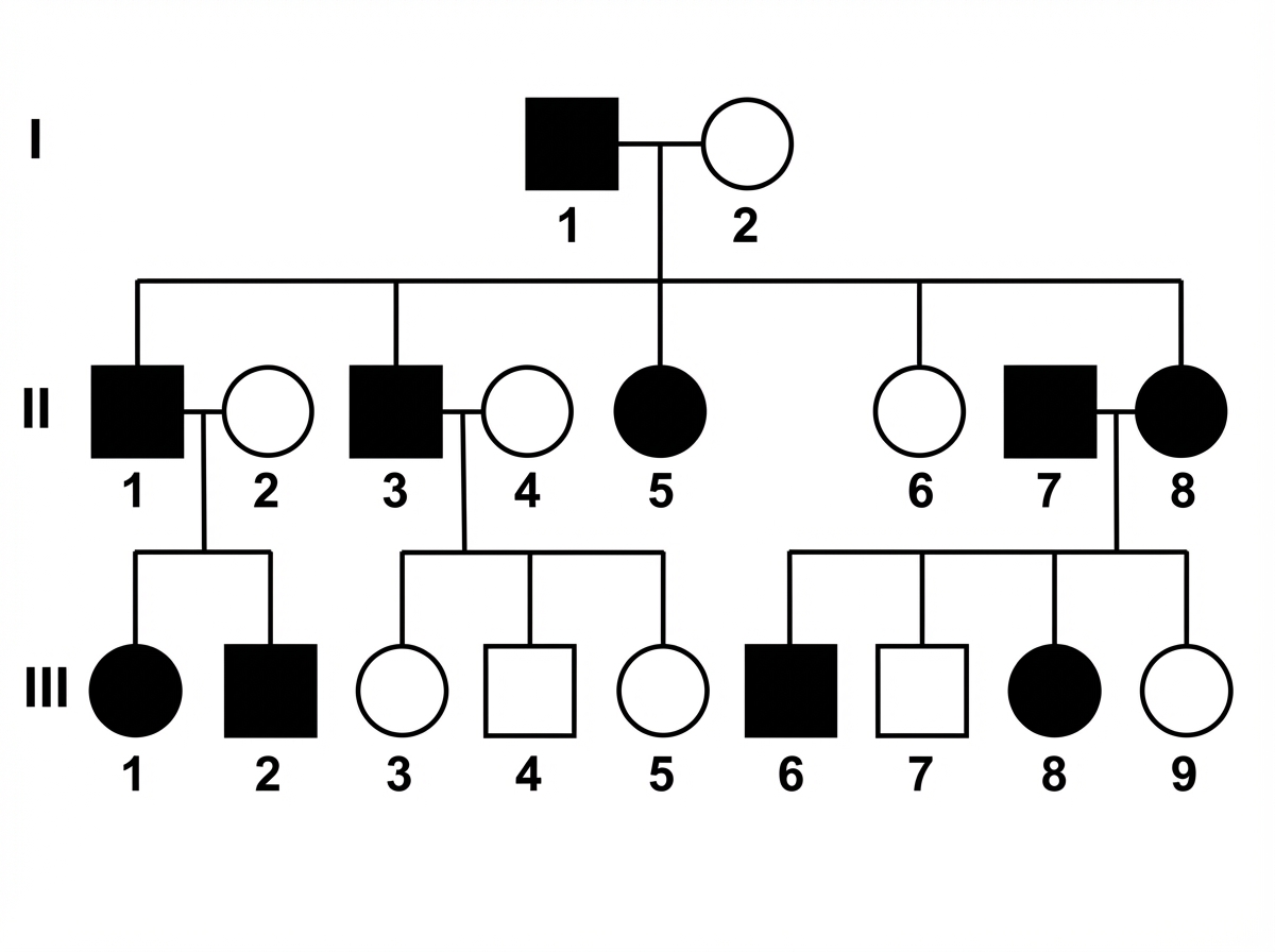
A 12-year-old girl is brought to an oncologist, as she was recently diagnosed with a rare form of cancer. Cytogenetic studies reveal that the tumor is responsive to vinblastine, which is a cell-cycle specific anticancer agent. It acts on the M phase of the cell cycle and inhibits the growth of cells. Which of the following statements best describes the regulation of the cell cycle?
A 4-year-old male presents with a 1-year history of swaying while walking and recent episodes of tripping when ambulating. He has trouble trying to sit and get up from chairs, as well as walking up the stairs to his bedroom. On physical exam, the pediatrician notices nystagmus, absent deep tendon reflexes, significant loss of vibratory and proprioceptive sensation in his extremities, pes cavus, and slight kyphoscoliosis. A blood sample is sent for DNA sequencing and the results show a significant expansion of the trinucleotide GAA on chromosome 9. Which of the following diseases displays a similar mode of inheritance as the disease affecting this patient?
A 1-year-old child who was born outside of the United States is brought to a pediatrician for the first time because she is not gaining weight. Upon questioning, the pediatrician learns that the child has had frequent pulmonary infections since birth, and on exam the pediatrician appreciates several nasal polyps. Genetic testing is subsequently ordered to confirm the suspected diagnosis. Testing is most likely to show deletion of which of the following amino acids from the protein involved in this child's condition?
A 19-year-old man presents to the office for a routine physical exam and a meningitis vaccination prior to attending college on a basketball scholarship. Also present at the appointment is his father who appears to be in his mid-sixties and is much shorter. The patient’s pulse is 70/min, respirations are 18/min, temperature is 37.0°C (98.6°F), and blood pressure is 120/80 mm Hg. He is 183 cm (6 ft 0 in) tall and weighs 79.4 kg (175 lb). His heart rate is regular with a mild diastolic murmur (II/VI) over the aortic valve and his lungs are clear to auscultation bilaterally. A scoliosis test shows mild deviation of his thoracic spine. A skin examination shows numerous red-to-white linear markings on the skin around his lower back. His fingers are long. Which of the following genes does this patient most likely have a mutation of?
An 11-year-old boy is brought to the physician for the evaluation of frequent falling. His mother reports that the patient has had increased difficulty walking over the last few months and has refused to eat solid foods for the past 2 weeks. He has met all developmental milestones. The patient has had multiple ear infections since birth. His temperature is 37°C (98.6°F), pulse is 90/min, and blood pressure is 120/80 mm Hg. Examination shows foot inversion with hammertoes bilaterally. His gait is wide-based with irregular and uneven steps. Laboratory studies show a serum glucose concentration of 300 mg/dL. Further evaluation of this patient is most likely to show which of the following findings?
A 9-year-old boy is admitted to the hospital for placement of halo gravitational traction in order to treat his previously observed kyphoscoliosis. Specifically, he has a previously diagnosed curve that has gotten worse over time and now threatens to compromise his thoracic cavity. His past medical history is significant for short stature, and he has consistently been below the 5th percentile for height since birth. On physical exam, he is found to have macrocephaly with frontal bossing, short arms and legs with disproportionate shortening of the proximal segments, and lumbar lordosis. Which of the following proteins are most likely mutated in this patient?
A mother brings her son to the pediatrician because she is concerned about his health. She states that throughout her child's life he has demonstrated aggressive behavior. However, he has recently begun biting himself causing injury and bleeding. The patient has a past medical history of mental retardation and episodes of severe joint pain. His temperature is 99.5°F (37.5°C), blood pressure is 87/48 mmHg, pulse is 90/min, respirations are 17/min, and oxygen saturation is 98% on room air. Physical exam reveals a child attempting to bite his arms. Which of the following is the inheritance pattern of the disease with which this patient presents?
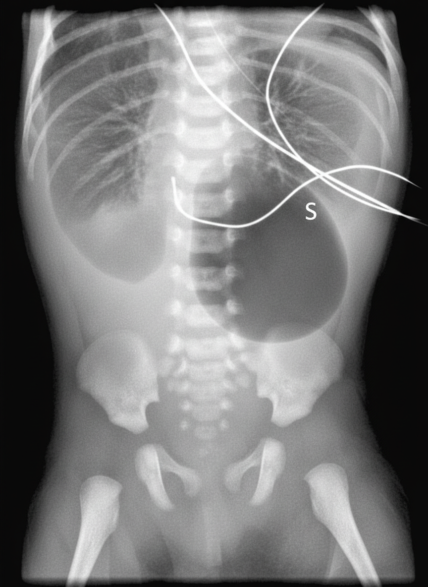
A 9-year-old boy is getting fitted for leg braces because he has become too weak to walk without them. He developed normally until age 3 but then he began to get tired more easily and fell a lot. Over time he started having trouble walking and would stand up by using the Gower maneuver. Despite this weakness, his neurologic development is normal for his age. On exam his calves appeared enlarged and he was sent for genetic testing. Sequence data showed that he had a mutation causing a shift in the reading frame, resulting in a severely truncated and non-functional protein. Which of the following types of mutations is most likely the cause of this patient's disorder?
A 2-year-old male is referred to a geneticist for developmental delay and intellectual disability. He was hypotonic at birth and his parents are concerned that he tries to eat everything, including erasers and chalk. Physical exam is remarkable for severe obesity and hypogonadism. Genetic analysis reveals that he has one mutated allele and one normal allele at the gene of interest. Which of the following is the most likely explanation for why this patient is affected despite having a normal allele?
Explanation: ***Fibrillin*** - The constellation of **tall stature**, **long arm span**, **pectus excavatum**, and **cardiac abnormalities** (e.g., aortic dissection or mitral valve prolapse) is classic for **Marfan syndrome**. - Marfan syndrome is caused by a defect in the *FBN1* gene, which codes for **fibrillin-1**, a glycoprotein essential for the formation of elastic fibers in connective tissue. *Elastin* - Defects in **elastin** are associated with conditions like **supravalvular aortic stenosis** (due to **Williams syndrome**) or cutis laxa, which have different phenotypic presentations. - While both elastin and fibrillin are components of elastic fibers, the specific features of Marfan syndrome point to fibrillin as the primary defect. *Collagen* - Defects in **collagen** (especially type I, III, or V) are associated with conditions such as **osteogenesis imperfecta** (brittle bones) or **Ehlers-Danlos syndrome** (hypermobility, skin hyperextensibility). - These conditions typically present with different clinical manifestations, lacking the specific combination of features seen in this patient. *Myosin* - **Myosin** is a fibrous protein primarily involved in **muscle contraction** and is not directly implicated in widespread connective tissue disorders affecting skeletal and cardiovascular systems in this manner. - Defects in myosin are more commonly associated with various forms of **cardiomyopathy** or skeletal muscle myopathies, not Marfan-like features. *Laminin* - **Laminin** is a major component of the **basement membrane** and plays a role in cell adhesion, migration, and tissue architecture. - Laminin defects are associated with certain forms of **muscular dystrophy** (e.g., congenital muscular dystrophy) and **epidermolysis bullosa**, not the specific skeletal and cardiovascular features of Marfan syndrome.
Explanation: ***Cyclin-dependent activation of CDK1 (CDC2) takes place upon the entry of a cell into M phase of the cell cycle.*** - The **M-phase promoting factor (MPF)**, composed of **CDK1 (CDC2)** and **cyclin B**, is activated at the G2/M transition, driving the cell into mitosis. - Activation of CDK1 by **cyclin B binding** and subsequent dephosphorylation of threonine 161 is crucial for initiation of mitosis. *Inhibitors of DNA synthesis act in the M phase of the cell cycle.* - **Inhibitors of DNA synthesis**, such as **hydroxyurea** and **methotrexate**, primarily act during the **S phase** of the cell cycle, when DNA replication occurs. - The M phase is characterized by **mitosis** (nuclear division) and **cytokinesis** (cytoplasmic division), not DNA synthesis. *The G0 phase is the checkpoint before G1.* - The **G0 phase** is a **resting state** where cells exit the cell cycle and cease to divide, not a checkpoint before G1. - The main checkpoint before G1 is typically referred to as the **restriction point** or **G1 checkpoint**, which determines if a cell will commit to division. *EGF from a blood clot stimulates the growth and proliferation of cells in the healing process.* - While **EGF (Epidermal Growth Factor)** does stimulate cell growth and proliferation in healing, it is not primarily associated with blood clots. - **Platelets** in blood clots release growth factors like **PDGF (Platelet-Derived Growth Factor)** and **TGF-β (Transforming Growth Factor-beta)**, which are critical for wound healing. *Replication of the genome occurs in the M phase of the cell cycle.* - **Replication of the genome** (DNA synthesis) occurs during the **S phase** (synthesis phase) of the cell cycle. - The **M phase** is dedicated to **mitosis** (separation of duplicated chromosomes) and **cytokinesis**, where the cell divides into two daughter cells.
Explanation: ***Von Gierke's disease*** - The patient's symptoms (ataxia, nystagmus, absent deep tendon reflexes, sensory loss, pes cavus, kyphoscoliosis) and genetic finding (GAA trinucleotide expansion on chromosome 9) are characteristic of **Friedreich's ataxia**. - Friedreich's ataxia is inherited in an **autosomal recessive** pattern, similar to Von Gierke's disease. *Osteogenesis imperfecta* - This condition is primarily inherited in an **autosomal dominant** pattern, although some severe forms can be recessive. - It is caused by defects in **collagen genes** and is characterized by brittle bones, blue sclera, and hearing loss, which are not present here. *Fragile X syndrome* - This is an **X-linked dominant** disorder characterized by a trinucleotide (CGG) repeat expansion on the X chromosome. - It presents with intellectual disability, distinctive facial features, and macroorchidism, which differ from the patient's presentation. *Leber hereditary optic neuropathy* - This condition is inherited via **mitochondrial inheritance**, meaning it is passed down exclusively from the mother. - It primarily causes bilateral vision loss due to optic nerve degeneration, which is not the patient's main complaint. *Menkes disease* - This is an **X-linked recessive** disorder, caused by a mutation in the ATP7A gene leading to copper deficiency. - It is characterized by sparse, kinky hair, developmental delay, and neurological degeneration, a different clinical picture than described.
Explanation: ***Phenylalanine*** - The patient's presentation with **failure to thrive**, **recurrent pulmonary infections**, and **nasal polyps** is highly suggestive of **cystic fibrosis (CF)**. - The most common mutation leading to CF is the **delta F508 mutation**, which involves a **deletion of a phenylalanine** residue at position 508 in the **CFTR protein**. *Leucine* - While other rare mutations in the **CFTR gene** can involve various amino acids, **leucine deletion** is not the most common or characteristic mutation associated with the clinical picture of CF. - This option does not represent the primary genetic defect seen in the majority of CF cases. *Histidine* - **Histidine deletion** is not a commonly described or significant mutation in the **CFTR gene** that would explain the overwhelming majority of cystic fibrosis cases. - The typical genetic defect in CF involves the malformation of the CFTR protein due to phenylalanine deletion. *Lysine* - **Lysine deletion** is not the most prevalent type of mutation found in patients with **cystic fibrosis** nor is it associated with the characteristic presentation mentioned. - The CFTR protein dysfunction primarily stems from the common phenylalanine deletion, affecting protein folding and function. *Valine* - Deletion of **valine** is not the most frequently observed mutation in the **CFTR gene** that causes cystic fibrosis. - The classic genetic basis for CF symptoms, particularly severe ones, relates to the absence of phenylalanine.
Explanation: ***FBN1*** - The patient exhibits features consistent with **Marfan syndrome**, including tall stature, long fingers (arachnodactyly), mild scoliosis, and a diastolic murmur indicative of **aortic root dilation** or valve insufficiency. Marfan syndrome is caused by a mutation in the **FBN1 gene**, which encodes **fibrillin-1**. - **Striae distensae** (red-to-white linear markings, or stretch marks) in a young, otherwise healthy individual are also a common cutaneous manifestation of Marfan syndrome due to connective tissue weakness. *COL5A1* - Mutations in **COL5A1** are associated with the **classical type of Ehlers-Danlos syndrome**, characterized by **skin hyperextensibility**, **joint hypermobility**, and **fragile tissues**, which are not the primary features described here. - While there can be joint issues, the prominent skeletal and cardiovascular findings in this patient point away from classical Ehlers-Danlos syndrome. *COL3A1* - Mutations in **COL3A1** are responsible for **vascular Ehlers-Danlos syndrome**, known for **arterial rupture**, **intestinal rupture**, and **fragile, translucent skin**. - This condition is particularly severe due to the risk of life-threatening vascular events, which are not suggested by the patient's presentation. *TGFBR1* - Mutations in **TGFBR1** (transforming growth factor beta receptor 1) cause **Loeys-Dietz syndrome**, another connective tissue disorder with overlapping features to Marfan syndrome, including aortic aneurysms and skeletal abnormalities. - However, Loeys-Dietz syndrome is typically distinguished by **hypertelorism** (widely spaced eyes), **bifid uvula**, and a higher risk of **early arterial dissection**, which are not present in this patient. *ELN* - Mutations in the **ELN gene** encode **elastin** and are associated with **supravalvular aortic stenosis** and Williams syndrome, which presents with distinct facial features, intellectual disability, and a particular type of heart defect. - The patient's diastolic murmur suggests aortic insufficiency or dilation, not typically supravalvular aortic stenosis, and other features of Williams syndrome are absent.
Explanation: ***Expansion of GAA trinucleotide repeats*** - The clinical presentation of **Friedreich ataxia** includes progressive **ataxia**, **dysarthria**, **hypertrophic cardiomyopathy**, and **diabetes mellitus** (indicated by serum glucose of 300 mg/dL), consistent with the patient's symptoms. - This condition is caused by an autosomal recessive **GAA trinucleotide repeat expansion** in the frataxin gene. *Duplication of PMP22 gene* - A duplication of the **PMP22 gene** is associated with **Charcot-Marie-Tooth disease type 1A**, which presents with progressive distal weakness, sensory loss, foot deformities (e.g., hammertoes, pes cavus), and a steppage gait. - While some features overlap, the severe **ataxia** and **diabetes mellitus** seen in this patient are not characteristic of Charcot-Marie-Tooth disease. *Mutation of type I collagen gene* - **Type I collagen gene mutations** are characteristic of **osteogenesis imperfecta**, a connective tissue disorder causing brittle bones, blue sclerae, and hearing loss. - The patient's symptoms of neurological decline and diabetes are not consistent with osteogenesis imperfecta. *Absence of dystrophin protein* - An absence of **dystrophin protein** is the hallmark of **Duchenne muscular dystrophy**, an X-linked recessive disorder causing progressive muscle weakness, Gowers' sign, and pseudohypertrophy of the calves. - While muscle weakness would cause falling, the patient's presentation with **ataxia** and a **wide-based gait** points away from a primary muscle disorder. *Defect of ATM protein* - A **defect in ATM protein** is responsible for **Ataxia-telangiectasia**, an autosomal recessive disorder characterized by cerebellar ataxia, telangiectasias, immunodeficiency, and an increased risk of cancer. - While ataxia is present, the patient's findings of **hammertoes** and **diabetes mellitus** are not typical features of Ataxia-telangiectasia.
Explanation: ***FGFR3 receptor*** - The constellation of **short stature**, **macrocephaly with frontal bossing**, **short limbs with disproportionate shortening of proximal segments (rhizomelia)**, and **lumbar lordosis** is classic for **achondroplasia**. - Achondroplasia is caused by an **activating mutation** in the **fibroblast growth factor receptor 3 (FGFR3)** gene, which inhibits endochondral ossification, leading to impaired bone growth. *RUNX2 transcription factor* - Mutations in **RUNX2** are associated with **cleidocranial dysplasia**, a disorder characterized by delayed closure of fontanelles, hypoplastic or absent clavicles, and dental abnormalities. - While it can cause short stature and scoliosis, the specific physical features like rhizomelia and frontal bossing point away from cleidocranial dysplasia. *Alpha-L-iduronidase* - Deficiency of **alpha-L-iduronidase** causes **Hurler syndrome (MPS I)**, a lysosomal storage disorder. - Patients present with coarse facial features, corneal clouding, hepatosplenomegaly, and skeletal deformities, but not typically the specific limb and head features described. *COL1A1 gene* - Mutations in the **COL1A1 gene** are associated with **osteogenesis imperfecta (OI)**, a disorder characterized by **brittle bones** and frequent fractures. - While OI can cause skeletal deformities and short stature, it doesn't typically present with macrocephaly, frontal bossing, or rhizomelia in the same way as achondroplasia. *SLC26A2 transporter* - Mutations in the **SLC26A2 gene** are associated with **diastrophic dysplasia**, another form of dwarfism. - While it causes short stature and skeletal issues, patients often have distinctive features like "hitchhiker's thumb," club feet, and highly mobile joints, which are not mentioned here.
Explanation: ***X-linked recessive*** - This patient's presentation of **self-mutilation**, **mental retardation**, and **aggressive behavior** is highly suggestive of **Lesch-Nyhan syndrome**. - Lesch-Nyhan syndrome is an **X-linked recessive disorder** caused by a defect in the gene for **hypoxanthine-guanine phosphoribosyltransferase (HGPRT)**, leading to a build-up of uric acid. *X-linked dominant* - X-linked dominant disorders affect both males and females, though males are often more severely affected or may have more prominent symptoms. - Affected fathers transmit the trait to all their daughters but no sons, which is not characteristic of Lesch-Nyhan syndrome. *Autosomal dominant* - Autosomal dominant disorders are inherited equally by males and females, and an affected individual has a 50% chance of passing the condition to each child. - This inheritance pattern does not fit the typical presentation of Lesch-Nyhan syndrome, which primarily affects males. *Autosomal recessive* - Autosomal recessive disorders typically require two copies of the mutated gene (one from each parent) for the disease to manifest, affecting males and females equally. - While some metabolic disorders are autosomal recessive, Lesch-Nyhan syndrome's sex-linked pattern of inheritance rules out this option. *Maternal* - Maternal inheritance refers to conditions passed down through **mitochondrial DNA**, affecting all children of an affected mother regardless of sex. - Affected fathers do not pass mitochondrial conditions to any of their children, since sperm contribute minimal mitochondria. - Lesch-Nyhan syndrome is associated with a nuclear gene on the X chromosome, not mitochondrial DNA, making maternal inheritance incorrect.
Explanation: ***Frameshift*** - A **frameshift mutation** is caused by the insertion or deletion of nucleotides not in multiples of three, leading to a shift in the reading frame of the mRNA. This results in altered codons downstream of the mutation, typically leading to a **premature stop codon** and a **severely truncated, non-functional protein**. - The description of a mutation causing "a shift in the reading frame, resulting in a severely truncated and non-functional protein" is characteristic of a frameshift mutation, which is the most common type of mutation in **Duchenne muscular dystrophy** (DMD). The clinical picture (onset around age 3-5, progressive proximal weakness, Gower maneuver, calf pseudohypertrophy) is classic for DMD. - In DMD, frameshift mutations in the dystrophin gene lead to complete loss of functional dystrophin protein, causing the severe progressive muscle weakness seen in this patient. *Splice site* - A **splice site mutation** affects the recognition sequences for intron-exon boundaries during mRNA splicing, potentially leading to exon skipping, intron retention, or use of cryptic splice sites. While splice site mutations can cause DMD (accounting for ~10% of cases), they are less common than frameshifts/deletions. - The specific description of a "shift in the reading frame" points more directly to a frameshift mutation rather than a splicing defect. *Missense* - A **missense mutation** results in a single nucleotide substitution that changes one codon to specify a different amino acid. This produces a full-length protein with a single amino acid substitution. - Missense mutations typically cause the milder **Becker muscular dystrophy** phenotype (with partially functional dystrophin), not the severe Duchenne phenotype described here. The description of a "severely truncated and non-functional protein" does not fit a missense mutation. *Nonsense* - A **nonsense mutation** introduces a premature stop codon directly by changing a codon that normally specifies an amino acid into a stop codon (UAG, UAA, or UGA). This results in a truncated protein. - While nonsense mutations can cause DMD and do produce truncated proteins, the specific wording "shift in the reading frame" is more characteristic of a frameshift mutation. Nonsense mutations don't shift the reading frame—they directly create a stop signal. *Silent* - A **silent (synonymous) mutation** is a nucleotide substitution that does not change the amino acid sequence due to the degeneracy of the genetic code (multiple codons can specify the same amino acid). - Silent mutations produce normal, full-length proteins and would not cause disease symptoms.
Explanation: ***Imprinting*** - The constellation of **hypotonia**, **developmental delay**, **hyperphagia** (eating everything), **obesity**, and **hypogonadism** is characteristic of **Prader-Willi syndrome**. - **Prader-Willi syndrome** is typically caused by the **loss of function of specific paternally inherited genes** on chromosome 15 (15q11-q13), a mechanism known as **genomic imprinting**, where only one parent's copy of a gene is active. - In this case, the patient has one mutated (paternal) allele and one normal but imprinted/silenced (maternal) allele, explaining why disease occurs despite the presence of a structurally normal allele. *Uniparental disomy* - **Maternal uniparental disomy** of chromosome 15 can also cause Prader-Willi syndrome if both copies of the chromosome are inherited from the mother, leading to no functional paternal genes. - However, the question explicitly states "one mutated allele and one normal allele," which describes a **deletion or mutation of the paternal copy** rather than UPD (where both chromosomes would be from the mother and appear structurally normal on standard testing). - The question asks for the *mechanism* explaining why the patient is affected despite having a normal allele - the answer is **imprinting**, which is the underlying mechanism in both deletion and UPD cases. *X-linked inheritance pattern* - **X-linked inheritance** typically affects males more severely with different phenotypic presentations and does not explain the combination of a normal and mutated allele resulting in the specific Prader-Willi phenotype. - The described syndrome (Prader-Willi) is associated with **chromosome 15** abnormalities, not the X chromosome. *Locus heterogeneity* - **Locus heterogeneity** describes a single disease phenotype caused by mutations in different gene loci, which is not relevant to explaining why a single mutated allele results in disease in the context of Prader-Willi syndrome. - The clinical picture strongly points to a specific genetic disorder with a known genetic cause on a single locus. *Autosomal dominant inheritance pattern* - While **autosomal dominant disorders** manifest with one mutated allele, the specific combination of symptoms with obesity, hypogonadism, and hyperphagia in this scenario is not typical for a general autosomal dominant pattern. - The question describes features characteristic of **Prader-Willi syndrome**, which is primarily an **imprinting disorder**, rather than a straightforward autosomal dominant condition where the presence of one normal allele would be expected to provide sufficient function in the absence of imprinting.
Explanation: ***Missense mutation*** - A missense mutation results in a **single nucleotide substitution** that changes the codon to code for a different amino acid, altering the protein. - In **sickle cell disease**, a missense mutation in the beta-globin gene (GAG to GTG) leads to the substitution of **glutamic acid for valine**, causing abnormal hemoglobin (HbS) that polymerizes under deoxygenated conditions. *Silent mutation* - A silent mutation is a **point mutation** that results in a new codon that still codes for the **same amino acid**, meaning there is no change in the protein sequence. - Therefore, it would not lead to an **abnormal hemoglobin** protein or the described disease phenotype. *Splice site* - A splice site mutation occurs at the **splice junctions** of introns and exons, leading to errors in mRNA processing. - This can result in **incorrect protein synthesis** due to exon skipping or intron retention, but it typically does not cause the specific amino acid substitution seen in sickle cell disease. *Nonsense mutation* - A nonsense mutation is a point mutation that results in a **premature stop codon**, leading to a **truncated, non-functional protein**. - While this can cause severe disease, it would typically lead to a complete absence or severe deficiency of functional hemoglobin rather than a structurally altered hemoglobin like HbS. *Frameshift mutation* - A frameshift mutation involves the **insertion or deletion of nucleotides** (not in multiples of three), which shifts the reading frame of the mRNA. - This typically leads to a completely **altered amino acid sequence** downstream of the mutation and usually results in a premature stop codon, leading to a non-functional protein rather than a specific single amino acid substitution.
Explanation: ***Sodium channels of respiratory epithelial cells*** - The patient's presentation with **failure to thrive**, genetic defect (deletion of codon 508 on chromosome 7), and **defective protein folding** are classic for **cystic fibrosis (CF)**. - In CF, the defective **CFTR protein** (a chloride channel) leads to reduced chloride secretion and increased **sodium absorption** in respiratory epithelial cells, causing thickened mucus. *Calcium channels of distal tubular cells* - Dysfunction of **calcium channels** in the distal tubules is not a primary feature of cystic fibrosis. - Renal calcium handling issues are typically associated with conditions like **Dent's disease** or various types of **renal tubular acidosis**, not CF. *ATP-sensitive potassium channels of pancreatic beta cells* - While CF can lead to pancreatic insufficiency and **CF-related diabetes**, the primary defect is not in the **ATP-sensitive potassium channels** of beta cells. - The insulin deficiency in CF diabetes is due to destruction of pancreatic islets secondary to duct obstruction and inflammation. *Bicarbonate channels of pancreatic ductal cells* - In cystic fibrosis, the **CFTR protein** is a chloride channel that also facilitates bicarbonate transport, and its dysfunction does impair **bicarbonate secretion** in pancreatic ductal cells. - However, the question specifically asks about an *increase* in channel activity, and bicarbonate channel activity is *decreased* in CF. *Chloride channels of epithelial cells in sweat glands* - The **CFTR protein** is indeed a **chloride channel** in sweat glands, and in CF, its *activity is decreased*, leading to reduced chloride reabsorption and high sweat chloride (the basis for the sweat test). - The question asks for an *increased* channel activity, which is seen with sodium channels due to the linked transport mechanisms.
Explanation: ***It is transmitted only through the mother.*** - The constellation of symptoms (muscle weakness, pain, vomiting, seizures, severe headache, hemiparesis) and the presence of **"ragged red fibers"** on muscle biopsy are classic findings in **Mitochondrial Encephalomyopathy, Lactic Acidosis, and Stroke-like episodes (MELAS) syndrome**. - **Mitochondrial disorders**, including MELAS, are inherited exclusively from the mother because ova contribute mitochondria to the zygote, while sperm contribute virtually none. *Mothers transmit to 50% of daughters and son* - This statement describes the inheritance pattern of an **X-linked recessive** or, in some cases, **autosomal dominant** trait with incomplete penetrance, which is not characteristic of mitochondrial inheritance. - In mitochondrial inheritance, all offspring (100%) of an affected mother will inherit the mitochondrial DNA, though the penetrance and expressivity can vary. *Skips generations* - "Skipping generations" is a hallmark of **recessive inheritance patterns**, where affected individuals may have unaffected parents who are carriers. - This is not typical for **mitochondrial inheritance**, where the disease is usually present in every generation through the maternal line. *Commonly more severe in males* - While some genetic conditions show sex-specific severity, such as X-linked disorders that are often more severe in males due to having only one X chromosome, this is not a general rule for **mitochondrial diseases**. - The severity of mitochondrial disorders is often related to the **proportion of mutated mitochondrial DNA (heteroplasmy)**, which can vary between sexes and tissues. *It can be transmitted through both parents.* - This statement generally describes **autosomal recessive** or **autosomal dominant** inheritance patterns, where genetic material from both parents contributes to the child's genotype. - **Mitochondrial inheritance** is exclusively maternal, as only the mother contributes mitochondria to the offspring.
Explanation: ***5' --> 3'*** - DNA polymerase can only add **nucleotides** to the 3' end of a growing strand, meaning synthesis always proceeds in a **5' to 3' direction**. - This is true for both the **leading strand** (synthesized continuously) and the **lagging strand** (synthesized discontinuously via Okazaki fragments). *3' --> 5'* - While the parental template strand is read in the 3' to 5' direction, the *newly synthesized* DNA strand is always built in the **opposite, antiparallel 5' to 3' direction**. - DNA polymerase lacks the ability to add new nucleotides to the **5' phosphate group** of the growing strand. *N terminus --> C terminus* - This directional notation refers to the synthesis of **proteins**, where amino acids are added to the C (carboxyl) terminus of the growing polypeptide chain. - It does not apply to the synthesis direction of **nucleic acids (DNA or RNA)**. *C terminus --> N terminus* - This directional notation is incorrectly applied; protein synthesis always proceeds from the **N (amino) terminus to the C (carboxyl) terminus**. - This has no relevance to the synthesis direction of **DNA**. *3' --> 5' & 5' --> 3'* - Although DNA replication involves two strands, one is synthesized continuously in the **5' → 3' direction (leading strand)** and the other discontinuously, but still *each fragment* is synthesized in the **5' → 3' direction (lagging strand)**. - No new DNA strand is synthesized in the **3' → 5' direction**.
Explanation: ***In-frame mutation*** - The deletion of three nucleotides, which together code for a single amino acid (phenylalanine), results in an **in-frame mutation** because the reading frame of the mRNA is maintained. - This specific mutation is **ΔF508**, the most common mutation in **cystic fibrosis (CF)**, consistent with the patient's symptoms of growth failure, thick secretions, chronic cough, and clubbed fingernails. *Triplet expansion* - This involves the **increase in the number of repeats of a trinucleotide sequence**, such as in Huntington's disease or fragile X syndrome. - The patient's mutation is a **deletion**, not an expansion of a trinucleotide repeat. *Nonsense mutation* - A nonsense mutation involves a **point mutation** that results in a **premature stop codon**, leading to a truncated protein. - The patient's mutation is a **deletion of an entire codon**, not a single base change leading to a stop codon. *Frameshift mutation* - A frameshift mutation occurs when the deletion or insertion of nucleotides is **not a multiple of three**, leading to a shift in the reading frame and alteration of all downstream amino acids. - In this case, the deletion of **three nucleotides** maintains the reading frame, so it is not a frameshift. *Silent mutation* - A silent mutation is a **point mutation** where a change in a single nucleotide does not alter the amino acid sequence due to the degeneracy of the genetic code. - Here, the **deletion of a codon** results in the loss of an amino acid, clearly altering the protein product, hence it is not silent.
Explanation: ***0%*** - This condition, **Leber's Hereditary Optic Neuropathy (LHON)**, is caused by a mutation in **mitochondrial DNA**. - **Mitochondrial DNA** is exclusively inherited from the mother, meaning the father cannot pass it on to his children. *33%* - This percentage does not align with any known pattern of Mendelian inheritance or mitochondrial inheritance. - It would be too low for a dominant mitochondrial disorder and too high for a recessive one if the mother were a carrier. *100%* - While all children of an affected mother would inherit the disease, the patient is a male, and mitochondria are inherited maternally. - Therefore, the father cannot pass on his **mitochondrial DNA** to his son. *25%* - This percentage is typically associated with **autosomal recessive inheritance**, where both parents are carriers. - LHON follows a **mitochondrial inheritance pattern**, which is distinct from autosomal inheritance. *50%* - This percentage is characteristic of **autosomal dominant inheritance** or X-linked recessive inheritance where the mother is a carrier. - Since this is a **mitochondrial disorder**, the father's genes are not involved in transmission to his son.
Explanation: ***Cadherin*** - **Cadherins** are calcium-dependent adhesion proteins that mediate cell-to-cell adhesion, particularly in epithelial tissues. - A decrease in calcium concentration would directly impair cadherin function, leading to reduced cell adhesion and increased intercellular gaps. *Actin* - **Actin** is a component of the cell's cytoskeleton, involved in cell shape, motility, and intracellular transport, but not directly responsible for calcium-dependent cell adhesion between epithelial cells. - While actin filaments interact with adhesion junctions, their primary role is structural and dynamic rather than adhesive. *Integrin* - **Integrins** are primarily involved in cell-to-extracellular matrix adhesion, connecting the cell to the surrounding matrix, not directly mediating calcium-dependent cell-to-cell adhesion between epithelial cells. - They can be affected by intracellular calcium signaling but do not directly bind calcium to mediate their adhesive function in the same way cadherins do. *Claudin* - **Claudins** are key components of **tight junctions**, which regulate paracellular permeability and form a barrier between cells, but they are not directly responsible for calcium-dependent cell-to-cell adhesion, which is characteristic of adherens junctions. - While tight junctions contribute to overall cell-cell contact, the observation of an *increased gap* suggests an issue with adhesive complexes like adherens junctions, where cadherins are prominent. *Cytokeratin* - **Cytokeratins** are intermediate filaments found in epithelial cells, providing structural support and mechanical strength. - They are linked to desmosomes and hemidesmosomes but are not directly involved in calcium-dependent cell-to-cell adhesion.
Explanation: ***Variable expressivity*** - This principle accounts for the **range of phenotypes** observed among individuals who carry the same genetic mutation, as seen in the siblings with Marfan syndrome. - Both siblings likely have the same genetic defect (e.g., in the *FBN1* gene), but express different combinations and severity of symptoms. *Incomplete penetrance* - This occurs when individuals with a specific genotype **do not always express** the associated phenotype. - In this case, both siblings *do* express some features of Marfan syndrome, indicating that the gene is penetrant in both. *Frameshift mutation* - A frameshift mutation is a type of **gene mutation** caused by indels (insertions or deletions) of a number of nucleotides not divisible by three. - While a frameshift mutation could be the underlying cause of Marfan syndrome, it does not explain the *phenotypical differences* between two individuals with the same underlying genetic defect. *Compound heterozygosity* - This describes a situation where an individual has **two different mutant alleles** at the same gene locus, one on each chromosome. - While relevant to some genetic disorders, it does not explain the phenotypic variability between two siblings who inherited the same underlying genetic susceptibility. *Chromosomal instability* - This refers to a high rate of **chromosome rearrangements** or aneuploidy (abnormal chromosome number) within cells. - While it can lead to various medical conditions, it is not the mechanism primarily responsible for the varying phenotypes of a single-gene disorder like Marfan syndrome in different individuals.
Explanation: ***Hemizygous; full penetrance*** - The condition described (**red color blindness**) is an **X-linked recessive** trait, meaning males have only one X chromosome and are thus **hemizygous** for genes on the X chromosome. - **Full penetrance** in X-linked recessive traits means that if a male inherits the affected X chromosome, he will express the phenotype of the condition. *Homozygote; reduced or incomplete penetrance* - **Homozygous** refers to having two identical alleles for a particular gene, which is not applicable to an X-linked gene in males as they only have one X chromosome. - **Reduced or incomplete penetrance** means that a person with the genotype may or may not express the phenotype, which is typically not the case for males with X-linked recessive color blindness. *Heterozygotes; reduced or incomplete penetrance* - **Heterozygous** means having two different alleles for a gene; this term is not applicable to males regarding X-linked genes. - Males are **hemizygous** for X-linked genes, meaning they only have one allele, not two different ones. *Hemizygous; reduced or incomplete penetrance* - While males are indeed **hemizygous** for X-linked genes like color blindness, the penetrance for red-green color blindness in males is generally considered to be **full**, meaning if they carry the mutated gene, they will express the trait. - **Reduced penetrance** would imply that some males with the genotype might not exhibit color blindness, which is rare for this condition. *Heterozygotes; full penetrance* - The term **heterozygous** applies to individuals with two different alleles for a gene, which is not the genetic state of males for X-linked genes. - While penetrance is full, the term **heterozygotes** is incorrect for defining the male genotype in this context.
Explanation: ***CGG on the sex chromosome X*** - The constellation of **intellectual disability**, a **long, thin face with a large jaw**, **protruding ears**, and **macroorchidism** are classic features of **Fragile X syndrome**. - Fragile X syndrome is caused by an expansion of the **CGG trinucleotide repeat** in the **FMR1 gene** on the **X chromosome**. The **high-pitched holosystolic murmur at the apex radiating to the axilla** suggests **mitral valve prolapse**, which is also frequently associated with Fragile X. *GAA on chromosome 9* - This describes the **GAA trinucleotide repeat expansion** associated with **Friedreich's ataxia**, affecting the **FXN gene** on **chromosome 9**. - Friedreich's ataxia is characterized by **progressive ataxia**, **dysarthria**, and **loss of vibratory/proprioceptive sensation**, not macroorchidism or the specific facial features seen here. *CTG on chromosome 19* - This describes the **CTG trinucleotide repeat expansion** associated with **myotonic dystrophy type 1**, affecting the **DMPK gene** on **chromosome 19**. - Myotonic dystrophy is characterized by **myotonia** (delayed muscle relaxation), **muscle weakness**, and **cataracts**, which are not consistently present in this case. *CTG on chromosome 8* - While **CTG repeats** are involved in some genetic conditions, the specific association with **chromosome 8** as a cause for the described symptoms (intellectual disability, specific facial features, macroorchidism, and mitral valve prolapse) is not a common trinucleotide repeat disorder. - This option does not correspond to a recognized trinucleotide repeat disorder that presents with the given clinical picture. *CAG on chromosome 4* - This describes the **CAG trinucleotide repeat expansion** associated with **Huntington's disease**, affecting the **HTT gene** on **chromosome 4**. - Huntington's disease typically presents with **chorea**, **psychiatric symptoms**, and **dementia** later in life, not with the childhood onset intellectual disability and physical features described.
Explanation: ***Alpha-galactosidase A*** - A deficiency in **alpha-galactosidase A** causes **Fabry disease**, which, like Hunter syndrome (iduronate sulfatase deficiency), is inherited in an **X-linked recessive** pattern. - Both conditions primarily affect males, with carrier females potentially exhibiting milder symptoms. *Sphingomyelinase* - A deficiency in sphingomyelinase leads to **Niemann-Pick disease types A and B**, which are inherited in an **autosomal recessive** pattern. - This mode of inheritance differs from the X-linked pattern of Hunter syndrome. *Glucocerebrosidase* - A deficiency in glucocerebrosidase causes **Gaucher disease**, inherited in an **autosomal recessive** pattern. - This is a common lysosomal storage disorder, but its inheritance pattern is distinct from X-linked disorders. *Galactocerebrosidase* - A deficiency in galactocerebrosidase causes **Krabbe disease (globoid cell leukodystrophy)**, which is inherited in an **autosomal recessive** pattern. - Krabbe disease is a severe neurodegenerative disorder, but its genetic transmission is not X-linked. *Alpha-L-iduronidase* - A deficiency in **alpha-L-iduronidase** causes **Hurler syndrome (MPS I)**, which is inherited in an **autosomal recessive** pattern. - While both Hunter and Hurler syndromes are mucopolysaccharidoses, their genetic inheritance patterns are different.
Explanation: ***LacI*** - The **LacI gene** encodes the **Lac repressor protein**, which is constitutively expressed (always produced) and binds to the operator region of the lac operon. - When bound, the **Lac repressor** blocks RNA polymerase from transcribing the structural genes (LacZ, LacY, LacA), thereby repressing the operon in the absence of lactose. *LacY* - The **LacY gene** encodes **lactose permease**, an enzyme responsible for transporting lactose into the bacterial cell. - Its expression is regulated by the lac operon and is not constitutively expressed; rather, it is induced in the presence of lactose. *LacZ* - The **LacZ gene** encodes **beta-galactosidase**, the enzyme that breaks down lactose into glucose and galactose. - Like LacY, its expression is part of the lac operon and is induced when lactose is available, not expressed constitutively. *CAP* - **CAP (Catabolite Activator Protein)** is a regulatory protein that, when bound to cAMP, activates transcription of the lac operon when glucose is absent. - While essential for lac operon regulation, CAP is not a gene whose constitutive expression leads to repression of the operon. *LacA* - The **LacA gene** encodes **thiogalactoside transacetylase**, an enzyme with a less clear role in lactose metabolism but is part of the lac operon. - Its expression is also regulated and induced along with LacZ and LacY, not constitutively expressed to repress the operon.
Explanation: ***0.2%*** - The brother's symptoms (infertility, right-sided heart, frequent infections) are characteristic of **Kartagener syndrome**, a form of **primary ciliary dyskinesia (PCD)**, which has an **autosomal recessive** inheritance pattern. - Since the patient's parents are obligate heterozygotes (carriers), the patient has a 2/3 chance of being a carrier. Given the population prevalence of 1/40,000 for an autosomal recessive disease, the carrier frequency (2pq) is approximately **2 x sqrt(1/40,000) = 2 x 1/200 = 1/100**. The chance of her child inheriting the disease is (2/3 chance of patient being carrier) x (1/100 chance of partner being carrier) x (1/4 chance of affected offspring) = 2/1200 ≈ **0.00166 or 0.166%**, which is closest to 0.2%. *25%* - This would be the risk if both parents were known carriers, and it represents the chance of an affected offspring from two heterozygotes. - In this scenario, the woman's partner's carrier status is unknown and based on population prevalence, making the overall risk much lower. *66%* - This is the probability that the patient (the healthy sister of an affected individual with an autosomal recessive disease) is a **carrier**. - This value alone does not account for the partner's carrier status or the final Mendelian inheritance probability (1/4) for an affected child. *0.7%* - This percentage is too high; it might result from incorrect calculation of the population carrier frequency or misapplication of probabilities. - The correct carrier frequency for the partner is 1/100, which is significantly lower than what would lead to a 0.7% final risk. *1%* - This value is also too high and likely results from a miscalculation of either the carrier frequency or the overall probability. - A 1% chance would suggest a much higher population carrier frequency or a different inheritance scenario.
Explanation: ***Abnormally decreased expression of the gene from the maternal chromosome*** - This patient's symptoms (non-verbal, severe developmental delays, cognitive disability, seizures, uncoordinated movements, wide-based gait, inappropriate laughter, and an abnormality in an E3 ubiquitin ligase gene) are characteristic of **Angelman syndrome**. - Angelman syndrome is typically caused by a deletion or mutation on the **maternally inherited copy of chromosome 15q11-q13**, specifically affecting the *UBE3A* gene, which is an E3 ubiquitin ligase. This leads to reduced or absent expression of the *UBE3A* gene in critical brain regions where only the maternal allele is expressed. *Abnormally increased expression of the gene from the maternal chromosome* - Angelman syndrome is caused by a **loss of function** of the maternally inherited *UBE3A* gene, not an increase in its expression. - Increased expression would not lead to the neurodevelopmental deficits seen in Angelman syndrome. *Abnormally decreased expression of the gene from both chromosomes* - While there is decreased expression of the functional *UBE3A* gene, the paternal allele is normally **silenced** in specific brain regions relevant to Angelman syndrome pathogenesis due to **genomic imprinting**. Therefore, the issue is with the maternal allele. - If both chromosomes had decreased expression, it would imply a different genetic mechanism or a more severe, potentially lethal, condition. *Abnormally decreased expression of the gene from the paternal chromosome* - In the brain regions relevant to Angelman syndrome, the paternal *UBE3A* allele is normally **silenced** due to genomic imprinting. Therefore, its decreased expression would not be an abnormal finding or contribute to the pathology. - Problems with the paternal allele in this region are associated with **Prader-Willi syndrome**, which has a different clinical presentation (e.g., hypotonia, hyperphagia, obesity). *Abnormally increased expression of the gene from the paternal chromosome* - The paternal *UBE3A* allele is normally **silenced** in the relevant brain regions; therefore, an increased expression would be abnormal but is not the genetic basis of Angelman syndrome. - Angelman syndrome is caused by the **loss or absence of functional maternal *UBE3A*** expression, not altered paternal expression.
Explanation: ***Mutation of gene on chromosome 15*** - The patient's presentation with **tall stature**, **long arms** (dolichostenomelia), **scoliosis**, and **bilateral upward lens subluxation** are classic features of **Marfan syndrome**. - Marfan syndrome is an autosomal dominant disorder caused by a mutation in the *FBN1* gene located on **chromosome 15**, which encodes for **fibrillin-1**, a glycoprotein essential for connective tissue formation. *Extra copy of sex chromosome* - An extra copy of a sex chromosome, such as in **Klinefelter syndrome (XXY)**, is associated with tall stature and disproportionately long limbs, but it typically presents with **hypogonadism**, infertility, and learning difficulties, not lens subluxation or significant scoliosis as the primary features. - Patients with Klinefelter syndrome often have a **eunuchoid body habitus** and gynecomastia, which are not described in this patient. *Mutation of COL5A1 or COL5A2* - Mutations in *COL5A1* or *COL5A2* are associated with **Ehlers-Danlos syndrome (classical type)**, which primarily features **skin hyperextensibility**, delayed wound healing, and **joint hypermobility**. - While some forms of Ehlers-Danlos can have ocular involvement (e.g., easy bruising, scleral fragility), **lens subluxation** and the specific tall, slender build with scoliosis are not characteristic clinical features. *Defective metabolism of methionine* - A defective metabolism of methionine is characteristic of **homocystinuria**, an autosomal recessive disorder. - Homocystinuria also causes **tall stature**, **scoliosis**, and **lens subluxation**, but the subluxation is typically **downward and inward**, differentiating it from the upward subluxation seen in Marfan syndrome. Patients also have an increased risk of **thromboembolic events** and **intellectual disability**. *Mutation of RET proto-oncogene* - Mutations of the *RET* proto-oncogene are associated with **Multiple Endocrine Neoplasia type 2 (MEN2)**. - MEN2 presents with specific endocrine tumors such as **medullary thyroid carcinoma**, **pheochromocytoma**, and **parathyroid hyperplasia**, and does not involve the skeletal or ocular abnormalities described in this patient.
Explanation: ***Deletion of Phe508 in husband*** - The husband's history of recurrent respiratory infections combined with **complete azoospermia** (zero sperm despite normal semen volume) is highly suggestive of **Cystic Fibrosis** due to **CFTR gene mutation**, with **deletion of Phe508 (ΔF508)** being the most common mutation. - CFTR mutations frequently cause **congenital bilateral absence of the vas deferens (CBAVD)**, resulting in obstructive azoospermia where sperm are produced but cannot be ejaculated due to absent vas deferens. - This provides a **unifying diagnosis** explaining both the male infertility and respiratory symptoms. *XO chromosome in wife* - **Turner syndrome (45,XO)** presents with **primary amenorrhea**, **streak gonads**, short stature, and absent secondary sexual characteristics. - The wife has **normal menstrual history** and unremarkable fertility evaluation, making this diagnosis incompatible with her presentation. - The semen analysis clearly identifies **male-factor infertility** as the cause. *Undescended testes in husband* - **Cryptorchidism** can impair spermatogenesis due to elevated testicular temperature, typically causing **oligospermia** (reduced sperm count) rather than complete azoospermia. - Physical examination of the husband was unremarkable, making undescended testes unlikely. - This diagnosis does not explain the recurrent respiratory infections. *XXY chromosome in husband* - **Klinefelter syndrome (47,XXY)** causes **primary hypogonadism** with azoospermia, small firm testes, gynecomastia, and often tall stature with eunuchoid proportions. - While it explains the azoospermia, it **does not account for the recurrent respiratory infections**, whereas CFTR mutation explains both features. - Physical exam was unremarkable, without typical Klinefelter stigmata. *Deletion of Phe508 in wife* - While the wife has asthma and nasal polyps (which can be seen in CF or overlap with asthma-related conditions), her **normal menstrual history** indicates she is likely fertile. - The **male-factor infertility** (complete azoospermia in the husband) is the direct cause of the couple's inability to conceive. - Even if the wife has CF, this would not explain the husband's azoospermia, which is the primary barrier to conception.
Explanation: **Caspase** - The shrinking of the thymus with age, known as **thymic involution**, is primarily driven by **apoptosis** (programmed cell death) of thymocytes. - **Caspases** are a family of proteases that play a central role in initiating and executing apoptosis, making them the most likely enzymes involved in this process. *Lipase* - **Lipases** are enzymes that catalyze the hydrolysis of fats (lipids). - While fat deposition occurs in the involuting thymus, lipases are not directly responsible for the **cell death** or tissue regression. *Collagenase* - **Collagenases** are enzymes that break down **collagen**, a major component of the extracellular matrix. - While there may be some remodeling of the extracellular matrix during thymic involution, collagenases are not the primary drivers of **thymocyte apoptosis**. *Metalloproteinase* - **Metalloproteinases (MMPs)** are enzymes that break down various components of the extracellular matrix and are involved in tissue remodeling. - While MMPs contribute to tissue restructuring, they are not the main enzymes responsible for the **programmed cell death** that underpins thymic involution. *NADPH oxidase* - **NADPH oxidase** is an enzyme complex that produces **reactive oxygen species (ROS)**, primarily for pathogen killing by phagocytes. - While excessive ROS can induce cell death, **NADPH oxidase** is not the primary or direct mechanism responsible for the physiological apoptosis during thymic involution.
Explanation: ***Eukaryotes have multiple origins of replication*** - Eukaryotic chromosomes are much larger than prokaryotic chromosomes and require multiple origins of replication to complete DNA synthesis within a reasonable timeframe. - Each origin of replication initiates simultaneously, allowing DNA synthesis to occur at many sites along the chromosome, effectively increasing the overall speed of replication. - This compensates for the slower rate of nucleotide addition by DNA polymerase in eukaryotes compared to prokaryotes. *Eukaryotes have helicase which can more easily unwind DNA strands* - While helicase activity is crucial for unwinding DNA, there is no evidence to suggest that eukaryotic helicases are significantly more efficient or faster at unwinding DNA compared to prokaryotic helicases in a way that would account for the large difference in overall replication time. - The rate of DNA unwinding by helicase is a factor in replication speed, but it does not overcome the fundamental limitation of a single origin of replication in prokaryotes. *Eukaryotes have fewer polymerase types* - Eukaryotic cells actually have **more** types of DNA polymerases than prokaryotic cells, each specialized for different functions like replication, repair, and mitochondrial DNA synthesis. - The number of polymerase types does not directly relate to the speed or efficiency of overall DNA replication in terms of replicating large amounts of DNA. *Eukaryotes have less genetic material to replicate* - Eukaryotic organisms typically have significantly **more** genetic material (a larger genome size) than prokaryotic organisms, not less. - If eukaryotes had less genetic material, the question itself about effective replication of "much larger amounts of DNA" would be contradictory. *Eukaryotes have a single, circular chromosome* - Eukaryotic cells have **multiple, linear chromosomes** within a membrane-bound nucleus, not a single circular chromosome. - Prokaryotic cells typically have a single, circular chromosome located in the nucleoid region. - The linear structure of eukaryotic chromosomes with multiple origins is actually what enables efficient replication of large genomes, making this statement both factually incorrect and contradictory to the mechanism in question.
Explanation: ***Frameshift*** - An insertion of four bases (TATC) is not a multiple of three, which will alter the **reading frame** of the codons downstream from the insertion. - This typically results in a completely different protein sequence and often an early stop codon, leading to a **non-functional or truncated protein**, consistent with the severe symptoms and enzyme deficiency described. *Missense* - A missense mutation involves a **single nucleotide substitution** that results in a codon coding for a different amino acid. - It does not involve an insertion of four bases and would not typically cause such a dramatic shift in protein structure as seen with a frameshift. *Nonsense* - A nonsense mutation is a **single nucleotide substitution** that results in a premature stop codon, leading to a truncated protein. - While it can lead to a non-functional protein, the described insertion of four bases is not a single nucleotide change and would not directly result in a nonsense mutation unless the frameshift coincidentally created one. *Splice site* - A splice site mutation occurs at the **exon-intron boundaries** and affects the proper removal of introns during mRNA processing. - While it can lead to abnormal proteins, the given mutation is an **insertion within an exon**, not at a splice site. *Silent* - A silent mutation is a **single nucleotide change** that does not alter the amino acid sequence of the protein due to the redundancy of the genetic code. - This mutation involves an insertion of four bases and would drastically change the protein sequence, making a silent mutation impossible in this scenario.
Explanation: ***Founder effect*** - The **founder effect** occurs when a new population is established by a small number of individuals, leading to a **reduced genetic diversity** and an increased frequency of certain alleles that were present in the founders. This is particularly relevant in populations like **Ashkenazi Jews**, who descended from a small, isolated group with certain allele frequencies. - In this scenario, the high prevalence of **Niemann-Pick disease** (and other genetic disorders) in the Ashkenazi Jewish population is due to their historical isolation and intermarriage within a relatively small gene pool, trapping and concentrating certain alleles. *Natural selection* - **Natural selection** typically describes the process by which traits that enhance survival and reproduction become more common in a population over time, or deleterious traits become less common. - While it can influence disease prevalence, it doesn't primarily explain the disproportionately high frequency of rare recessive disorders in specific isolated populations in the manner described. *Imprinting* - **Genomic imprinting** refers to the phenomenon where certain genes are expressed in a **parent-of-origin-specific manner**, meaning that only the allele inherited from either the mother or the father is expressed. - This mechanism explains certain genetic conditions but does not account for the increased prevalence of a recessive disorder due to population history and isolation. *De novo mutations* - **De novo mutations** are new genetic alterations that appear for the first time in an individual and are not inherited from either parent. - While de novo mutations are a source of genetic variation, they do not explain the high prevalence of a specific ancestral allele within an entire population. *Gene flow* - **Gene flow** (or migration) is the transfer of genetic material from one population to another, which tends to **decrease genetic differences** between populations and introduce new alleles. - This principle would suggest a *reduction* in the prevalence of specific rare alleles over time as populations mix, rather than an *increase* in isolated groups.
Explanation: ***Chromosomal translocations*** - The described process, involving gene expression analysis using a **DNA chip** (microarray), is effective at identifying translocations indirectly by detecting abnormal **fusion transcripts** or altered **gene expression patterns** resulting from the translocation. - Many leukemias, especially **acute lymphoblastic leukemia (ALL)**, are characterized by specific chromosomal translocations that lead to the creation of **oncogenic fusion genes**, thereby altering gene expression. *Trisomies* - Trisomies are **numerical chromosomal abnormalities** involving an extra copy of an entire chromosome, which are typically detected by **karyotyping** or **fluorescence in situ hybridization (FISH)**, not by gene expression arrays directly unless they cause a widespread, quantifiable change in dosage. - While gene expression might be altered, a microarray designed for gene expression profiling is not the primary or most sensitive tool for identifying the presence of an entire extra chromosome. *Large scale chromosomal deletions* - Large-scale deletions would lead to a **reduced expression** of a large number of genes in the affected region, but direct detection of the deletion itself is usually done with **comparative genomic hybridization (CGH)** or **karyotyping**, which are designed to identify copy number variations. - While gene chips can show altered expression, they are less precise for delineating the exact boundaries of a deletion compared to genetic-level analyses. *Frame-shift mutations* - **Frame-shift mutations** are small insertions or deletions within a gene that alter the reading frame, leading to a truncated or non-functional protein. - These are typically detected by **DNA sequencing**, not broadly by gene expression microarrays, which measure the abundance of mRNA transcripts rather than sequence changes. *Single nucleotide polymorphisms* - **Single nucleotide polymorphisms (SNPs)** are variations in a single nucleotide at a specific position in the genome. - While specialized SNP arrays exist, general gene expression microarrays are designed to quantify mRNA levels and are not the primary method for identifying individual SNPs, which require **genotyping** techniques.
Explanation: ***RNA-dependent synthesis of DNA*** - The TTAGGG sequence repeats are **telomeric sequences**, which are maintained by **telomerase**, an enzyme that synthesizes DNA from an RNA template. - **Spermatogonia** are germline stem cells that express high levels of telomerase to maintain telomere length across generations. *Ligation of Okazaki fragments* - This function is carried out by **DNA ligase**, which joins discontinuous DNA fragments during replication on the lagging strand. - This process is essential for general DNA replication but is not specific to the formation or maintenance of telomeric repeats. *Proofreading of synthesized daughter strands* - This is a function of **DNA polymerase exonuclease activity**, which corrects errors during DNA replication. - While important for genetic fidelity, it does not explain the presence or increase of specific TTAGGG repeat sequences at telomeres. *Production of short RNA sequences* - This function is performed by **primase**, which synthesizes RNA primers necessary to initiate DNA synthesis during replication. - These RNA primers are later removed and replaced with DNA, and this process is not directly responsible for generating or extending telomeric repeats. *Hemimethylation of DNA strand* - Hemimethylation occurs during **DNA replication** when new DNA strands are unmethylated while parental strands are methylated. - This phenomenon is involved in DNA repair and gene regulation but is unrelated to the synthesis or regulation of telomeric sequences.
Explanation: ***Chromosome 16*** - This patient's presentation with bilateral flank pain, hypertension, hematuria (dark urine with 3+ blood on dipstick), and palpable bilateral irregular abdominal masses is highly suggestive of **Autosomal Dominant Polycystic Kidney Disease (ADPKD)**. - The most common form of ADPKD, comprising about 85% of cases, is caused by mutations in the **PKD1 gene** located on **chromosome 16**. *Chromosome 6* - Mutations on chromosome 6 are associated with conditions such as **hemochromatosis (HFE gene)** and certain types of **human leukocyte antigen (HLA) linked diseases**, neither of which fits the patient's primary symptoms. - There is no direct link between chromosome 6 mutations and the classic presentation of ADPKD. *Chromosome 7* - Mutations on chromosome 7 are linked to conditions like **Cystic Fibrosis (CFTR gene)** and **Williams-Beuren Syndrome**. - While CFTR mutations can cause renal cysts in some atypical cases, it does not typically present with the extensive renal manifestations and palpable masses seen in ADPKD. *Chromosome 4* - Chromosome 4 harbors the **PKD2 gene**, which is responsible for approximately 15% of ADPKD cases (ADPKD type 2). - While PKD2 mutations can cause ADPKD, they generally present with a milder phenotype and later onset compared to PKD1 mutations. Given this patient's classic presentation with significant bilateral masses and relatively younger age, PKD1 (chromosome 16) is more likely. - Chromosome 4 is also associated with **Huntington's disease**. *Chromosome 15* - Mutations on chromosome 15 are linked to conditions such as **Marfan syndrome** and **Prader-Willi/Angelman syndromes**. - These conditions have distinct clinical features that do not align with the patient's symptoms of significant renal pathology.
Explanation: ***Southern blot with DNA probes*** - The patient's presentation with developmental delay, repetitive behaviors, flat feet, hypotonia, elongated face, prominent ears, and macroorchidism (*enlarged testicles*) is highly suggestive of **Fragile X syndrome**. - **Fragile X syndrome** is caused by an abnormal expansion of a **CGG trinucleotide repeat** in the *FMR1* gene on the X chromosome, which leads to hypermethylation and silencing of the gene. **Southern blot analysis with DNA probes** is the gold standard for detecting these expansions, as it can measure the size of the *CGG* repeat and the methylation status of the gene. *Northern blot with RNA probes* - **Northern blot analysis** is used to detect and quantify specific **RNA molecules**. - While the *FMR1* gene's mRNA expression is affected in Fragile X syndrome, northern blot is not the primary diagnostic test for detecting the underlying **DNA repeat expansion** and methylation. *Two-dimensional gel electrophoresis* - **Two-dimensional gel electrophoresis** is a technique used to separate **proteins** based on their **isoelectric point** and then by **molecular weight**. - This method is used for proteomic studies and is not relevant for diagnosing a genetic disorder caused by a DNA repeat expansion. *Western blot* - **Western blot analysis** is used to detect specific **proteins** in a sample. - In Fragile X syndrome, the *FMR1* gene product, **FMRP (Fragile X Mental Retardation Protein)**, is absent or reduced, which could be theoretically detected by Western blot. However, the definitive diagnosis relies on identifying the genetic mutation (CGG expansion and methylation) in the DNA, for which Southern blot is superior. *PCR followed by northern blot with RNA probes* - **PCR (Polymerase Chain Reaction)** is used to amplify specific **DNA sequences**. While *PCR* can detect smaller *CGG* expansions, it often fails to accurately size the full mutations found in Fragile X syndrome due to the large repeat numbers. - Combining *PCR* with **northern blot (RNA detection)** would still not be the primary diagnostic approach for the DNA-based *CGG* repeat expansion and methylation status, which is essential for diagnosing Fragile X.
Explanation: ***Fibrillin-1*** - The clinical presentation of tall stature with **disproportionately long limbs**, **arachnodactyly (long, slender fingers)**, **joint laxity**, and a **high-arched palate** is highly suggestive of **Marfan syndrome**. - **Marfan syndrome** is an **autosomal dominant connective tissue disorder** caused by a mutation in the *FBN1* gene, which codes for **fibrillin-1**, a glycoprotein essential for the formation of elastic fibers. - Classic cardiovascular manifestations of Marfan syndrome include **aortic root dilatation**, **mitral valve prolapse**, and **aortic dissection**. While the patient has a history of **patent ductus arteriosus** repair, this is not a typical feature of Marfan syndrome but may represent a concurrent finding or diagnostic consideration in the workup. *α2-laminin (merosin)* - Mutations in the gene encoding **α2-laminin** are associated with **congenital muscular dystrophy (Merosin-deficient CMD)**, a condition characterized by **muscle weakness**, hypotonia, and **white matter abnormalities** in the brain, none of which are described here. - This condition does not typically present with the skeletal and cardiovascular features seen in the patient. *Caveolin and the sarcoglycan proteins* - Defects in **caveolin-3** are associated with **limb-girdle muscular dystrophy type 1C**, and defects in **sarcoglycan proteins (α, β, γ, δ)** are linked to **sarcoglycanopathies**, which are forms of **limb-girdle muscular dystrophy (LGMD)**. - These conditions are characterized by **progressive muscle weakness and wasting**, not the connective tissue abnormalities observed in this patient. *Lamin A* - Mutations in the *LMNA* gene, which encodes **lamin A**, are associated with a spectrum of disorders called **laminopathies**, including **Emery-Dreifuss muscular dystrophy**, **dilated cardiomyopathy**, and **Hutchinson-Gilford progeria syndrome**. - While some forms can affect cardiac function (**dilated cardiomyopathy**), they do not present with the characteristic skeletal and connective tissue features of Marfan syndrome. *Emerin* - **Emerin** is a protein localized to the inner nuclear membrane, and mutations in its gene (*EMD*) cause **X-linked Emery-Dreifuss muscular dystrophy**. - This disorder is characterized by a **clinical triad of contractures (elbow, ankles, spine)**, **progressive muscle weakness and wasting**, and **cardiac conduction defects**, which are not the primary features presented in this case.
Explanation: ***Polymerase chain reaction*** - The process described, including **denaturation** by heating, **annealing** of primers upon cooling, and **extension** by a heat-stable DNA polymerase (like from *Thermus aquaticus*), are the hallmark steps of **Polymerase Chain Reaction (PCR)**. - PCR is used to **amplify specific DNA sequences** exponentially, making it possible to detect and analyze even minute amounts of genetic material. *Northern blot* - **Northern blot** is a laboratory technique used to detect specific **RNA molecules** among a mixture of RNA. It involves electrophoresis, transfer to a membrane, and hybridization with a probe. - It does not involve repetitive heating, cooling, or the use of DNA primers and heat-stable polymerases for amplification. *Western blot* - **Western blot** is a widely used analytical technique in molecular biology and immunogenetics to detect specific **proteins** in a sample of tissue homogenate or extract. - This method separates proteins by size using gel electrophoresis, transfers them to a membrane, and then detects the target protein using specific antibodies. It does not involve DNA denaturation or amplification. *Immunohistochemistry* - **Immunohistochemistry (IHC)** is a histological technique that uses the principle of specific antibody-antigen binding to **detect specific antigens (proteins) in cells or tissues**. - It involves staining tissues with antibodies labeled with a chromogenic reporter or fluorophore to visualize the location and distribution of target proteins within preserved tissue sections. *Fluorescence in-situ hybridization* - **Fluorescence in-situ hybridization (FISH)** is a cytogenetic technique used to detect and **localize specific DNA or RNA sequences within cells or tissues** using fluorescent probes that bind to parts of the chromosome. - While it involves hybridization, it is primarily for visualizing genetic material within its cellular context, not for amplifying DNA like PCR.
Explanation: ***Anaphase I*** - Nondisjunction during **Anaphase I** occurs when homologous chromosomes fail to separate properly, meaning both homologs of a chromosome pair go to the same pole. - This results in two secondary gametocytes with abnormal chromosome numbers: one with n+1 chromosomes (24) and one with n-1 chromosomes (22). - When meiosis II proceeds normally, each abnormal secondary gametocyte divides to produce 2 identical gametes, resulting in **all 4 gametes being abnormal** in a 2:2 ratio (two n+1 and two n-1), matching the observed pattern. *Telophase I* - **Telophase I** is the final stage of meiosis I where chromosomes arrive at the poles and the cell divides, but it's not where the initial separation error (nondisjunction) occurs. - Nondisjunction happens due to a failure of **chromosome segregation**, which is a process of anaphase, not telophase. *Metaphase II* - **Metaphase II** involves the alignment of sister chromatids at the metaphase plate in secondary gametocytes. Nondisjunction at this stage would involve sister chromatids failing to separate. - Nondisjunction in Metaphase II (or Anaphase II) would lead to 2 normal gametes (23 chromosomes), one gamete with n+1 (24 chromosomes), and one gamete with n-1 (22 chromosomes), which differs from the given ratio. *Anaphase II* - **Nondisjunction in Anaphase II** would involve the failure of sister chromatids to separate in one of the secondary gametocytes. - This would produce two normal gametes (23 chromosomes), one gamete with 24 chromosomes (n+1), and one gamete with 22 chromosomes (n-1), which is not the 2:2 ratio observed. *Metaphase I* - **Metaphase I** is characterized by the alignment of homologous chromosome pairs at the metaphase plate. While an issue here could precede nondisjunction, the actual event of failed separation occurs during anaphase. - No separation of chromosomes occurs in Metaphase I; it is the stage of **chromosome alignment** before segregation.
Explanation: ***CTG trinucleotide expansion in the DMPK gene*** - The patient's symptoms, including **myotonia** (evidenced by the slow relaxation after percussion of the thenar eminence), muscle weakness (especially in the face and hands), ptosis, and intellectual disability, are classic features of **myotonic dystrophy type 1 (DM1)**. - DM1 is caused by a **CTG trinucleotide repeat expansion** in the 3' untranslated region of the **_DMPK_ (dystrophia myotonica protein kinase) gene**. *Defect of voltage-gated sodium channels of the sarcolemmal membrane* - This description is characteristic of **nondystrophic myotonias**, such as **paramyotonia congenita** or **potassium-aggravated myotonia**. - While these present with myotonia, they typically lack the systemic features of DM1, such as the intellectual disability, ptosis, and characteristic facial weakness. *Complete impairment of the dystrophin protein* - **Complete impairment of dystrophin** is the underlying cause of **Duchenne muscular dystrophy (DMD)**. - DMD presents with progressive **proximal muscle weakness**, Gowers' sign, and calf pseudohypertrophy, and typically manifests much earlier with significant motor developmental delays, which are not the primary complaints here. *Apoptosis of lower motor neurons* - **Apoptosis of lower motor neurons** is characteristic of conditions like **spinal muscular atrophy (SMA)**. - SMA causes progressive weakness and atrophy but typically presents as a **flaccid paralysis** and does not involve myotonia, ptosis, or intellectual disability in the same manner as described. *Humoral immune attack against the endomysial blood vessels* - A **humoral immune attack against endomysial blood vessels** is the hallmark of **dermatomyositis**, a type of inflammatory myopathy. - Dermatomyositis presents with **proximal muscle weakness**, characteristic skin rashes (e.g., heliotrope rash, Gottron papules), and systemic inflammation, which are not described in this patient.
Explanation: ***CGG trinucleotide repeat expansion*** - The patient presents with key features of **Fragile X Syndrome**: developmental delay ("behind on his alphabet," 2-3 word sentences), poor eye contact, prominent jaw, and **bilaterally enlarged testicles (macroorchidism)**. - Fragile X Syndrome is caused by an expansion of a **CGG trinucleotide repeat** in the *FMR1* gene on the X chromosome. *Microdeletion of the long arm of chromosome 7* - This mechanism is associated with **Williams Syndrome**, characterized by elfin facies, an outgoing personality, and cardiovascular anomalies (especially supravalvular aortic stenosis). - These features differ significantly from the patient's presentation; specifically, macroorchidism and poor eye contact are not typical of Williams Syndrome. *Meiotic nondisjunction of chromosome 21* - This leads to **Down Syndrome**, which presents with intellectual disability, distinctive facial features such as epicanthal folds and a flat nasal bridge, a single palmar crease, and hypotonia. - While a single palmar crease is present in this patient, the prominent jaw, poor eye contact, and macroorchidism are not characteristic of Down Syndrome. *CTG trinucleotide repeat expansion* - A **CTG trinucleotide repeat expansion** is associated with **Myotonic Dystrophy**, a multisystem disorder characterized by myotonia, muscle wasting, cataracts, and frontal baldness. - This condition typically presents with muscle weakness and myotonia, which are not described in the patient's symptoms, and does not cause macroorchidism or prominent jaw. *Microdeletion of the short arm of chromosome 5* - This microdeletion causes **Cri-du-chat syndrome**, characterized by a distinctive high-pitched cry resembling a cat's meow, microcephaly, intellectual disability, and widely spaced eyes. - The patient's symptoms (e.g., prominent jaw, macroorchidism, no mention of a characteristic cry) do not align with the typical presentation of Cri-du-chat syndrome.
Explanation: ***See a genetic counselor; risk of having another child with FA is 25%*** - **Friedreich ataxia (FA)** is an **autosomal recessive** disorder. For a child to inherit an autosomal recessive disorder, both parents must be carriers of the recessive allele, and the child must inherit one copy from each parent. - Since their first child has FA, both parents must be **heterozygous carriers**. In this scenario, the probability of each subsequent child inheriting two recessive alleles (one from each carrier parent) is **25%** (1 in 4). A genetic counselor can provide precise risk assessment and discuss options. *Proceed with conception; risk of having another child with FA is unpredictable* - The risk is **predictable** as FA follows an autosomal recessive inheritance pattern. - Parents who are both carriers have a consistent 25% chance of having an affected child with each pregnancy. *See a genetic counselor; risk of having another child with FA is 66%* - A 66% risk is not associated with an autosomal recessive inheritance pattern for subsequent children once carrier status is established for both parents. - This percentage might refer to the probability of an unaffected sibling being a carrier (2/3 chance), but not the risk of having an affected child. *Proceed with conception; risk of having another child with FA is 0%* - This statement is incorrect because both parents are confirmed carriers, meaning there is always a **25% chance** of having another child with FA. - The disease has already manifested in a previous child, confirming the genetic risk. *See a genetic counselor; risk of having another child with FA is 50%* - A **50% risk** is characteristic of an autosomal dominant inheritance pattern, where only one affected parent passes on the gene, or if one parent is a carrier for a recessive disease and the other parent is affected. - This scenario does not apply to two healthy parents who are both carriers for an **autosomal recessive** condition.
Explanation: ***Hereditary angioedema*** - The patient's history of **recurrent angioedema** following minor procedures, coupled with a **low C1 esterase inhibitor level**, is highly characteristic of hereditary angioedema. Swelling can affect the **lips, airways, and gastrointestinal tract**. - **Triggers** such as dental procedures, trauma, and stress are common, and the condition is not mediated by histamine, explaining the lack of prominent urticaria. *Allergic reaction* - While allergic reactions can cause angioedema, they are typically accompanied by **urticaria (hives)** and often respond to antihistamines and epinephrine. The patient's symptoms **lack urticaria** and are linked to C1 inhibitor deficiency. - The onset and progression, as well as the specific laboratory finding of **low C1 inhibitor**, rule out a typical IgE-mediated allergic reaction. *Drug-induced angioedema* - Drug-induced angioedema, most commonly associated with **ACE inhibitors**, can present similarly. However, the explanation for this angioedema, a low C1 esterase inhibitor level, points to an underlying genetic disorder. - While drug-induced angioedema can cause isolated swelling without urticaria, the **recurrent nature** and **specific C1 inhibitor deficiency** are more consistent with hereditary angioedema. *Hypothyroidism* - Severe hypothyroidism can cause **myxedema**, leading to facial swelling and macroglossia, but this swelling is **non-pitting**, generalized, and develops gradually. - It would not typically present as acute, localized angioedema with inspiratory stridor after a dental procedure, nor would it involve a **low C1 esterase inhibitor level**. *Contact dermatitis* - Contact dermatitis is an **inflammatory skin reaction** caused by direct contact with an allergen or irritant, typically manifesting as a **rash with erythema, pruritus, and vesicles**. - It would not cause rapid onset lip edema and airway compromise without prominent skin lesions, nor is it associated with a **low C1 esterase inhibitor level**.
Explanation: ***Deficiency of type 1 collagen*** - This patient presents with a history of **multiple fractures**, **blue sclera**, and **discolored teeth**, which are classic signs of **osteogenesis imperfecta (OI)**. - OI is most commonly caused by **mutations in COL1A1 or COL1A2 genes**, leading to **quantitative or qualitative defects in type I collagen synthesis**, which is a major structural component of bone, sclera, and dentin. - This represents the primary molecular defect in the majority of OI cases. *Defect in the glycoprotein that forms a sheath around elastin* - This defect is characteristic of **Marfan syndrome**, which typically presents with **tall stature**, **arachnodactyly**, and **cardiovascular abnormalities** (e.g., aortic root dilation). - The clinical presentation with multiple fractures and blue sclera does not align with Marfan syndrome. *Deficiency of type 3 procollagen* - A deficiency in type III procollagen is associated with **Ehlers-Danlos syndrome, vascular type**, which primarily involves **fragile blood vessels**, **rupture of internal organs**, and **easy bruising**. - This does not explain the patient's severe bone fragility or blue sclera. *Defect in the hydroxylation step of collagen synthesis* - Defects in collagen hydroxylation can occur due to **vitamin C deficiency (scurvy)** or mutations in hydroxylase enzymes (e.g., **LEPRE1** in some rare OI types). - While certain rare forms of OI can involve hydroxylation defects, the **classic presentation** with blue sclera and dentinogenesis imperfecta is most characteristic of **primary structural mutations in type I collagen genes** (COL1A1/COL1A2), making "deficiency of type 1 collagen" the more precise answer for this patient's etiology. *Deficiency of type 5 collagen* - Deficiency of type V collagen is associated with the **classical type of Ehlers-Danlos syndrome**, characterized primarily by **skin hyperextensibility** and **joint hypermobility**. - While it is a collagen disorder, it does not typically present with the severe bone fragility and blue sclera seen in this patient.
Explanation: ***Loss of paternal gene expression on chromosome 15*** - The patient's symptoms, including **hyperphagia**, **obesity**, behavioral issues, short stature, and **hypogonadism** (undescended testes), are characteristic of **Prader-Willi syndrome**. - Prader-Willi syndrome is most commonly caused by the **loss of paternal gene expression** from the **q11-q13 region of chromosome 15**, either due to a paternal deletion, maternal uniparental disomy, or a defect in the imprinting center. *Microdeletion of long arm of chromosome 7* - A microdeletion on the long arm of chromosome 7 (7q11.23) is associated with **Williams syndrome**, characterized by an **elfin facial appearance**, supravalvular aortic stenosis, and intellectual disability. - This does not match the patient's symptoms of obesity, hyperphagia, or hypogonadism. *Deletion of Phe508 on chromosome 7* - A deletion of phenylalanine at position 508 (**ΔF508**) on chromosome 7 is the most common mutation in the **cystic fibrosis transmembrane conductance regulator (CFTR)** gene, causing **cystic fibrosis**. - Cystic fibrosis is an **autosomal recessive disorder** requiring mutations in both alleles (inherited from both parents), and primarily affects the exocrine glands, leading to lung disease, pancreatic insufficiency, and infertility, which are unrelated to the patient's presentation. *Mutation of FBN-1 gene on chromosome 15* - A mutation in the **FBN1 gene** on chromosome 15 (15q21.1) causes **Marfan syndrome**, which is a connective tissue disorder. - Marfan syndrome presents with tall stature, long limbs (**arachnodactyly**), lens dislocation, and aortic root dilation, none of which are described in this patient. *Mitotic nondisjunction of chromosome 21* - Mitotic nondisjunction of chromosome 21 can lead to **mosaic Down syndrome**, but **trisomy 21** (due to meiotic nondisjunction) is the most common cause of Down syndrome. - Down syndrome is associated with characteristic facial features, intellectual disability, and congenital heart defects, which are distinct from the symptoms presented.
Explanation: ***Relaxation of DNA coiling*** - Histone deacetylase (HDAC) inhibitors block the removal of **acetyl groups** from **histones**, leading to increased histone acetylation. - Increased acetylation **reduces the positive charge** of histones, loosening their grip on the negatively charged DNA and causing **relaxation of DNA coiling**. *Prevention of DNA strand reannealing* - This process is primarily influenced by factors affecting **hydrogen bonding** between DNA strands, such as **temperature** or **DNA denaturing agents**, not directly by histone acetylation. - DNA reannealing is the reformation of a **double helix** from single strands, a different mechanism than chromatin structure. *Increased heterochromatin formation* - **Heterochromatin** is characterized by **tightly coiled DNA** and is associated with **deacetylated histones** and gene silencing. - Increased acetylation, as caused by HDAC inhibitors, would lead to less heterochromatin and more **euchromatin**. *Suppression of gene transcription* - **Relaxation of DNA coiling** makes the DNA more accessible to transcription factors and RNA polymerase, thereby generally **promoting gene transcription**, not suppressing it. - **HDAC inhibitors** primarily promote gene expression by increasing the accessibility of DNA to the transcriptional machinery. *Tighter coiling of DNA* - **Deacetylation of histones** leads to stronger interaction between histones and DNA, resulting in **tighter coiling** and chromatin condensation. - HDAC inhibitors, by preventing deacetylation, promote the opposite effect: **DNA uncoiling** and relaxation.
Explanation: ***Polymerase chain reaction*** - **PCR is the essential first step** for detecting Huntington's disease carrier status by **amplifying the CAG trinucleotide repeat region** in the *HTT* gene. - After PCR amplification, **fragment analysis** (capillary electrophoresis or gel electrophoresis) is used to determine the exact number of CAG repeats, which distinguishes carriers (≥36 repeats) from non-carriers (<27 repeats). - Among the options listed, **PCR is the critical enabling technology** without which carrier testing cannot proceed. - The complete diagnostic test is called "CAG repeat analysis" or "trinucleotide repeat analysis," which uses PCR as its foundation. *Gel electrophoresis* - **Gel electrophoresis** can be used to visualize the size of PCR-amplified DNA fragments and may help distinguish expanded repeats from normal-sized alleles. - However, modern laboratories typically use **capillary electrophoresis** (automated fragment analysis) for more precise repeat counting. - Gel electrophoresis alone, without prior PCR amplification, cannot detect the CAG repeat expansion. *Restriction enzyme digestion products* - Huntington's disease is caused by a **CAG trinucleotide repeat expansion**, not a point mutation that creates or abolishes restriction enzyme sites. - **Restriction fragment length polymorphism (RFLP)** analysis is not the primary method for detecting dynamic repeat expansions. - This approach was historically used for linkage analysis before direct repeat testing became available. *Pyrosequencing* - **Pyrosequencing** is designed for **sequencing short DNA stretches** and detecting single nucleotide polymorphisms (SNPs). - It is **not suitable for quantifying long trinucleotide repeat expansions** like those in Huntington's disease, where repeat numbers can range from 10 to over 100. - **Fragment analysis** is the standard method for measuring repeat length, not sequencing. *DNA isolation and purification* - **DNA isolation** is a necessary preliminary step for any genetic testing but provides no diagnostic information by itself. - It simply extracts genomic DNA from blood or tissue samples, which must then be analyzed using specific molecular techniques like PCR and fragment analysis.
Explanation: ***Heteroplasmy*** - **Heteroplasmy** refers to the presence of more than one type of mitochondrial DNA (mtDNA) within the same cell or individual. The varying proportions of mutated mtDNA can lead to a wide spectrum of disease severity, explaining why the daughter is severely affected while her brother is unaffected. - The daughter's symptoms (developmental regression, seizures), combined with the muscle biopsy showing **red ragged fibers**, are characteristic of **mitochondrial disorders**, which are often maternally inherited and can manifest with variable expressivity due to heteroplasmy. *Allelic heterogeneity* - **Allelic heterogeneity** occurs when different mutations at the *same gene locus* can cause the same disease. - This term does not explain the differential severity or presence of disease in siblings with a maternally inherited mitochondrial disorder, as it focuses on different mutations within a single gene rather than varying proportions of mutated mitochondria. *Locus heterogeneity* - **Locus heterogeneity** describes situations where mutations in *different genes* can cause the same disease phenotype. - This concept is not applicable here because the clinical picture and red ragged fibers point towards a specific mitochondrial disorder, and the siblings would typically be expected to share the same genetic locus underlying their condition if it were present. *Phenotypic heterogeneity* - **Phenotypic heterogeneity** refers to different clinical manifestations or phenotypes resulting from mutations in the *same gene*. - While there is varying severity of the disease (different phenotypes) between the siblings, this term doesn't specifically explain the underlying *genetic mechanism* of varying mitochondrial mutation loads, which is crucial for mitochondrial disorders. *Genetic heterogeneity* - **Genetic heterogeneity** is a broad term encompassing both allelic and locus heterogeneity, meaning that a single phenotype can be caused by mutations at different loci (locus heterogeneity) or by different mutations within the same gene (allelic heterogeneity). - While mitochondrial disorders can exhibit genetic heterogeneity, this general term doesn't specifically address the mechanism of variable penetrance and expressivity seen in mitochondrial inheritance due to the varying proportions of mutated mtDNA within cells, which is uniquely explained by heteroplasmy.
Explanation: ***67%*** - The sister's symptoms of **abdominal distension** without emesis and lack of stool passage, along with genetic testing identifying a **deletion of an amino acid in a membrane channel**, strongly suggest **Cystic Fibrosis (CF)**. CF is an **autosomal recessive disorder**. - Since the affected twin sister has CF (genotype **aa**), and both parents are healthy, both parents must be **heterozygous carriers (Aa)**. When two carriers (Aa x Aa) have offspring, the probability of any child being a carrier (Aa) is **2/3** among the unaffected offspring. The twin brother is currently unaffected (phenotypically healthy), so the probability of him being a carrier is 2/3, or approximately 67%. *100%* - This would only be true if one or both parents were **homozygous affected (aa)**, or if the disease inheritance was **dominant** and the parents were carriers, which is not the case for this autosomal recessive disorder where the parents are healthy carriers and the brother is phenotypically unaffected. - While both parents *are* carriers, the brother, being unaffected, has a chance of being **homozygous dominant (AA)**, meaning he is not a carrier. *0%* - This is incorrect because we know both parents are **obligate carriers** (heterozygous, Aa) for the recessive allele, given their affected child (aa). Therefore, their children have a 75% chance of inheriting at least one disease allele (50% carrier, 25% affected). - The twin brother being unaffected means he has a 2/3 chance of being a carrier, not 0%. *50%* - This probability (1/2) is the chance of a child inheriting a specific allele from one parent, or the chance of being a carrier if one parent is affected and the other is homozygous dominant. - In an **autosomal recessive** inheritance pattern where both parents are carriers (Aa x Aa) and the offspring is unaffected, the probability of being a carrier is **2/3**, not 1/2. *25%* - This is the probability of a child being **homozygous dominant (AA)** from two carrier parents (Aa x Aa), meaning they would neither have the disease nor be carriers. - It is also the probability of a child being affected (aa) if both parents are carriers. Neither of these scenarios matches the question asking for the probability of the *unaffected* twin brother being a carrier.
Explanation: ***25%*** - The familial history (brother with a genetic liver condition, father-in-law with cirrhosis and tremor) and the **ATP7B gene mutation** indicate **Wilson's disease**, which is typically inherited in an **autosomal recessive** pattern. - If both parents are carriers (heterozygous for the mutation), the probability that their offspring will inherit two copies of the mutated gene (one from each parent) and, therefore, develop the condition is **25%** as per Mendelian inheritance. *0%* - This is incorrect because both parents are identified as carriers, meaning there is a definite risk of passing on the mutated genes to their offspring. - For the risk to be 0%, at least one parent would need to be completely free of the mutation or the inheritance pattern would need to be dominant with no penetrance. *100%* - This would only be the case if both parents had the disease (were homozygous for the mutation) or if the condition were dominant and at least one parent had the disease and passed on the dominant allele. - Since both are carriers, the chance of inheriting two mutated alleles is not 100%. *50%* - A 50% chance would apply if one parent had the disease (homozygous recessive) and the other was a carrier, or if it were an autosomal dominant condition with one affected heterozygous parent. - This does not reflect the inheritance pattern for two carrier parents in an autosomal recessive condition. *75%* - A 75% chance is not typical for a single genetic outcome in standard Mendelian inheritance patterns from carrier parents. - In the context of two carriers for an autosomal recessive trait, 75% represents the chance of the offspring either being a carrier (50%) or being completely unaffected (25%), but not the chance of developing the condition.
Explanation: ***Paternal deletion of 15q11-q13*** - This is the **most common genetic mechanism** (occurring in about 70-75% of cases) for Prader-Willi syndrome, involving the loss of genetic material from the paternally inherited chromosome 15 in the specified region - The deletion affects genes that are **normally expressed only from the paternal chromosome** due to genomic imprinting, leading to the characteristic features of hypotonia, feeding difficulties in infancy, subsequent hyperphagia with obesity, hypogonadism, and developmental delays *Incorrect: Anticipation* - Anticipation describes a genetic phenomenon where a disorder appears earlier or symptoms become more severe with each successive generation - This is typically seen in disorders caused by expanding **trinucleotide repeats** (e.g., Huntington's disease, myotonic dystrophy), not applicable to Prader-Willi syndrome *Incorrect: Heteroplasmy* - Heteroplasmy refers to the presence of more than one type of mitochondrial DNA within a cell or individual - This concept is relevant to **mitochondrial genetic disorders** which are maternally inherited, not to Prader-Willi syndrome which is a nuclear chromosomal imprinting disorder *Incorrect: Incomplete penetrance* - Incomplete penetrance occurs when individuals carrying a pathogenic mutation do not express the associated clinical phenotype - Prader-Willi syndrome typically presents with a **consistent set of features** when the genetic defect is present; incomplete penetrance is not the mechanism of disease development *Incorrect: Maternal uniparental disomy* - Maternal uniparental disomy (UPD) of chromosome 15 is the **second most common mechanism** for Prader-Willi syndrome (occurring in about 20-25% of cases) - This involves inheriting **both copies of chromosome 15 from the mother** and none from the father, leading to absence of paternal gene expression in the critical 15q11-q13 region - While less common than paternal deletion, this is still a significant cause of the syndrome
Explanation: ***Primase*** - **Primase** is an **RNA polymerase** that synthesizes short **RNA primers** required for DNA replication. The new growth medium lacks **ribonucleotides**, which are the building blocks for RNA. - Without **ribonucleotides**, primase cannot synthesize RNA primers, thereby directly affecting its function and subsequently inhibiting DNA replication. *DNA polymerase II* - **DNA polymerase II** is primarily involved in **DNA repair** and translesion synthesis, not in synthesizing the main leading and lagging strands of DNA replication. - Its function is not directly dependent on the availability of **ribonucleotides** for primer synthesis during normal replication. *DNA polymerase I* - **DNA polymerase I** is crucial for removing **RNA primers** and filling in the resulting gaps with DNA nucleotides. - While it acts on the primers made by primase, its direct catalytic activity does not involve synthesizing RNA primers from **ribonucleotides**. *Telomerase* - **Telomerase** is a specialized reverse transcriptase that extends telomeres at the ends of eukaryotic chromosomes. - **Campylobacter jejuni** is a prokaryote and therefore lacks linear chromosomes and **telomeres**, making telomerase irrelevant to its DNA replication. *Ligase* - **Ligase** is an enzyme that joins **Okazaki fragments** and other DNA breaks by forming phosphodiester bonds. - Its function involves sealing nicks in the DNA backbone and does not directly rely on the presence of **ribonucleotides** for creating new primers.
Explanation: ***DMPK*** - The patient's symptoms, including **myotonia** (difficulty releasing grip, slow return from smile), **muscle atrophy** (hands, feet, neck), and **frontal balding**, are classic for **myotonic dystrophy type 1** (DM1). - DM1 is an autosomal dominant disorder caused by a **trinucleotide repeat expansion** (CTG) in the **dystrophia myotonica protein kinase (DMPK) gene**. - The **family history of early cardiac death** is also consistent, as cardiac conduction abnormalities and cardiomyopathy are common complications of DM1. *Dystrophin* - Mutations in the **dystrophin gene** cause **Duchenne and Becker muscular dystrophies**. - These conditions typically present with **progressive muscle weakness** and **atrophy**, but not with myotonia, frontal balding, or the characteristic delayed grip release seen in this patient. *SMN1* - Mutations in the **survival motor neuron 1 (SMN1) gene** cause **spinal muscular atrophy (SMA)**. - SMA is characterized by **progressive muscle weakness** and **atrophy** due to degeneration of anterior horn cells, but it does not present with myotonia or frontal balding. *Frataxin* - Mutations in the **FXN (frataxin) gene** cause **Friedreich ataxia**. - This condition primarily affects the **nervous system** (ataxia, dysarthria, sensory loss) and heart (cardiomyopathy), but it does not cause myotonia or frontal balding. - The **negative Romberg's test** and **normal gait** make Friedreich ataxia unlikely. *DPC* - The gene symbol **DPC** is not commonly associated with a known muscular dystrophy or neuromuscular disorder. - It may refer to **deleted in pancreatic carcinoma locus 4** (DPC4/SMAD4), which is involved in cancer pathways, not neuromuscular diseases.
Explanation: ***Autosomal dominant*** - The clinical presentation with **short 4th and 5th fingers**, **round face**, **short stature** (height 135 cm, which is below average for a 13-year-old girl), **hypocalcemia** (4.0 mg/dL), and **elevated PTH** (930 pg/mL) is characteristic of **pseudohypoparathyroidism (PHP)**, specifically **Albright hereditary osteodystrophy (AHO)**. - PHP type 1A (AHO) is typically inherited in an **autosomal dominant** pattern, often due to mutations in the **GNAS gene**. *X-linked recessive* - This mode of inheritance primarily affects males, with females usually being asymptomatic carriers. The patient is a female, making X-linked recessive less likely for a symptomatic presentation like this. - Conditions like **Duchenne muscular dystrophy** or **fragile X syndrome** are X-linked recessive and have different clinical features. *Mitochondrial inheritance* - This inheritance pattern involves genes located in the mitochondria and is passed down from the mother to all her children; however, the clinical picture here does not align with typical mitochondrial disorders such as **MELAS syndrome** or **Leber's hereditary optic neuropathy**. - Mitochondrial disorders often present with neurological or muscular symptoms that are progressive and distinct from the endocrine and skeletal features seen in this patient. *X linked dominant* - X-linked dominant inheritance would typically cause affected fathers to pass the trait to all their daughters, but not to their sons. Affected mothers have a 50% chance of passing it to each child. While females can be affected, the specific constellation of symptoms (PHP type 1A with AHO features) does not primarily follow an X-linked dominant pattern. - Examples include **Rett syndrome** and **vitamin D-resistant rickets**, which have different clinical manifestations. *Autosomal recessive* - Autosomal recessive conditions require two copies of a mutated gene for the disease to manifest, meaning both parents are usually carriers. This mode of inheritance is seen in conditions like **cystic fibrosis** or **sickle cell anemia**, which have distinct presentations and are not consistent with the patient's symptoms of AHO and PHP. - While some forms of hypoparathyroidism can be autosomal recessive, the classic features of AHO with brachydactyly and osteodystrophy strongly point away from autosomal recessive inheritance for this specific syndrome.
Explanation: ***Meiotic non-disjunction*** - The combination of **midface hypoplasia**, **upslanting palpebral fissures**, **flat nasal bridge**, and a **holosystolic murmur** (suggesting a **ventricular septal defect**) in an infant born to an older mother is highly characteristic of **Down syndrome (Trisomy 21)**. - **Trisomy 21** is most commonly caused by **meiotic non-disjunction**, where homologous chromosomes fail to separate during meiosis I or sister chromatids fail to separate during meiosis II, resulting in a gamete with an extra chromosome 21. *Uniparental disomy* - **Uniparental disomy** occurs when an individual receives both copies of a chromosome from a single parent, rather than one from each parent. - While it can lead to various genetic disorders, it does not typically cause **Trisomy 21** or the specific constellation of features described. *Genomic imprinting* - **Genomic imprinting** is an epigenetic phenomenon where certain genes are expressed in a parent-of-origin specific manner. - While relevant to conditions like Prader-Willi or Angelman syndromes, it is not the mechanism responsible for **Trisomy 21**. *Partial deletion* - A **partial deletion** refers to the loss of a segment of a chromosome. - While chromosomal deletions cause various syndromes (e.g., Cri-du-chat syndrome), they would result in a **monosomy or partial monosomy**, not the extra chromosome seen in Trisomy 21. *Trinucleotide repeat* - **Trinucleotide repeat disorders** involve an abnormal expansion of a three-nucleotide sequence within a gene, leading to conditions like Huntington's disease or fragile X syndrome. - This mechanism is not associated with the etiology of **Down syndrome**.
Explanation: ***UAU -> UAA*** - A **nonsense mutation** results in a prematurely truncated protein due to the introduction of a **stop codon** where a coding codon previously existed. - Changing UAU (tyrosine) to UAA creates a **stop codon**, leading to premature termination and a dysfunctional muscle glycogen phosphorylase, consistent with **McArdle's disease**. *UGU -> CGC* - This change from UGU (cysteine) to CGC (arginine) is a **missense mutation**, where one amino acid is substituted for another. - While it could potentially affect protein function, it does not introduce a **stop codon** and therefore is not a nonsense mutation. *CUG -> AUG* - This change from CUG (leucine) to AUG (methionine) is a **missense mutation**. - Although AUG functions as a start codon at the beginning of mRNA, within the coding sequence it simply codes for methionine. - This does not create a **stop codon** and therefore is not a nonsense mutation. *UGA -> UAG* - Both UGA and UAG are **stop codons** (UGA = opal, UAG = amber). - Since both codons already terminate translation, this change would not create a **new** nonsense mutation in a coding sequence. - A nonsense mutation requires changing a **coding codon** to a **stop codon**. *AUG -> UCA* - This change from AUG (methionine) to UCA (serine) is a **missense mutation**. - It results in the substitution of one amino acid for another, rather than the creation of a **stop codon**.
Explanation: ***Uniparental disomy of chromosome 15*** - The presented symptoms—developmental delay, recurrent tonic-clonic seizures, mandibular protrusion, strabismus, paroxysmal laughter, unsteady gait, and speech impairment—are all characteristic features of **Angelman syndrome**. - **Angelman syndrome** is typically caused by the loss of function of the maternal copy of the *UBE3A* gene on chromosome 15q11-q13. This can occur due to a maternal deletion, a paternal **uniparental disomy** (inheriting both copies of chromosome 15 from the father), or a mutation in the *UBE3A* gene. - In this case, the normal karyotype without chromosomal deletions points to **paternal uniparental disomy** as the mechanism. *Trinucleotide repeat in FMR1 gene* - This describes the genetic basis of **Fragile X syndrome**, which is characterized by intellectual disability, behavioral problems (e.g., hyperactivity, autism-like features), and characteristic physical features such as a **long face**, large ears, and **macro-orchidism in males**. - While there is developmental delay and hyperactivity, the specific facial features (mandibular protrusion, laughing expression) and unsteady gait are not typical for Fragile X syndrome. *De novo mutation of MECP2 on the X chromosome* - This describes the genetic basis of **Rett syndrome**, which almost exclusively affects females and is characterized by a period of normal development followed by regression in communication and motor skills, **stereotypic hand movements**, microcephaly, and seizures. - The patient is a male and presents with features inconsistent with Rett syndrome. *Chromosome 22q11 microdeletion* - This is associated with **DiGeorge syndrome** or velocardiofacial syndrome, characterized by cardiac defects, abnormal facies, thymic hypoplasia, cleft palate, and hypocalcemia. - The symptoms described in the patient (seizures, laughing expression, gait ataxia) are not typical for 22q11 deletion syndrome. *Nondisjunction of chromosome 21 during meiosis I* - This is the most common cause of **Down syndrome** (Trisomy 21), characterized by intellectual disability, characteristic facial features (upslanting palpebral fissures, epicanthic folds, flat nasal bridge), and associated medical problems like congenital heart defects and hypotonia. - The patient's symptoms, particularly the prominent mandible, strabismus, and laughing expression, do not align with the typical presentation of Down syndrome.
Explanation: ***Imprinting defect*** - **Angelman syndrome** in this case results from a **primary imprinting defect** at the 15q11-q13 region, causing abnormal methylation patterns without deletions, mutations, or uniparental disomy. - The key diagnostic findings are: **normal karyotype** (ruling out deletions), **no mutations in family members** (ruling out UBE3A mutations), and **abnormal methylation studies** demonstrating the imprinting center defect. - An **imprinting defect** refers to an error in the establishment or maintenance of methylation patterns at imprinted genes, which in this case leads to silencing of the maternally inherited *UBE3A* gene expression. - This mechanism accounts for approximately **3-5%** of Angelman syndrome cases and is diagnosed specifically through methylation studies when other causes are excluded. *Uniparental disomy* - While **paternal uniparental disomy (UPD)** of chromosome 15 can cause Angelman syndrome, it would typically be detected through genetic testing and represents a different mechanism. - UPD involves inheriting **two copies of a chromosome from one parent** and none from the other, which **leads to** an imprinting defect as a secondary consequence. - The question stem emphasizes that methylation studies were the key finding after ruling out mutations and karyotype abnormalities, suggesting a **primary imprinting center defect** rather than UPD. *Variable expressivity* - This refers to individuals with the **same genotype** exhibiting **different phenotypes** ranging from mild to severe. - While Angelman syndrome can show variable severity, this term describes **phenotypic variation**, not the underlying genetic mechanism causing the disorder. *Incomplete penetrance* - This occurs when only a **portion of individuals** with a particular genotype actually **express the associated phenotype**. - In Angelman syndrome due to imprinting defects, the condition is fully penetrant when the genetic abnormality is present, so this is not the relevant mechanism. *Anticipation* - **Anticipation** describes symptoms becoming **more severe** and/or appearing at **earlier ages** in succeeding generations. - This phenomenon is associated with **trinucleotide repeat expansion disorders** (e.g., Huntington disease, myotonic dystrophy), not imprinting disorders like Angelman syndrome.
Explanation: ***Intron*** - A point mutation at an **intron-exon boundary** (splice donor or acceptor site) can disrupt normal splicing, leading to **exon skipping** during mRNA processing. If the skipped exon encodes exactly 5 amino acids, the resulting protein will be missing this specific sequence while all flanking amino acids remain intact. - This is the classic mechanism for many **thalassemias**. The clinical presentation of **microcytic, hypochromic anemia with target cells** plus **cholecystitis** (from chronic hemolysis causing pigmented gallstones) strongly suggests a hemoglobinopathy caused by a splice site mutation. - Splice site mutations (located in introns at exon-intron boundaries) are among the most common causes of beta-thalassemia and can result in precise, predictable deletions of amino acid sequences. *Exon* - A point mutation within the coding sequence of an **exon** typically causes a **single amino acid substitution** (missense), a **premature stop codon** (nonsense), or if it's an insertion/deletion, a **frameshift mutation**. - A point mutation within an exon **cannot** cause the deletion of exactly 5 consecutive amino acids while preserving the flanking sequences. This pattern is characteristic of exon skipping, not intra-exonic mutations. *Kozak consensus sequence* - The **Kozak sequence** surrounds the start codon and affects **translation initiation efficiency**. Mutations here would reduce the amount of protein produced but would not cause internal deletions of specific amino acid sequences. - It does not explain the deletion of 5 consecutive amino acids from the middle of the protein. *Transcriptional promoter* - Mutations in the **promoter region** affect the **rate of transcription**, leading to increased or decreased mRNA levels. - They do not alter the amino acid sequence of the protein or cause specific internal deletions. *Polyadenylation sequence* - The **polyadenylation signal** is important for **mRNA stability and 3' end processing**. - Mutations here affect mRNA stability and abundance but do not change the amino acid sequence or cause internal deletions within the protein.
Explanation: ***Regulation of the G1-S transition*** - This patient's symptoms (bilateral **leukocoria**, **nystagmus**, family history of eye tumor) are characteristic of **retinoblastoma**, which is often caused by a mutation in the **RB1 gene**. - The **RB1 gene** product (retinoblastoma protein) is a key **tumor suppressor** that regulates the G1-S cell cycle transition, and its dysfunction leads to uncontrolled cell proliferation. *Base excision repair* - This process is primarily involved in repairing damaged bases in DNA, often due to oxidation or alkylation. - Defects in base excision repair are typically associated with conditions such as **MUTYH-associated polyposis**, not retinoblastoma. *DNA mismatch repair* - This system corrects errors that occur during DNA replication, such as incorrect base pairings or small insertions/deletions. - Impairment of mismatch repair is a hallmark of **Lynch syndrome** (hereditary nonpolyposis colorectal cancer), which does not present with retinoblastoma. *Stem cell self-renewal* - While uncontrolled self-renewal can contribute to cancer, retinoblastoma is specifically linked to defects in the **RB1 gene**, which is a cell cycle regulator, not directly a primary regulator of stem cell self-renewal itself. - Loss of G1-S checkpoint control is a more direct and proximal cause of the tumor formation in retinoblastoma. *Nucleotide excision repair* - This pathway is responsible for repairing bulkier DNA lesions, such as those caused by UV radiation. - Deficiencies in nucleotide excision repair lead to diseases like **xeroderma pigmentosum**, characterized by extreme sensitivity to sunlight and increased skin cancer risk, which is unrelated to the presented case.
Explanation: ***TP53*** - This patient's presentation with **early-onset ovarian cancer**, a history of childhood **osteosarcoma**, and a family history of early-onset cancers (maternal breast cancer, grandfather with bronchioloalveolar carcinoma) is highly suggestive of **Li-Fraumeni syndrome**, which is caused by a germline mutation in the **TP53 tumor suppressor gene**. - The combination of a **sarcoma** (osteosarcoma), **breast cancer**, and other early-onset malignancies points strongly to a **TP53 mutation**. *MLH1* - **MLH1** mutations are associated with **Lynch syndrome** (Hereditary Nonpolyposis Colorectal Cancer), which primarily predisposes to **colorectal** and **endometrial cancers**, not typically osteosarcoma or ovarian cancer in this pattern. - While Lynch syndrome can increase the risk of ovarian cancer, the presence of childhood osteosarcoma and the specific family cancer spectrum are not characteristic of MLH1 mutations. *STK11* - **STK11** mutations cause **Peutz-Jeghers syndrome**, characterized by **gastrointestinal hamartomatous polyps** and mucocutaneous pigmentation. - While it increases the risk of various cancers, including breast and ovarian, it does not typically present with osteosarcoma or the specific constellation of cancers seen in this patient. *BRCA2* - **BRCA2** mutations are primarily associated with an increased risk of **breast cancer** (in both males and females), **ovarian cancer**, and some other cancers like prostate and pancreatic cancer. - While ovarian and breast cancer are present in this case, a history of childhood osteosarcoma is not typically linked to BRCA2 mutations. *BRCA1* - **BRCA1** mutations are strongly associated with **hereditary breast and ovarian cancer syndrome**, leading to a significantly increased risk of developing these cancers at an earlier age. - Similar to BRCA2, the presence of an **osteosarcoma** in childhood is not a typical feature of BRCA1-associated conditions.
Explanation: ***Pyrimidine 5'-nucleotidase deficiency*** - This condition is associated with a specific type of **hemolytic anemia** characterized by prominent **basophilic stippling** on the peripheral blood smear. - The patient's history of **prolonged neonatal jaundice**, chronic anemia, recurrent jaundice without fever, and splenomegaly are all consistent with a chronic hemolytic process. *Pyruvate kinase deficiency* - While it causes **chronic hemolytic anemia** and can result in jaundice and splenomegaly, it typically does not present with prominent **basophilic stippling**. - Instead, the peripheral smear usually shows **echinocytes** or spiculated red cells. *Glucose-6-phosphate dehydrogenase deficiency* - This disorder causes **episodic hemolytic anemia** triggered by **oxidative stress**, not a continuous chronic anemia. - The characteristic finding on blood smear during hemolytic episodes is **Heinz bodies**, not basophilic stippling. *Cytochrome b5 reductase deficiency* - This deficiency causes **congenital methemoglobinemia**, leading to **cyanosis** and a "chocolate-brown" appearance of blood, not hemolytic anemia with jaundice and splenomegaly. - It does not cause basophilic stippling. *Lead poisoning* - Lead poisoning can cause **microcytic hypochromic anemia** and **basophilic stippling** due to inhibition of heme synthesis enzymes. - However, the clinical picture would typically include other signs of lead toxicity (e.g., abdominal pain, neurological symptoms, developmental delay in children) and not primarily a long-standing hemolytic anemia with episodic jaundice.
Explanation: ***Defective type V collagen*** - The constellation of **easy bruising**, **skin hyperextensibility** and **fragility**, **joint hypermobility**, and **tenderness of the temporomandibular joints (TMJ)** in a young boy is highly suggestive of **Ehlers-Danlos syndrome (EDS)**, particularly the **classical type**. - **Classical EDS** is primarily caused by defects in **type V collagen** (COL5A1 or COL5A2 genes), which plays a crucial role in maintaining the integrity of connective tissues. *Defective type III collagen* - Defects in **type III collagen** are associated with the **vascular type of Ehlers-Danlos syndrome**, which is characterized by fragile blood vessels and visceral rupture. - While patients may have easy bruising and skin fragility, **extensive joint hypermobility** and TMJ pain are less prominent than in the classical type. *Factor VIII deficiency* - **Factor VIII deficiency** causes **Hemophilia A**, a bleeding disorder characterized by deep tissue bleeds, hemarthrosis, and easy bruising. - It does **not explain the skin hyperextensibility and fragility** or the joint hypermobility seen in this patient. *Impaired copper absorption* - **Impaired copper absorption** (e.g., in Menkes disease) leads to **defective lysyl oxidase activity**, which is essential for collagen cross-linking. - Symptoms include **kinky hair, developmental delay**, and arterial rupture, but not the specific collagen-related skin and joint findings typical of EDS. *Defective type I collagen* - Defects in **type I collagen** are primarily associated with **osteogenesis imperfecta**, a condition characterized by **bone fragility**, multiple fractures, and blue sclera. - While some mild joint laxity can occur, it **does not typically present with the pronounced skin hyperextensibility and fragility** seen in this patient.
Explanation: **Correct: Hydrogen bonds** - **Hydrogen bonds** are the primary forces responsible for stabilizing the regular secondary structures of proteins, such as **α-helices**. - In an α-helix, hydrogen bonds form between the **carbonyl oxygen** of one peptide bond and the **amide hydrogen** of a peptide bond four residues away, creating a stable coiled structure. - This backbone hydrogen bonding pattern is the defining characteristic of the α-helix conformation. *Incorrect: Electrostatic side chain attraction* - While important for **tertiary and quaternary structures**, electrostatic interactions between charged **R-groups** are not the primary forces defining the backbone conformation of an α-helix. - These forces arise from charged side chains, which may influence helix stability but do not form the fundamental helical structure. *Incorrect: Hydrophobic interactions* - **Hydrophobic interactions** are crucial for maintaining **tertiary and quaternary protein structures**, especially in an aqueous environment, by driving nonpolar residues to the interior of the protein. - They do not directly stabilize the backbone structure of the α-helix itself. *Incorrect: Disulfide bonds* - **Disulfide bonds** are **covalent bonds** between two cysteine residues and are involved in stabilizing the **tertiary and quaternary structure** of proteins. - They are not typically found within α-helices and are not the primary force responsible for forming the helical shape. *Incorrect: Peptide bonds* - **Peptide bonds** are the **covalent bonds** that link amino acids together to form the primary sequence of a polypeptide chain. - While essential for the very existence of the protein sequence, they do not dictate the specific **α-helical conformation**; rather, the rotation around these bonds (phi and psi angles) allows for various secondary structures, which are then stabilized by hydrogen bonds.
Explanation: ***Chromosome 3*** - The patient's symptoms (**headache, palpitations, excessive sweating**) and **hypertension** (181/124 mmHg, pulse 105/min) suggest a **pheochromocytoma**, which is a catecholamine-secreting tumor often found in the adrenal medulla. The abdominal CT finding of a mass supports this diagnosis. - The family history of **clear cell renal cell carcinoma** in the father and **retinal hemangioblastomas** in the sister, combined with the pheochromocytoma, points to **Von Hippel-Lindau (VHL) disease**. VHL disease is caused by a germline mutation in the **VHL tumor suppressor gene** located on **chromosome 3p25-26**. *Chromosome 11* - Mutations on chromosome 11 are associated with **Multiple Endocrine Neoplasia (MEN) type 1**, which includes tumors of the **parathyroid, pituitary, and pancreatic islet cells**, but typically not pheochromocytomas or renal cell carcinoma in this familial pattern. - While other conditions like **Beckwith-Wiedemann syndrome** and some **leukemias** are linked to chromosome 11, they do not fit the presented clinical picture of pheochromocytoma and VHL-associated cancers. *Chromosome 2* - No major familial cancer syndromes or endocrine disorders that would present with the combination of pheochromocytoma, renal cell carcinoma, and retinal hemangioblastomas are primarily linked to chromosome 2. - While various genetic conditions involve chromosome 2, they do not align with the specific presentation of **VHL disease**. *Chromosome 17* - Mutations on chromosome 17 are notably associated with **Neurofibromatosis type 1 (NF1)**, which can present with pheochromocytoma, but typically also involves **café-au-lait spots, neurofibromas, optic gliomas**, and Lisch nodules. The patient's presentation does not describe these characteristic NF1 features. - **TP53 gene mutations** on chromosome 17 are linked to **Li-Fraumeni syndrome**, predisposing to various cancers, but not typically with the VHL-specific combination described. *Chromosome 10* - Mutations on chromosome 10 are associated with **Multiple Endocrine Neoplasia (MEN) type 2**, which includes medullary thyroid cancer and pheochromocytoma, but not renal cell carcinoma or retinal hemangioblastomas. - The specific array of familial cancers (clear cell renal cell carcinoma, retinal hemangioblastoma) strongly deviates from typical MEN2 presentation.
Explanation: ***STK11*** - The patient's presentation with **abdominal pain**, **bowel obstruction symptoms** (vomiting, no bowel movement, abdominal distension), a **palpable sausage-shaped abdominal mass** (suggesting intussusception), and **mucocutaneous blue-gray macules** (perioral and buccal hyperpigmentation) is highly characteristic of **Peutz-Jeghers syndrome (PJS)**. - PJS is an autosomal dominant disorder caused by a germline mutation in the **STK11 (serine/threonine kinase 11)** gene, which acts as a tumor suppressor. *APC* - The **APC gene** is associated with **Familial Adenomatous Polyposis (FAP)**, a condition characterized by thousands of colonic polyps and a high risk of colorectal cancer. - FAP does not typically present with mucocutaneous pigmentation or intussusception in childhood as prominently as Peutz-Jeghers syndrome. *TP53* - The **TP53 gene** is a tumor suppressor gene associated with **Li-Fraumeni syndrome**, which increases the risk of various cancers including sarcomas, breast cancer, brain tumors, and adrenocortical carcinoma. - While it can lead to childhood cancers, it does not typically present with the specific dermatologic and gastrointestinal features seen in this patient. *NF1* - The **NF1 gene** is responsible for **Neurofibromatosis type 1**, characterized by **café-au-lait spots**, neurofibromas, optic gliomas, and Lisch nodules. - While some gastrointestinal manifestations can occur, the mucocutaneous macules in neurofibromatosis are typically café-au-lait spots, not the blue-gray macules of PJS, nor does it commonly cause intussusception. *C-KIT* - Mutations in the **C-KIT gene** are primarily associated with **Gastrointestinal Stromal Tumors (GISTs)** and some forms of mastocytosis. - These conditions do not present with the characteristic mucocutaneous pigmentation or typical intussusception presentation seen in a 13-year-old with Peutz-Jeghers syndrome.
Explanation: ***GAA*** - The patient's symptoms of progressive gait disturbance, poor balance, pes cavus, and positive family history of early cardiac deaths and kyphoscoliosis are highly suggestive of **Friedreich's ataxia**. - **Friedreich's ataxia** is caused by an autosomal recessive **GAA trinucleotide repeat expansion** in the intron of the *FXN* gene on chromosome 9, leading to reduced frataxin protein production. *CAG* - **CAG trinucleotide repeats** are associated with conditions like **Huntington's disease**, which presents with chorea, psychiatric symptoms, and cognitive decline, not the cerebellar and spinal symptoms seen here. - Other disorders with CAG repeats include **spinocerebellar ataxias (SCA)**, but the specific presentation and associated features (pes cavus, cardiomyopathy) are more consistent with Friedreich's ataxia. *GAC* - **GAC trinucleotide repeats** are not a standard or recognized cause of any major trinucleotide repeat disorder. - Genetic mutations responsible for neurological conditions typically involve specific, well-characterized repeat sequences. *CTG* - **CTG trinucleotide repeats** are characteristic of **myotonic dystrophy type 1** (DM1 or Steinert's disease), which typically presents with myotonia, muscle weakness, cataracts, and cardiac conduction defects. - While myotonic dystrophy has cardiac involvement, its neuromuscular features (e.g., myotonia) and lack of prominent ataxia and pes cavus differentiate it from this patient's presentation. *CGG* - **CGG trinucleotide repeats** are associated with **Fragile X syndrome**, the most common inherited cause of intellectual disability. - While Fragile X can have neurological features, it typically presents with intellectual disability, behavioral issues, and physical characteristics like large ears and macroorchidism, which are not described in this patient.
Explanation: ***XIST*** - The **XIST gene** (X-inactive specific transcript) is a key player in **X-chromosome inactivation** in females, leading to the transcriptional silencing of one of the two X chromosomes. - This gene produces a **long non-coding RNA** that coats and silences an entire chromosome, making it a perfect model for studying whole-chromosome gene silencing. *NF1* - The **NF1 gene** encodes neurofibromin, a tumor suppressor protein, and is associated with **neurofibromatosis type 1**. - Its role is primarily in cell growth regulation, not in the large-scale silencing of an entire chromosome. *Hox* - **Hox genes** are a group of related genes that control the body plan of an embryo along the **anterior-posterior (head-tail) axis** during early developmental stages. - While critical for development, they do not function in general chromosome silencing mechanisms. *SRY* - The **SRY gene** (sex-determining region Y) is responsible for initiating **male sex determination** in mammals. - It triggers the development of testes but does not play a role in silencing entire chromosomes; its function is specific to sexual differentiation. *Hedgehog* - The **Hedgehog signaling pathway** is an important regulator of development, cell differentiation, and tissue patterning. - It is involved in various cellular processes and diseases like cancer but does not have a role in the epigenetic silencing of an entire chromosome.
Explanation: ***Lysine and arginine*** - DNA is **negatively charged** due to its phosphate backbone. Proteins that compact DNA (like **histones**) must be **positively charged** to electrostatically interact with and bind to DNA. - **Lysine** and **arginine** are positively charged amino acids that are abundant in histones, facilitating this interaction. *Phosphate* - **Phosphate** groups are negatively charged and are a major component of the **DNA backbone** itself, not the proteins that compact DNA. - Proteins rich in phosphate would be negatively charged, which would inhibit DNA binding due to **electrostatic repulsion**. *Disulfide-bonded cysteine* - **Cysteine residues** can form disulfide bonds, which are important for maintaining the **tertiary and quaternary structure** of many proteins. - However, disulfide bonds do not primarily contribute to the basicity or positive charge required for DNA binding; rather, they play a crucial role in protein **folding and stability**. *Heparan sulfate* - **Heparan sulfate** is a **glycosaminoglycan** that is negatively charged and found on cell surfaces and in the extracellular matrix. - It plays roles in cell signaling and adhesion but is not a component of the core histone proteins that compact DNA. *Proline and alanine* - **Proline** and **alanine** are common amino acids, but they are **nonpolar** or **neutral** at physiological pH. - They do not contribute a significant **positive charge** to proteins, which is essential for binding to the negatively charged DNA.
Explanation: ***Overactivation of FGFR3*** - This presentation describes **achondroplasia**, a form of dwarfism characterized by **shortened limbs** (micromelia) due to impaired endochondral ossification, with a relatively normal-sized trunk and head. - Achondroplasia is caused by a **gain-of-function mutation** in the **fibroblast growth factor receptor 3 (FGFR3)** gene, leading to its overactivation and inhibition of chondrocyte proliferation. *FBN1 gene mutation* - A mutation in the **FBN1 gene** is associated with **Marfan syndrome**, which is characterized by disproportionately long limbs (arachnodactyly), not short limbs. - Marfan syndrome also typically involves skeletal (pectus abnormalities), ocular (lens dislocation), and cardiovascular (aortic root dilation) manifestations, which are not described. *GAA repeat* - A **GAA trinucleotide repeat expansion** in the Frataxin gene causes **Friedreich's ataxia**, a neurodegenerative disorder. - Its symptoms include progressive ataxia, dysarthria, and cardiomyopathy, which are entirely different from the skeletal disproportion described. *Deletion of DMD* - A deletion in the **DMD gene** causes **Duchenne muscular dystrophy**, an X-linked recessive disorder characterized by progressive muscle weakness and degeneration. - This condition primarily affects muscle function and is not associated with disproportionate limb shortening as the primary developmental issue. *Underactivation of FGFR3* - **Underactivity** or loss of function of **FGFR3** would lead to effects opposite to achondroplasia, such as excessive bone growth or craniosynostosis syndromes, rather than limb shortening. - The phenotype of achondroplasia is specifically due to the constant activation and inhibitory signaling of FGFR3 on cartilage growth.
Explanation: ***Male: 50% Female: 0%*** - The symptoms (light purple eyes, gradual vision loss, nystagmus, defective melanin production) are characteristic of **ocular albinism**. This condition is typically **X-linked recessive**. - If the patient's mother is a **carrier** (XAXa) and the father is unaffected (XAY), there is a **50% chance** that a male child will inherit the affected X chromosome (XaY) and thus be affected, and a **0% chance** for a female child to be affected if the father is unaffected (all female children would either be carriers XAXa or unaffected XAXA). *Male: 25% Female: 25%* - This probability pattern would typically suggest an **autosomal recessive** inheritance pattern, where both parents are carriers (Aa x Aa), and there is a 25% chance for any child to be affected regardless of sex. - However, ocular albinism most commonly follows an X-linked recessive pattern, and the described clinical features (e.g., light purple eyes due to melanin defect in the iris) are highly suggestive of ocular albinism. *Same as general population* - This would only be true if the disorder was not genetic or if the parents' carrier status did not increase the risk for subsequent children. - Given the heritable nature of albinism and the specific family history (parents unaffected, one affected child), the risk for subsequent children is significantly higher than the general population. *Male: 50% Female: 50%* - This pattern would occur in an **autosomal dominant** disorder with 100% penetrance, where one parent is affected (Aa x aa), or in some specific scenarios of X-linked inheritance if the father was affected and the mother was a carrier. - Ocular albinism is X-linked recessive, not autosomal dominant, and the father is stated to be unaffected. *Male: 100% Female: 0%* - This genetic pattern is highly unlikely unless the mother was fully mosaic for the condition or an extremely rare and specialized inheritance pattern was at play. - In a typical X-linked recessive inheritance with an unaffected father and a carrier mother, there is always a 50% chance for a male child to be unaffected.
Explanation: ***Mutation in the TCOF1 gene*** - This clinical presentation, characterized by craniofacial abnormalities such as **micrognathia**, **cleft lip/palate**, **downward-slanting palpebral fissures**, and **malformed ears**, is highly consistent with **Treacher Collins syndrome**. - **Treacher Collins syndrome** is an autosomal dominant disorder usually caused by a **mutation in the TCOF1 gene**, which affects cranial neural crest cell development. *Microdeletion at chromosome 22q11.2* - This describes **DiGeorge syndrome** (also known as 22q11.2 deletion syndrome), which typically presents with **cardiac defects** (e.g., tetralogy of Fallot), **abnormal facies** (e.g., short palpebral fissures, bulbous nose), **thymic hypoplasia** (T-cell immunodeficiency), **cleft palate**, and **hypocalcemia**. - While cleft palate is present, the absence of cardiac or immunodeficiency signs makes Treacher Collins syndrome a more likely fit given the specific constellation of craniofacial findings. *Retinoic acid use during gestation* - **Retinoic acid embryopathy** (isotretinoin teratogenicity) can cause a range of birth defects, including **craniofacial dysmorphism** (e.g., microtia, micrognathia), **cardiac malformations**, and **central nervous system abnormalities** (e.g., hydrocephalus). - While some features overlap, the specific pattern of severe craniofacial abnormalities without mention of CNS or cardiac issues makes Treacher Collins syndrome a stronger diagnosis. *Mutation of the SOX9 gene* - A **mutation in the SOX9 gene** is associated with **Campomelic Dysplasia**, a severe skeletal dysplasia characterized by **bowing of long bones**, short stature, and **sex reversal** in XY individuals. - Although it can present with some facial anomalies (e.g., flat face, micrognathia), the absence of skeletal bowing and the specific combination of craniofacial defects make another diagnosis more likely. *Trisomy 18* - **Trisomy 18** (Edwards syndrome) typically presents with severe **growth retardation**, **developmental delay**, **micrognathia**, **rocker-bottom feet**, **clenched hands with overlapping fingers**, and often serious **cardiac defects**. - While micrognathia is present, the full spectrum of features for Trisomy 18 is not described, and the specific craniofacial abnormalities strongly point away from this diagnosis.
Explanation: ***Rb*** - The **retinoblastoma (Rb)** gene is a classic example of a **tumor suppressor gene**. Its protein product, Rb, plays a critical role in regulating the **cell cycle** by preventing uncontrolled cell division. - When **Rb is mutated or inactivated**, cells can divide without proper checks, leading to tumor formation, particularly in cases like retinoblastoma. *Her2/neu* - **Her2/neu** (also known as ERBB2) is an **oncogene** that encodes a receptor tyrosine kinase involved in cell growth and differentiation. - Its overexpression or amplification is associated with certain cancers, notably **breast cancer**, but it is not a tumor suppressor. *BRAF* - **BRAF** is an **oncogene** that codes for a serine/threonine kinase involved in the RAS/MAPK signaling pathway, which regulates cell growth. - **Gain-of-function mutations** in BRAF are frequently found in melanoma, thyroid cancer, and colorectal cancer, promoting uncontrolled cell proliferation. *BCL-2* - **BCL-2** is an **anti-apoptotic gene**, meaning it prevents programmed cell death. While its overexpression can contribute to cancer by allowing abnormal cells to survive, it is not classified as a tumor suppressor gene. - Instead, BCL-2 is considered an **oncogene** because mutations or overexpression promote cell survival and inhibit apoptosis. *JAK2* - **JAK2** (Janus Kinase 2) is a **proto-oncogene** encoding a tyrosine kinase involved in cytokine receptor signaling, which regulates hematopoiesis. - **Gain-of-function mutations**, such as JAK2 V617F, are frequently found in **myeloproliferative neoplasms** (e.g., polycythemia vera, essential thrombocythemia, myelofibrosis), leading to uncontrolled blood cell production.
Explanation: ***Primers complementary to target DNA sequences*** - **Primers** are short, synthetic single-stranded DNA sequences that **bind specifically** to the flanking regions of the target DNA sequence to be amplified. - In PCR, these primers define the **start and end points** of the DNA segment that will be copied, allowing for the exponential amplification of a specific region of interest. *Single-stranded binding proteins* - **Single-stranded binding proteins (SSBs)** are crucial in **DNA replication** to stabilize unwound single-stranded DNA and prevent re-annealing or degradation. - They are generally **not required** in standard PCR as the DNA strands are separated by heat denaturation, and the rapid cooling in primer annealing prevents re-annealing of the entire template. *Ligation of Okazaki fragments* - **Okazaki fragments** are short DNA segments synthesized on the **lagging strand** during **DNA replication**. - Their ligation by **DNA ligase** is a key step in DNA replication, but it is **not part of the PCR process**, which synthesizes DNA continuously from primers. *Complete genome DNA sequence* - While knowing the **complete genome sequence** of an organism would be helpful for understanding the entire genetic makeup, it is **not a prerequisite** for performing PCR. - PCR only requires knowledge of the **short flanking sequences** where the primers will bind to amplify a specific gene or region. *RNA-dependent DNA polymerase* - **RNA-dependent DNA polymerase**, also known as **reverse transcriptase**, is used to synthesize DNA from an RNA template in **reverse transcription PCR (RT-PCR)**. - While RT-PCR is a variant of PCR, standard PCR, as described for identifying a genetic trait in DNA, **does not require this enzyme**; instead, it uses a **DNA-dependent DNA polymerase** (e.g., Taq polymerase).
Explanation: ***Anticipation*** - **Anticipation** describes a genetic phenomenon where the severity of a genetic disorder increases and/or the age of onset decreases in successive generations. This is typical of disorders caused by **trinucleotide repeat expansions**, such as myotonic dystrophy. - The patient's severe symptoms (third-degree AV block, foot drop, muscle wasting) occurring at a young age, compared to his father's milder symptoms (cataracts, early balding) without cardiac disease, are a classic presentation of anticipation in **myotonic dystrophy type 1 (DM1)**. *Penetrance* - **Penetrance** refers to the proportion of individuals with a particular genotype that express the associated phenotype. - While it explains whether or not a trait is expressed, it does not explain the increasing severity or earlier onset across generations. *Codominance* - **Codominance** occurs when two different alleles for a gene are both expressed, and both phenotypes are observable (e.g., AB blood type). - This concept does not apply to the increasing severity or earlier onset of symptoms observed in this family. *Loss of heterozygosity* - **Loss of heterozygosity** is a genetic event where an individual inheriting one mutated allele for a tumor suppressor gene loses the normal allele, leading to disease (e.g., retinoblastoma). - This mechanism primarily relates to tumor formation and is not relevant to the progressive worsening of symptoms across generations in the context of myotonic dystrophy. *Pleiotropy* - **Pleiotropy** refers to a single gene affecting multiple phenotypic traits (e.g., the FBN1 gene in Marfan syndrome affecting skeletal, ocular, and cardiovascular systems). - While myotonic dystrophy exhibits pleiotropy (affecting multiple systems), pleiotropy itself does not explain the intergenerational increase in severity or decrease in age of onset, which is specifically attributed to anticipation.
Explanation: ***Amino acid substitution*** - This patient's symptoms (bone pain requiring emergency treatment, need for chronic exchange transfusions, hydroxyurea therapy, African American ethnicity, family history) are characteristic of **sickle cell disease** - Sickle cell disease is caused by a **single point mutation** (GAG → GTG) in the beta-globin gene, resulting in substitution of **glutamic acid with valine at position 6** of the beta-globin chain - This amino acid substitution causes hemoglobin to polymerize under low oxygen conditions, leading to **sickling of red blood cells** and vaso-occlusive crises *Nonsense mutation* - A nonsense mutation creates a **premature stop codon**, resulting in a **truncated, nonfunctional protein** - This mechanism causes conditions like **beta-thalassemia major** (some cases), but is not the mechanism of sickle cell disease - Sickle cell involves a missense mutation (amino acid substitution), not a nonsense mutation *Enzyme deficiency* - Enzyme deficiencies such as **glucose-6-phosphate dehydrogenase (G6PD) deficiency** or **pyruvate kinase deficiency** can cause hemolytic anemias - However, these typically present with episodic hemolysis triggered by oxidative stress, not chronic vaso-occlusive crises requiring regular exchange transfusions - The mechanism in sickle cell disease is a **structural hemoglobin defect**, not an enzyme deficiency *Amino acid deletion* - An amino acid deletion involves **removal of one or more amino acids** from the protein sequence - This mechanism causes conditions like **cystic fibrosis** (ΔF508 deletion in CFTR protein) - Sickle cell disease involves **substitution**, not deletion of an amino acid *Trinucleotide repeat* - Trinucleotide repeat disorders involve **expansion of a three-nucleotide sequence** within a gene, showing anticipation across generations - Examples include **Huntington disease (CAG repeats)**, **myotonic dystrophy (CTG repeats)**, and **fragile X syndrome (CGG repeats)** - These affect neurological or muscular function and are unrelated to hemoglobinopathies
Explanation: ***Nonreceptor tyrosine kinase*** - This patient's symptoms (blurry vision, fatigue, gum bleeding, deep vein thrombosis, splenomegaly, **elevated hemoglobin, hematocrit, leukocytes, and platelets**, and **decreased erythropoietin**) are highly suggestive of **polycythemia vera**. - Polycythemia vera is a myeloproliferative neoplasm characterized by a mutation in the **JAK2 gene**, which encodes a **nonreceptor tyrosine kinase**. This mutation leads to constitutive activation of the JAK-STAT pathway, resulting in uncontrolled proliferation of myeloid cells independent of growth factors. *Serine/threonine kinase* - While serine/threonine kinases are involved in various cellular signaling pathways, their constitutive activation is not the primary underlying cause of polycythemia vera. - Mutations in serine/threonine kinases are more commonly associated with other conditions, such as certain cancers, but not specifically with the **JAK2 V617F mutation** characteristic of PV. *Antiapoptotic molecule* - Activation of antiapoptotic molecules plays a role in the survival of cancer cells, but it is a downstream effect rather than the primary initiating event in polycythemia vera. - The **JAK2 mutation** leads to increased cell proliferation and reduced apoptosis indirectly by enhancing survival signals. *Transcription factor* - Transcription factors regulate gene expression, and their dysregulation can contribute to various diseases, including cancers. However, the direct activation of a transcription factor is not the root cause of polycythemia vera. - The **JAK-STAT pathway** ultimately affects transcription factors, but the initial genetic defect is in the JAK2 kinase. *Cytokine receptor* - Cytokine receptors bind cytokines and initiate signaling cascades, often involving JAK kinases. While cytokine receptor signaling is hyperactive in polycythemia vera, the primary defect is not in the receptor itself but in the downstream **JAK2 kinase**. - The **JAK2 V617F mutation** causes **cytokine-independent activation** of the signaling pathway, meaning the cells don't need external cytokines to proliferate.
Explanation: ***Lamin*** - The clinical presentation with **accelerated aging** symptoms (wrinkled skin, prominent veins, abnormal growth percentiles, lagophthalmos/difficulty closing eyes) combined with a defect in a **scaffold protein** of the **inner nuclear membrane** is diagnostic of **Hutchinson-Gilford Progeria Syndrome (HGPS)**. - **Lamins** (specifically Lamin A/C) are intermediate filaments that form the **nuclear lamina**, the primary structural scaffold underlying the inner nuclear membrane, and mutations in the **LMNA gene** cause progeria and other laminopathies. - The mutation typically produces progerin, an abnormal lamin protein that destabilizes the nuclear envelope leading to premature cellular senescence. *Vimentin* - **Vimentin** is an intermediate filament primarily found in **mesenchymal cells** and plays a role in cell shape, integrity, and motility within the **cytoplasm**. - Defects in vimentin are not associated with disorders of the nuclear membrane or premature aging syndromes. *Plectin* - **Plectin** is a **cytoskeletal linker protein** that cross-links intermediate filaments to each other, to microtubules, and to actin filaments, reinforcing cellular stability. - While important for cellular integrity, plectin is a **cytoplasmic protein**, not a component of the inner nuclear membrane scaffold. *Nesprin* - **Nesprins** (Nuclear Envelope Spectrin-repeat Proteins) are components of the **Linker of Nucleoskeleton and Cytoskeleton (LINC) complex**, bridging the nuclear lamina to the cytoskeleton at the **outer nuclear membrane**. - While nesprins interact with the nuclear envelope, they are not the primary scaffold protein of the **inner nuclear membrane** itself (that role belongs to lamins), and mutations in nesprins are associated with muscular dystrophies, not progeria. *Desmin* - **Desmin** is an intermediate filament found predominantly in **muscle cells** (cardiac, skeletal, and smooth muscle), forming a scaffold that connects myofibrils to each other and to the sarcolemma. - Mutations in desmin are associated with **myopathies** and **cardiomyopathies**, not with defects in the inner nuclear membrane or premature aging.
Explanation: ***Fibrillin*** - The patient's clinical features, including **scoliosis**, being in the **99th percentile for height**, having **abnormally long extremities** (arachnodactyly), and **upward lens dislocation**, are classic signs of **Marfan syndrome**. - **Marfan syndrome** is an autosomal dominant disorder caused by a mutation in the *FBN1* gene, which codes for **fibrillin-1**, a glycoprotein essential for the formation of elastic fibers in connective tissue. *Type I collagen* - Defects in **Type I collagen** are primarily associated with **osteogenesis imperfecta**, characterized by **bone fragility**, multiple fractures, blue sclera, and hearing loss. - While it can present with skeletal abnormalities, it does not typically cause the extreme height, arachnodactyly, or lens dislocation seen in this patient. *Type IV collagen* - Defects in **Type IV collagen** are linked to conditions like **Alport syndrome**, which primarily affects the kidneys (glomerulonephritis), ears (hearing loss), and eyes (ocular defects including lenticonus), but not typically the skeletal features described. - It is a major component of **basement membranes**, important for filtration and structural support in various organs. *Elastin* - Mutations in **elastin** are associated with conditions like **supravalvular aortic stenosis** (Williams syndrome) or cutis laxa, which affect the skin and cardiovascular system. - It does not explain the characteristic skeletal and ocular findings of Marfan syndrome. *ATP7A* - A mutation in the *ATP7A* gene, which codes for an ATPase involved in copper transport, is responsible for **Menkes disease**. - **Menkes disease** is characterized by **sparse, kinky hair**, failure to thrive, neurological degeneration, and connective tissue abnormalities due to copper deficiency, which does not align with the patient's presentation.
Explanation: ***Polygenic inheritance*** - **Type 2 diabetes mellitus** is a classic example of a **polygenic disorder**, where multiple genes interact with environmental factors (like obesity) to increase disease risk. - The physician's statement that "the development of diabetes mellitus cannot be explained by a single genetic mutation" directly supports a polygenic model. *Synergistic epistasis* - This describes a specific type of **gene interaction** where the combined effect of two or more genes is greater than the sum of their individual effects. While epistasis can contribute to complex diseases, it's a specific mechanism within polygenic inheritance, not the overarching genetic mechanism for the described condition. - The question asks for the best explanation of the *genetics* of the condition (T2DM), and polygenic inheritance provides a broader, more accurate classification for multifactorial diseases. *Anticipation* - This phenomenon is observed in certain genetic disorders where the disease onset occurs earlier and/or symptoms become more severe in successive generations. - **Anticipation** is typically seen in disorders caused by expansion of trinucleotide repeats (e.g., Huntington's disease, myotonic dystrophy), which is not characteristic of Type 2 Diabetes Mellitus. *Natural selection* - **Natural selection** is an evolutionary process by which organisms that are better adapted to their environment tend to survive and produce more offspring. - While evolutionary pressures can influence genetic predispositions to diseases, natural selection is a mechanism of evolution over populations and generations, not the direct genetic inheritance pattern for a disorder in an individual. *Genomic imprinting* - This is an epigenetic phenomenon where certain genes are expressed in a parent-of-origin-specific manner, meaning only the allele inherited from either the mother or the father is expressed. - Conditions like **Prader-Willi** and **Angelman syndromes** are examples of imprinting disorders; it does not explain the inheritance pattern of Type 2 Diabetes Mellitus.
Explanation: ***CAG*** - The clinical presentation of **worsening tics**, **difficulty walking** (suggesting motor dysfunction), and the MRI finding of **caudate nucleus atrophy** are classic signs of **Huntington's disease**. - **Huntington's disease** is an autosomal dominant neurodegenerative disorder caused by an unstable expansion of **CAG trinucleotide repeats** within the *HTT* gene. *CCG* - Expansions of **CCG repeats** are associated with conditions like **fragile X-associated tremor/ataxia syndrome (FXTAS)**. - While FXTAS involves neurological symptoms, the specific presentation of prominent tics and caudate atrophy points more strongly to Huntington's. *CTG* - **CTG trinucleotide repeat** expansions are characteristic of **myotonic dystrophy type 1 (DM1)**. - Myotonic dystrophy primarily manifests with muscle weakness, myotonia, and cataracts, which are not the primary presenting symptoms here. *CGG* - Expansions of **CGG repeats** are the genetic basis of **Fragile X syndrome**, the most common inherited cause of intellectual disability. - While Fragile X syndrome can have neurological features, it typically presents with developmental delay and distinctive physical features, rather than adult-onset tics and caudate atrophy. *GAA* - An expansion of **GAA trinucleotide repeats** is responsible for **Friedreich's ataxia**. - Friedreich's ataxia is characterized by progressive ataxia, dysarthria, and loss of proprioception, which differ from the motor tics and specific caudate atrophy seen in this patient.
Explanation: ***Nonsense mutation*** - A **nonsense mutation** leads to the formation of a **premature stop codon**, resulting in a truncated, non-functional protein, which aligns with the total absence of iduronidase. - This type of mutation can severely impair protein function, leading to the severe phenotype described with **Hurler syndrome**, which is caused by a complete lack of **alpha-L-iduronidase** activity due to mutations in the *IDUA* gene. *Interstitial deletion* - An **interstitial deletion** involves the loss of a segment of a chromosome; while it can cause genetic disorders, it typically results in the **complete absence of a gene** or multiple genes, not specific protein truncation from a gene sequence. - Though a deletion in the *IDUA* gene could cause Hurler syndrome, the specific finding of **no production of the protein iduronidase** suggests a point mutation affecting protein synthesis rather than a large chromosomal deletion. *Silent mutation* - A **silent mutation** is a change in a single nucleotide that does not alter the **amino acid sequence** of the protein due to the redundancy of the genetic code. - This type of mutation would **not cause any change** in protein function or expression, as seen in this patient with complete absence of iduronidase. *Missense mutation* - A **missense mutation** involves a change in a single nucleotide that results in a **different amino acid** being incorporated into the protein. - While a missense mutation can impair protein function, it typically results in a **partially functional** or altered protein, not the complete absence of protein product as described. *Chromosomal translocation* - A **chromosomal translocation** involves the rearrangement of parts between non-homologous chromosomes. - While translocations can lead to genetic disorders by disrupting gene function or dosage, they are less likely to cause a **complete absence of a specific enzyme** unless the translocation directly disrupts the gene's coding region or regulatory elements in a way that prevents any transcription or translation.
Explanation: ***Pleiotropy*** - **Pleiotropy** refers to a single gene affecting multiple, seemingly unrelated phenotypic traits, which is the hallmark of **phenylketonuria (PKU)** - In this patient, a single genetic defect in the **phenylalanine hydroxylase enzyme** leads to: seizures, developmental delay, growth retardation, fair skin/blue eyes (hypopigmentation), and musty-smelling urine (phenylacetic acid) - This demonstrates how one mutated gene can have widespread effects across multiple organ systems *Incomplete penetrance* - **Incomplete penetrance** describes when individuals with a disease-causing genotype do not always express the associated phenotype - This patient clearly *expresses* the phenotype with multiple manifestations, making incomplete penetrance incorrect - PKU shows complete penetrance - all untreated homozygotes develop the condition *Variable expressivity* - **Variable expressivity** refers to different degrees of severity of the *same* phenotypic feature among individuals with the same genotype - While PKU can show variable expressivity in symptom *severity*, this concept does not explain the existence of multiple *distinct, unrelated* symptoms from a single gene defect - The question specifically asks about the *variety* of different traits, not severity variation *Anticipation* - **Anticipation** is a phenomenon where genetic disorders become more severe and/or have earlier onset in successive generations - This occurs primarily in trinucleotide repeat expansion disorders (e.g., Huntington's disease, myotonic dystrophy, fragile X syndrome) - PKU is an autosomal recessive enzyme deficiency without trinucleotide repeats and does not demonstrate anticipation *Loss of heterozygosity* - **Loss of heterozygosity (LOH)** occurs when a heterozygous individual loses the functional copy of a gene in somatic cells, typically affecting tumor suppressor genes - LOH is a mechanism in cancer development (e.g., retinoblastoma, Li-Fraumeni syndrome), not metabolic disorders - PKU is a germline autosomal recessive condition requiring two mutated alleles from birth, not a somatic mutation event
Explanation: ***Fibroblast growth factor receptor 3*** - The constellation of **short stature**, prominent forehead, and **shortened upper and lower extremities** with a normal vertebral column in a child with normal intelligence is characteristic of **achondroplasia**. - Achondroplasia is caused by a gain-of-function mutation in the **fibroblast growth factor receptor 3 (FGFR3)** gene, which inhibits chondrocyte proliferation and differentiation, leading to impaired endochondral ossification. *Runt-related transcription factor 2* - Mutations in **Runt-related transcription factor 2 (RUNX2)** are associated with **cleidocranial dysplasia**, a condition characterized by absent or hypoplastic clavicles, delayed closure of fontanelles, and dental abnormalities, which are not described in this patient. - While it affects bone development, the specific features of achondroplasia, such as rhizomelic dwarfism and a prominent forehead, are not typical of RUNX2 mutations. *Alpha-1 type I collagen* - Mutations in **collagen genes**, particularly type I collagen (COL1A1, COL1A2), are linked to **osteogenesis imperfecta**, characterized by **fragile bones**, blue sclera, and hearing loss. - The patient's presentation does not include these features, and the primary issue is disproportionate short stature due to impaired cartilage growth, not bone fragility. *Insulin-like growth factor 1 receptor* - Mutations in the **insulin-like growth factor 1 receptor (IGF1R)** can lead to **pre- and postnatal growth retardation** and microcephaly, often associated with developmental delay and feeding difficulties. - While IGF1R mutations cause short stature, the specific skeletal dysmorphology (e.g., prominent forehead, shortened limbs) and normal intelligence are much more suggestive of achondroplasia. *Fibrillin-1* - Mutations in **fibrillin-1** are responsible for **Marfan syndrome**, which typically presents with **tall stature**, long limbs (dolichostenomelia), joint hypermobility, and cardiovascular abnormalities such as aortic root dilation. - The patient's short stature and shortened limbs directly contradict the clinical picture of Marfan syndrome.
Explanation: ***Metaphase*** - The infant's features (flat face, upslanting palpebral fissures, epicanthal folds) are **characteristic of Down syndrome** (Trisomy 21). - Karyotyping, which visualizes chromosomes to detect trisomies, is best performed during **metaphase** because chromosomes are maximally condensed and aligned at the metaphase plate, making them easily distinguishable. *S-phase* - During the **S-phase**, DNA replication occurs, and chromosomes are not yet condensed, making them unsuitable for microscopic visualization and karyotyping. - This phase is primarily for **DNA synthesis**, not chromosomal analysis. *Anaphase* - In **anaphase**, sister chromatids separate and move to opposite poles of the cell. - While chromosomes are visible, they are in motion, making it challenging to **accurately count and analyze their structure**. *Prophase* - **Prophase** involves the condensation of chromosomes and the breakdown of the nuclear envelope. - Chromosomes are still condensing in prophase and not yet fully aligned, making them less ideal for detailed **karyotype analysis compared to metaphase**. *Telophase* - In **telophase**, chromosomes decondense, the nuclear envelope reforms, and the cell divides into two. - Chromosomes are no longer discrete or condensed enough for **accurate visualization and karyotyping** during this phase.
Explanation: ***Prophase I*** - **Crossing over** (genetic recombination) occurs specifically during **Prophase I** of meiosis, particularly during the pachytene stage - During this phase, homologous chromosomes pair up (synapsis) and exchange genetic material through recombination - Inhibiting this exchange means targeting the phase where this critical genetic recombination takes place *Prophase II* - Prophase II is a stage in meiosis II where chromosomes condense again after a brief interkinesis - **Crossing over does not occur** in Prophase II - genetic recombination has already been completed in Prophase I - Homologous chromosomes are no longer paired at this stage *Metaphase II* - During Metaphase II, individual chromosomes (not homologous pairs) align along the metaphase plate - There is **no exchange of genetic material** between homologous chromosomes at this stage - This phase prepares for the separation of sister chromatids *Telophase I* - Telophase I involves decondensation of chromosomes and reformation of nuclear envelopes around the separated homologous chromosomes - This marks the end of meiosis I, **after** genetic exchange has already occurred in Prophase I - No crossing over occurs during this phase *Anaphase I* - In Anaphase I, **homologous chromosomes separate** and move to opposite poles of the cell - This phase is characterized by segregation of chromosomes, **not genetic exchange** - Crossing over has already been completed by this stage
Explanation: ***Orotate to uridine 5'-monophosphate*** - The patient presents with **macrocytic anemia**, as evidenced by an elevated MCV (108 μm3) and the presence of **hypersegmented neutrophils** on the peripheral smear, along with symptoms of poor weight gain and irritability. - The failure of **orotate to uridine 5'-monophosphate** conversion is characteristic of **hereditary orotic aciduria**, a defect in pyrimidine synthesis that leads to the accumulation of orotic acid and megaloblastic anemia refractory to folate and cobalamin supplementation. *Hypoxanthine to inosine monophosphate* - This reaction is part of the **purine salvage pathway**, catalyzed by **hypoxanthine-guanine phosphoribosyltransferase (HGPRT)**. - Deficiencies in HGPRT lead to **Lesch-Nyhan syndrome**, characterized by hyperuricemia, self-mutilation, and neurological symptoms, not megaloblastic anemia. *Glucose-6-phosphate to 6-phosphogluconate* - This is the initial step in the **pentose phosphate pathway**, catalyzed by **glucose-6-phosphate dehydrogenase (G6PD)**. - G6PD deficiency can cause **hemolytic anemia** (often microcytic or normocytic), especially under oxidative stress, but not megaloblastic anemia with hypersegmented neutrophils. *Ornithine and carbamoylphosphate to citrulline* - This reaction is catalyzed by **ornithine transcarbamylase (OTC)**, a key enzyme in the **urea cycle**. - OTC deficiency leads to **hyperammonemia**, but not megaloblastic anemia. The patient's ammonia level is within the normal range, ruling out a urea cycle disorder. *Phosphoenolpyruvate to pyruvate* - This is the final step in **glycolysis**, catalyzed by **pyruvate kinase**. - Pyruvate kinase deficiency typically causes **chronic hemolytic anemia** (often normocytic or macrocytic with reticulocytosis), but without hypersegmented neutrophils or a defect in DNA synthesis as seen in megaloblastic anemia.
Explanation: ***1/400*** - For X-linked recessive disorders, the incidence in males equals the allele frequency (q). Given that 1/200 males are affected, **q = 1/200**. - In Hardy-Weinberg equilibrium, the frequency of carrier females is **2pq ≈ 2q** (since p ≈ 1). Therefore, carrier female frequency = 2 × (1/200) = **1/100**. - A phenotypically normal couple from the general population can only have an affected child if the mother is a carrier (probability 1/100). - When a normal male (X⁺Y) crosses with a carrier female (X⁺Xᶜ), the offspring probabilities are: - 1/4 affected son (XᶜY) - **has color blindness** - 1/4 normal son (X⁺Y) - 1/4 carrier daughter (X⁺Xᶜ) - 1/4 normal daughter (X⁺X⁺) - **Total probability = P(mother is carrier) × P(affected child from carrier mother) = (1/100) × (1/4) = 1/400** *1/200* - This represents the allele frequency (q) or the incidence in males, not the probability of a random phenotypically normal couple having an affected child. - It fails to account for the probability that the mother is a carrier (1/100) and the 1/4 chance of an affected child from a carrier mother. *1/100* - This value represents the frequency of carrier females (2pq ≈ 2q) in the population. - It does not account for the additional 1/4 probability of having an affected child when the mother is a carrier. *199/200* - This represents the probability of a male NOT being affected (1 - q = 1 - 1/200). - It is irrelevant to calculating the probability of a couple having an affected child. *99/100* - This likely represents the probability of a female NOT being a carrier. - It does not address the question of producing an affected offspring from a phenotypically normal couple.
Explanation: ***Pleiotropy*** - **Pleiotropy** refers to a single gene affecting multiple seemingly unrelated phenotypic traits, which is evident in this case of **phenylketonuria (PKU)** where a single enzyme deficiency (phenylalanine hydroxylase) leads to **seizures**, developmental delay, **microcephaly**, hypopigmentation, and a **"musty" odor**. - The diverse clinical manifestations arise from the accumulation of phenylalanine and its metabolites, which are toxic to various organ systems, primarily the brain and also interfering with melanin synthesis. *Incomplete penetrance* - **Incomplete penetrance** describes a situation where individuals with a specific genotype do not always express the associated phenotype, meaning some might carry the gene but show no symptoms. - This principle usually describes "all or nothing" manifestation of the disease, not varied manifestations of one disease. *Multiple gene mutations* - This principle applies when a disease is caused by defects in **several different genes** working together or independently, leading to a complex inheritance pattern. - While many complex diseases involve multiple genes, the constellation of symptoms described (seizures, developmental delay, hypopigmentation, odor) in PKU points to a single primary defect with widespread effects. *Anticipation* - **Anticipation** describes a genetic phenomenon where the severity of disease increases and/or age of onset decreases in successive generations, typically seen in diseases like Huntington's or myotonic dystrophy due to unstable trinucleotide repeats. - The clinical presentation in this child does not suggest a pattern of worsening symptoms across generations within the family. *Variable expressivity* - **Variable expressivity** describes how individuals with the same genotype can exhibit different clinical features or varying degrees of disease severity. - While PKU can have variable expressivity (depending on dietary control or residual enzyme activity), the core concept explaining *why* so many *different types* of symptoms exist from a single gene defect is pleiotropy, not just variability in one type of symptom.
DNA structure and organization
Practice Questions
Chromosomal structure
Practice Questions
DNA replication
Practice Questions
Cell cycle and mitosis
Practice Questions
Meiosis and genetic recombination
Practice Questions
Mutation types and consequences
Practice Questions
Mendelian inheritance patterns
Practice Questions
Non-Mendelian inheritance
Practice Questions
Genetic linkage and mapping
Practice Questions
Population genetics principles
Practice Questions
Genetic polymorphisms
Practice Questions
Gene therapy approaches
Practice Questions
Get full access to all questions, explanations, and performance tracking.
Start For Free