Which of the following foods should be consumed to prevent thiamine deficiency?
A patient with homocystinuria presents with ectopia lentis (dislocation of the lens). Which vitamin should be supplemented?
What is the primary function of IL-8?
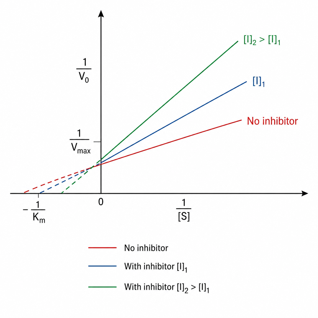
A 45-year-old patient presents with joint pain and weakness and is known to have homocystinuria. Which vitamin is required in the treatment?
A neonate was brought to the hospital with chief complaints of poor feeding, vomiting, acidosis, and cataract. Benedict's test on urine was positive, but urinary glucose was negative. What is the defective enzyme in the above-mentioned disorder?
A patient's relatives sent a message on social media to the consulting doctor, mentioning that the patient's urinary coproporphyrin test is positive. What is the probable cause?
A 5-year-old girl was washing her doll with shampoo containing rotenone. Her mother noticed her in an unconscious state. Which enzyme is inhibited by the above chemical?
A patient presents with symptoms of dermatitis, dementia, and cognitive decline. Which micronutrient deficiency is most likely responsible?
A mother brings her 14 -year-old mentally retarded son to the OPD, who is suffering from joint pain and has an irresistible urge to bite his fingers and lips. His serum uric acid level was found to be elevated. What is the disorder?
An infant presents with vomiting after feeding. Benedict's test was positive for a non-glucose reducing substance. What is the most likely diagnosis?
Explanation: ***Whole grain cereals and legumes*** - **Whole grain cereals** (brown rice, oats, wheat germ, fortified cereals) and **legumes** (beans, lentils, peas) are **excellent natural sources of thiamine (vitamin B1)** - They retain the **bran and germ** layers where thiamine is concentrated - Regular consumption effectively prevents **thiamine deficiency** and associated conditions like beriberi and Wernicke-Korsakoff syndrome - This is the **primary dietary recommendation** for thiamine adequacy *Polished white rice* - Polished white rice has the **bran and germ removed** during processing, eliminating most of the thiamine content - Consuming polished white rice as a staple **causes thiamine deficiency**, leading to **beriberi** (common in populations with rice-based diets) - Unless fortified with thiamine, polished white rice contributes to deficiency rather than preventing it *Dairy products and eggs* - While nutritious, dairy products and eggs contain **relatively low amounts of thiamine** - Not reliable sources for meeting daily thiamine requirements - Contribute to overall nutrition but insufficient alone to prevent thiamine deficiency *Fresh fruits and vegetables* - Most fruits and vegetables contain **modest amounts of thiamine** compared to whole grains - Some exceptions include peas, asparagus, and Brussels sprouts, but typical servings provide limited thiamine - Important for overall health but not primary thiamine sources *Red meat and poultry* - Pork is actually a **good source of thiamine**, particularly organ meats - However, **whole grains and legumes** remain the most reliable and accessible plant-based sources - Red meat and poultry provide moderate thiamine but are not the best answer for preventing deficiency in general populations
Explanation: ***Vitamin B6*** - Many cases of **homocystinuria** are due to a deficiency in **cystathionine beta-synthase (CBS)**, an enzyme that requires **pyridoxal phosphate (active B6)** as a cofactor. - Supplementing with **high doses of vitamin B6** can significantly improve outcomes in patients with **B6-responsive homocystinuria** by increasing residual CBS enzyme activity. *Vitamin B12* - While vitamin B12 (cobalamin) is a cofactor for **methionine synthase**, an enzyme involved in homocysteine metabolism, it is not the primary treatment for **homocystinuria due to CBS deficiency**. - B12 deficiency can lead to increased homocysteine levels, but B12 supplementation alone will not address the underlying enzyme defect in most cases of homocystinuria. *Thiamine* - **Thiamine (Vitamin B1)** is essential for carbohydrate metabolism and nerve function, but it has no direct role in the metabolic pathway involved in homocystinuria. - Thiamine deficiency causes **beriberi** and **Wernicke-Korsakoff syndrome**, symptoms distinctly different from homocystinuria. *Vitamin B9 (Folate)* - **Folate** is a cofactor for **methionine synthase**, working alongside vitamin B12 to convert homocysteine back to methionine. - While folate deficiency can contribute to hyperhomocysteinemia, it is not the primary therapeutic intervention for homocystinuria caused by **CBS deficiency**. *Vitamin C* - **Vitamin C (ascorbic acid)** is important for collagen synthesis and acts as an antioxidant, but it has no role in homocysteine metabolism or the treatment of homocystinuria. - Vitamin C deficiency causes **scurvy**, which presents with bleeding gums, petechiae, and poor wound healing—completely unrelated to homocystinuria.
Explanation: ***Chemotaxis*** - **IL-8** (also known as **CXCL8**) is a potent **chemokine** that primarily functions to recruit and activate **neutrophils** to sites of inflammation. - It guides these immune cells by forming a chemical gradient, allowing them to extravasate from blood vessels and migrate to the infected or injured tissue. *Lymphocyte proliferation* - Lymphocyte proliferation is primarily stimulated by **IL-2**, which acts as a growth factor for T cells. - While IL-8 contributes to the inflammatory environment, its direct role in inducing lymphocyte cell division is minor compared to its chemotactic function. *TH1 activation* - **TH1 cell activation** is mainly driven by **IL-12** and **IFN-γ**, which promote the differentiation and function of T helper 1 cells, crucial for fighting intracellular pathogens. - IL-8's role is unrelated to guiding TH1 cell differentiation. *Fever* - Fever is primarily induced by **pyrogenic cytokines** such as **IL-1**, **IL-6**, and **TNF-α**, which act on the hypothalamus to raise body temperature. - While IL-8 is part of the inflammatory response that can sometimes coincide with fever, it does not directly act as a pyrogen. *B cell activation* - **B cell activation** and differentiation are primarily regulated by interleukins such as **IL-4**, **IL-5**, **IL-6**, and **IL-21**, which promote antibody production and class switching. - IL-8 does not play a significant role in B cell function; its action is focused on innate immunity, particularly neutrophil recruitment.
Explanation: ***Vitamin B6*** - **Homocystinuria** is often caused by a deficiency in the enzyme **cystathionine beta-synthase**, which requires **pyridoxal phosphate (active form of B6)** as a cofactor. - Supplementation with high-dose **vitamin B6** can help some patients by increasing the residual activity of the enzyme, thereby reducing **homocysteine levels**. - This is the **primary treatment** for **B6-responsive homocystinuria** (approximately 50% of cases respond to B6 therapy). *Vitamin B12* - Vitamin B12 is a cofactor for the enzyme **methionine synthase**, which converts homocysteine back to methionine. - While it plays a role in homocysteine metabolism, **vitamin B6** is typically the primary treatment for homocystinuria caused by **cystathionine beta-synthase deficiency**. *Vitamin B9* - Vitamin B9 (folic acid) works together with **vitamin B12** as a cofactor in the **remethylation pathway** via methionine synthase. - While folate supplementation may help lower homocysteine levels, it is **not the primary treatment** for classical homocystinuria due to cystathionine beta-synthase deficiency. - **Vitamin B6** remains the first-line vitamin therapy for enzyme deficiency-related homocystinuria. *Vitamin B7* - Vitamin B7, or **biotin**, is a cofactor for carboxylase enzymes and is involved in fatty acid synthesis and gluconeogenesis. - It has no direct role in the metabolism of **homocysteine** or the treatment of homocystinuria. *Vitamin B1* - Vitamin B1, or **thiamine**, is essential for carbohydrate metabolism and nerve function. - It is not involved in the metabolic pathways that regulate **homocysteine levels** or the treatment of homocystinuria.
Explanation: ***Galactose 1-phosphate uridyl transferase*** - This enzyme deficiency leads to **classic galactosemia**, characterized by the accumulation of **galactose-1-phosphate**, which is toxic. - Clinical features like **poor feeding, vomiting, acidosis, and cataracts** are typical, and a positive **Benedict's test** (detecting reducing sugars like galactose) with negative urinary glucose confirms the presence of another reducing sugar. *Fructokinase* - Deficiency of fructokinase causes **essential fructosuria**, a benign condition where **fructose** accumulates in the urine. - Unlike classic galactosemia, it does not present with severe symptoms like **acidosis** or **cataracts**. *Aldolase B* - **Aldolase B deficiency** causes hereditary fructose intolerance, presenting with **vomiting, hypoglycemia, and hepatomegaly** after fructose ingestion. - It does not cause **cataracts**, and Benedict's test would detect fructose, but the clinical context (symptoms with fructose/sucrose intake) differs from galactosemia. *Lactase* - **Lactase deficiency** (lactose intolerance) results in gastrointestinal symptoms such as **bloating, diarrhea, and abdominal pain** upon lactose consumption. - It does not typically cause **acidosis, cataracts**, or a positive Benedict's test in urine unless secondary bacterial fermentation leads to other reducing substances. *Sucrase* - **Sucrase-isomaltase deficiency** leads to the malabsorption of sucrose, causing symptoms similar to lactose intolerance like **diarrhea and abdominal cramping**. - It does not result in the systemic, severe metabolic derangements or signs like **cataracts** seen in classic galactosemia.
Explanation: ***Lead poisoning*** - **Lead poisoning** is associated with an increase in **urinary coproporphyrin III**, as lead inhibits the enzyme **coproporphyrinogen oxidase** in the heme synthesis pathway. - This leads to the accumulation and excretion of **coproporphyrin III** in the urine, making it a valuable biomarker for lead exposure. *Asbestosis* - **Asbestosis** is a chronic lung disease caused by inhaling **asbestos fibers**, leading to diffuse interstitial fibrosis. - It does not directly affect the **heme synthesis pathway** or cause an increase in urinary coproporphyrins. *Silicosis* - **Silicosis** is a chronic occupational lung disease caused by inhaling **crystalline silica dust**, resulting in pulmonary fibrosis. - It is not associated with alterations in **porphyrin metabolism** or increased urinary coproporphyrin levels. *Mercury poisoning* - While **mercury poisoning** can affect various organ systems, including renal and neurological, it is not primarily associated with disturbances in the **heme synthesis pathway** or elevated urinary coproporphyrins. - **Mercury poisoning** often manifests with symptoms like **tremors**, **neurological deficits**, and **kidney damage**. *Arsenic poisoning* - **Arsenic poisoning** causes a variety of systemic effects including gastrointestinal symptoms, peripheral neuropathy, and skin changes (hyperpigmentation, hyperkeratosis). - Unlike lead poisoning, **arsenic does not significantly elevate urinary coproporphyrin III** levels, making it distinguishable from lead toxicity through this biomarker.
Explanation: ***NADH dehydrogenase*** - **Rotenone** is a potent **inhibitor of mitochondrial complex I (NADH dehydrogenase)**, preventing the transfer of electrons from NADH to ubiquinone. - This inhibition disrupts the **electron transport chain**, leading to a halt in ATP synthesis and cellular energy failure, causing symptoms like unconsciousness. *Succinate dehydrogenase* - **Succinate dehydrogenase** (Complex II) is involved in both the **Krebs cycle and electron transport chain**, but is not directly inhibited by rotenone. - Only severe compromise of the electron transport chain can cause a secondary effect, but not direct enzyme inhibition. *Cytochrome C* - **Cytochrome C** is a mobile electron carrier in the electron transport chain, but it is not directly inhibited by rotenone. - **Cytochrome C** transfers electrons from complex III to complex IV. *Cytochrome oxidase* - **Cytochrome oxidase** (Complex IV) is responsible for the final transfer of electrons to oxygen, which is not inhibited by rotenone. - Inhibitors like **cyanide and carbon monoxide** specifically target **cytochrome oxidase**. *Cytochrome b-c1 complex* - **Cytochrome b-c1 complex** (Complex III) catalyzes electron transfer from ubiquinol to cytochrome C, but is not inhibited by rotenone. - This complex is specifically inhibited by **antimycin A**, not rotenone.
Explanation: ***Niacin*** - The classic triad of symptoms known as the "3 Ds"—**dermatitis, dementia, and diarrhea**—is characteristic of **pellagra**, a severe deficiency of **niacin (vitamin B3)**. - **Cognitive decline** is a common manifestation of the neurological symptoms associated with dementia in pellagra. *Thiamine* - **Thiamine (vitamin B1)** deficiency causes **beriberi**, leading to cardiovascular (wet beriberi) or neurological (dry beriberi/Wernicke-Korsakoff syndrome) symptoms. - While **fatigue and cognitive impairment** can occur, the constellation of prominent dermatitis and severe dementia is not typical of primary thiamine deficiency. *Tryptophan* - **Tryptophan** is an **essential amino acid** and a precursor to niacin. A deficiency in tryptophan could indirectly lead to niacin deficiency. - However, the direct deficiency of tryptophan as the primary cause of the "3 Ds" is less common than a direct niacin deficiency or impaired niacin synthesis. *Retinol* - **Retinol (vitamin A)** deficiency primarily affects **vision**, leading to **night blindness** and xerophthalmia. - It also plays a role in immune function and epithelial cell integrity, but it does not cause the specific triad of dermatitis, dementia, and cognitive decline seen here. *Riboflavin* - **Riboflavin (vitamin B2)** deficiency causes **angular stomatitis, cheilosis, and glossitis** along with seborrheic dermatitis. - While it can cause skin manifestations, it does not typically present with the severe dementia and cognitive decline characteristic of pellagra.
Explanation: ***Lesch-Nyhan syndrome*** - This syndrome is characterized by **X-linked recessive inheritance** and a deficiency of the enzyme **hypoxanthine-guanine phosphoribosyltransferase (HGPRT)**, leading to an overproduction of uric acid. - Clinical manifestations include **hyperuricemia**, **gout-like arthritis**, **neurological dysfunction** (mental retardation, dystonia), and a distinctive feature of **self-mutilating behaviors** such as biting fingers and lips. *Xanthine oxidase deficiency* - This condition leads to an accumulation of **hypoxanthine** and **xanthine** due to impaired conversion to uric acid. - While it can cause kidney stones and some joint pain, it typically results in *low* serum uric acid levels and does not present with the characteristic self-mutilating behavior or severe neurological deficits seen in Lesch-Nyhan. *CPS II defect* - **Carbamoyl phosphate synthetase II (CPS II)** is involved in *pyrimidine synthesis*, not purine metabolism. - A defect in CPS II would lead to issues with pyrimidine production and typically presents with megaloblastic anemia or other symptoms related to pyrimidine deficiency, not hyperuricemia or self-mutilation. *Thymidylate synthetase* - **Thymidylate synthetase (TS)** is an enzyme critical for the synthesis of **thymidylate**, a precursor for DNA synthesis. - Inhibition or deficiency of TS is primarily associated with DNA replication issues, megaloblastic anemia, and is a target for chemotherapy, not with hyperuricemia or the neurological and behavioral symptoms described. *Von Gierke disease* - **Von Gierke disease** (Glycogen storage disease type Ia) is caused by deficiency of **glucose-6-phosphatase**, leading to impaired conversion of glucose-6-phosphate to glucose. - While it can cause **hyperuricemia** due to increased uric acid production from accelerated purine degradation, the clinical presentation is distinct with **hepatomegaly**, **hypoglycemia**, **lactic acidosis**, and growth retardation. - It does **not** present with self-mutilating behavior or the severe neurological dysfunction characteristic of Lesch-Nyhan syndrome.
Explanation: ***Galactosemia due to GAL-1-P Uridyl Transferase enzyme deficiency*** - Vomiting after feeding in an infant, coupled with a **positive Benedict's test** for a **non-glucose reducing substance**, is highly indicative of galactosemia. The accumulation of **galactose-1-phosphate** and **galactitol** leads to toxicity and symptoms. - This enzyme deficiency, causing **classic galactosemia**, prevents the proper metabolism of **galactose**, leading to its buildup. *Fructosuria due to Fructokinase deficiency* - This condition is a **benign metabolic disorder** with no significant clinical symptoms. - While it would lead to fructose in the urine, the infant would not typically present with **vomiting after feeding**. *Hereditary fructose intolerance due to Aldolase B deficiency* - Symptoms usually appear after the introduction of **fructose-containing foods** into the diet, causing severe hypoglycemia and vomiting. - The positive Benedict's test in this scenario would typically indicate a reducing substance in the urine, while fructose intolerance is characterized by **hypoglycemia** and metabolic crises upon fructose ingestion. *Glycogen storage disease due to Glucose-6-phosphatase deficiency* - This disorder primarily causes **hypoglycemia** and liver enlargement, not primarily vomiting after feeding due to a **non-glucose reducing substance**. - Glucose-6-phosphatase deficiency (Von Gierke's disease) leads to an inability to release **glucose from glycogen** and causes severe hypoglycemia, often requiring frequent feeding. *Primary lactose intolerance* - While lactose intolerance can cause vomiting and gastrointestinal symptoms, it is **extremely rare in infants** (primary lactose intolerance is a late-onset condition). - Lactose would be a reducing sugar, but the key differentiator is that **galactose** (from galactosemia) is the non-glucose reducing substance detected in this case, along with the typical **toxic presentation** in neonates.
Explanation: ***Copper*** - **Copper deficiency** can lead to anemia due to its role in iron metabolism, **depigmented hair** (achromotrichia) due to impaired melanin synthesis, and **myelopathy** due to its involvement in maintaining the myelin sheath. - Symptoms often mimic those of **vitamin B12 deficiency**, including neurological manifestations like ataxia and spasticity. *Iron* - **Iron deficiency** is the most common cause of anemia but does not typically cause **depigmented hair** or **myelopathy**. - Its neurological symptoms are usually limited to **restless legs syndrome** and **pica**, not demyelination. *Fluoride* - **Fluoride deficiency** is primarily associated with an increased risk of dental caries and does not cause anemia, hair depigmentation, or myelopathy. - Excessive intake (fluorosis) can lead to **bone and tooth abnormalities**. *Zinc* - **Zinc deficiency** can cause immune dysfunction, **dermatitis**, impaired wound healing, and growth retardation. - It may rarely cause anemia in severe cases but does not typically cause hair depigmentation or myelopathy as primary symptoms. *Selenium* - **Selenium deficiency** is associated with **Keshan disease** (cardiomyopathy) and **Kashin-Beck disease** (osteoarthropathy). - While it can cause muscle weakness and fatigue, it does not typically present with the specific triad of anemia, hair depigmentation, and myelopathy seen in copper deficiency.
Explanation: ***Cysteine*** - In **cystathionine synthase deficiency** (homocystinuria), the body cannot convert **homocysteine to cystathionine**, and subsequently to **cysteine**. - **Cysteine** therefore becomes an **essential amino acid** in these patients and must be supplemented. *Tyrosine* - **Tyrosine** is a non-essential amino acid synthesized from **phenylalanine**, and its deficiency is not directly related to cystathionine synthase deficiency. - It is not involved in the **methionine or homocysteine metabolic pathway** that is disrupted in homocystinuria. *Serine* - **Serine** is a substrate for the **cystathionine synthase enzyme**, which combines with **homocysteine** to form **cystathionine**. - Supplementation with serine alone would not bypass the enzyme deficiency or provide the essential product, **cysteine**. *Methionine* - **Methionine** is the precursor to **homocysteine**, and in cystathionine synthase deficiency, there's often an accumulation of methionine and homocysteine. - Therefore, **methionine restriction** is typically part of the treatment, not supplementation. *Glycine* - **Glycine** is involved in one-carbon metabolism but is not directly involved in the **transsulfuration pathway** affected by cystathionine synthase deficiency. - Glycine supplementation would not address the inability to synthesize **cysteine** from homocysteine.
Explanation: ***Niacin deficiency*** - The symptoms described (dermatitis that worsens with sun exposure, glossitis, and a diet high in maize) are characteristic of **pellagra**, which is caused by a **niacin (Vitamin B3) deficiency**. - **Maize** is a poor source of niacin, and the niacin it contains is largely bound and unavailable, leading to higher rates of deficiency in populations relying on it as a staple. - The classic **3 D's of pellagra** are: **Dermatitis** (photosensitive), **Diarrhea**, and **Dementia**. *Riboflavin deficiency* - **Riboflavin (Vitamin B2) deficiency** causes **angular stomatitis**, **cheilosis**, and **glossitis**, but the dermatitis is typically **seborrheic** (not photosensitive). - The **photosensitive** nature of the rash in this case is the key differentiating feature pointing to niacin deficiency. *Vitamin C deficiency* - **Vitamin C deficiency** leads to **scurvy**, characterized by **gingival bleeding**, **poor wound healing**, and **petechiae**, none of which are mentioned here. - While it can manifest with fatigue, it does not typically cause a photosensitive rash or glossitis as described. *Vitamin K deficiency* - **Vitamin K deficiency** primarily affects **blood clotting**, leading to **bleeding disorders** and **easy bruising**. - It is not associated with skin rashes or glossitis. *Folic acid deficiency* - **Folic acid deficiency** causes **macrocytic anemia**, fatigue, and potentially **glossitis**, but it does not cause a photosensitive rash. - The distinctive combination of photosensitive dermatitis and glossitis points away from folic acid deficiency.
Explanation: ***Homogentistae oxidase*** - The presentation of a child with **black urine** (alkaptonuria) in the absence of other symptoms is characteristic of a deficiency in **homogentisate oxidase**. - This enzyme is crucial in the catabolism of **tyrosine**, and its deficiency leads to the accumulation of **homogentisic acid**, which oxidizes upon exposure to air, turning urine black. *Homogentisate dehydrogenase* - This enzyme is not a recognized component of the **tyrosine degradation pathway** in humans. - The correct enzyme involved in the breakdown of **homogentisate** is an oxidase, not a dehydrogenase, in this context. *Tyrosine Transaminase* - A deficiency in **tyrosine transaminase** (tyrosinemia type II) would lead to elevated tyrosine levels and typically presents with symptoms affecting the eyes, skin, and intellectual disability, not primarily black urine. - This condition is characterized by **ocular findings** (corneal ulcers), **skin lesions**, and **neurological symptoms**. *Tryptophan Hydroxylase* - This enzyme is involved in the synthesis of **serotonin** and **melatonin** from tryptophan, a different amino acid pathway. - A deficiency or abnormality in **tryptophan hydroxylase** would not cause black urine but could lead to neurological or mood disorders. *Phenylalanine Hydroxylase* - A deficiency in **phenylalanine hydroxylase** causes **phenylketonuria (PKU)**, which affects phenylalanine metabolism, not tyrosine metabolism directly. - PKU typically presents with **intellectual disability**, **musty odor**, **fair skin**, and **seizures** if untreated, not black urine.
Explanation: ***Liver Glycogen*** - After an overnight fast (approximately 11 hours in this scenario), the primary mechanism for maintaining blood glucose levels is the breakdown of **liver glycogen** stores. - The liver is crucial for glucose homeostasis as it can release glucose directly into the bloodstream, a function muscle glycogen cannot perform. *Muscle Glycogen* - **Muscle glycogen** serves as an energy reserve primarily for the muscle itself and cannot be directly released into the bloodstream to maintain blood glucose levels. - It is utilized for physical activity and local energy demands within muscle cells. *Gluconeogenesis* - **Gluconeogenesis**, the synthesis of glucose from non-carbohydrate precursors, becomes increasingly important for glucose production after prolonged fasting (typically *after* liver glycogen stores are depleted). - While it contributes during an overnight fast, **liver glycogenolysis** is the dominant source initially. *Dietary Carbohydrate* - **Dietary carbohydrates** from the previous dinner (8 PM) would have been absorbed and utilized or stored as glycogen much earlier than 7 AM the next morning. - By 7 AM, the direct impact of the previous night's meal on circulating glucose is negligible, having been processed hours before. *Ketone Bodies* - **Ketone bodies** are alternative fuel sources produced during prolonged fasting or starvation, but they are **not glucose**. - While they can be used by tissues (brain, heart, muscle) for energy during extended fasting, they do not contribute to blood glucose levels and are metabolically distinct from glucose.
Explanation: ***Cysteine*** - In **cystathionine beta synthase (CBS) deficiency**, the conversion of **homocysteine** to **cystathionine** (and subsequently to cysteine) is impaired. - This makes **cysteine** an **essential amino acid** for these patients, requiring dietary supplementation. *Tryptophan* - **Tryptophan** is an **essential amino acid** and a precursor for **serotonin** and **niacin**, but its metabolism is not directly affected by CBS deficiency. - Its supplementation is not specifically indicated for this condition. *Serine* - **Serine** is a **non-essential amino acid** that provides the **carbon skeleton** for the synthesis of cysteine from homocysteine in the presence of CBS. - While important in the pathway, CBS deficiency specifically disrupts the downstream conversion of homocysteine, making **cysteine** the deficient product, not serine. *Methionine* - **Methionine** is an **essential amino acid** that is a precursor to **homocysteine**; in CBS deficiency, homocysteine levels are already elevated due to impaired conversion to cystathionine. - Restricting methionine intake is typically recommended in CBS deficiency to reduce homocysteine accumulation, not supplementing it. *Tyrosine* - **Tyrosine** is a **non-essential amino acid** derived from **phenylalanine** and serves as a precursor for catecholamines and thyroid hormones. - Its metabolism is not affected by CBS deficiency, and supplementation is not indicated for this condition.
Explanation: ***Niacin*** - The classic presentation of **pellagra**, caused by a deficiency of **niacin (Vitamin B3)**, is characterized by the "**3 Ds**": **dermatitis**, **diarrhea**, and **dementia**. In severe cases, a fourth 'D' for death can also occur. - A **maize (corn)** staple diet is a known risk factor for niacin deficiency because maize contains niacin in a bound, non-bioavailable form (niacytin) and is low in tryptophan, a precursor to niacin. *Riboflavin* - **Riboflavin (Vitamin B2)** deficiency leads to **ariboflavinosis**, which can cause **cheilosis**, **angular stomatitis**, **glossitis**, and **seborrheic dermatitis**, but not the constellation of diarrhea, dementia, and dermatitis seen in pellagra. - It does not typically manifest with neurological or gastrointestinal symptoms as severe as those described in the question. *Thiamine* - **Thiamine (Vitamin B1)** deficiency causes **beriberi**, characterized by **neurological (dry beriberi)** or **cardiovascular (wet beriberi)** symptoms. - It can lead to **Wernicke-Korsakoff syndrome** in severe cases, which includes neurological deficits, but not the specific "3 Ds" of pellagra. *Pyridoxine* - **Pyridoxine (Vitamin B6)** deficiency can cause **neurological symptoms** such as **peripheral neuropathy**, **seizures**, and **depression**, as well as **dermatitis** and **glossitis**. - It does not present with the characteristic triad of dermatitis, diarrhea, and dementia seen in pellagra. *Cobalamin* - **Cobalamin (Vitamin B12)** deficiency causes **megaloblastic anemia** and **neurological symptoms** including **subacute combined degeneration** of the spinal cord, **peripheral neuropathy**, and **cognitive changes**. - While it can cause neurological symptoms, it does not present with the classic dermatitis and diarrhea combination seen in pellagra.
Explanation: ***Homogentisic acid oxidase*** - The patient's symptoms, including **black urine (alkaptonuria)**, **scleral darkening (ochronosis)**, and progressive **arthralgia** in multiple joints, are classic manifestations of **alkaptonuria**. - **Alkaptonuria** is an autosomal recessive disorder caused by a deficiency of **homogentisic acid oxidase**, an enzyme important in the metabolism of **tyrosine**. *Cystathionine synthase deficiency* - This deficiency causes **homocystinuria**, characterized by Marfanoid habitus, mental retardation, and premature atherosclerosis. - It does not present with black urine or joint pain in this manner. *Branched-chain alpha-ketoacid dehydrogenase* - A deficiency in this enzyme leads to **maple syrup urine disease**, an inborn error of metabolism affecting branched-chain amino acids. - Symptoms include neurological deterioration, seizures, and a distinctive maple syrup odor in urine, not black urine or joint issues. *Phenylalanine hydroxylase* - This deficiency causes **phenylketonuria (PKU)**, characterized by intellectual disability, seizures, and fair skin/hair if untreated. - Patients typically have a mousy odor to their urine, but it does not turn black upon exposure to air, nor do they develop ochronosis or specific arthralgia. *Propionyl-CoA carboxylase* - A deficiency in this enzyme leads to **propionyl-CoA carboxylase deficiency**, a type of organic acidemia. - It presents with metabolic acidosis, hyperammonemia, and developmental delay, not with black urine or the specific musculoskeletal symptoms described.
Explanation: ***Avoid meat*** - This patient's symptoms are highly suggestive of **Phenylketonuria (PKU)** a genetic disorder causing an inability to metabolize **phenylalanine**. Meat is a high source of phenylalanine and must be avoided. - The distinctive **musty odor** is due to the accumulation of **phenylacetate** which is a metabolite of phenylalanine. *Avoid fresh fruits* - **Fresh fruits** are generally low in protein and phenylalanine, making them safe to consume for individuals with PKU. - There is no medical basis for avoiding fresh fruits in the management of PKU or any related conditions. *Increase intake of dairy products* - **Dairy products** such as milk, cheese, and yogurt are rich in protein, and therefore high in phenylalanine. - Increasing their intake would exacerbate the accumulation of **phenylalanine** in individuals with PKU. *Increase intake of bread* - While some breads might be lower in protein than meat or dairy, regular bread still contains a significant amount of **protein** and thus **phenylalanine**. - A special **low-protein bread** might be used, but increasing intake of typical bread is not advised for PKU patients. *Switch to diet soda* - Many diet sodas contain the artificial sweetener **aspartame**, which is metabolized in the body into **phenylalanine**. - Consumption of aspartame-containing products is specifically contraindicated in patients with PKU.
Explanation: ***Methylmalonic acid*** - In **vitamin B12 deficiency**, **methylmalonyl-CoA mutase** (which requires **adenosylcobalamin**, a form of B12) is impaired, leading to accumulation of **methylmalonyl-CoA**, which is converted to **methylmalonic acid (MMA)**. - **Elevated MMA** is a **sensitive and specific marker** for **vitamin B12 deficiency** because this metabolic pathway is **exclusively dependent on B12**. - MMA levels remain **normal in folate deficiency**, making it useful for distinguishing between these two causes of megaloblastic anemia. *Methionine* - **Methionine** is synthesized from **homocysteine** via **methionine synthase**, which requires **methylcobalamin** (vitamin B12) as a cofactor. - In B12 deficiency, this reaction is impaired, so **methionine levels decrease** or remain normal, not elevated. *Succinyl-CoA* - **Succinyl-CoA** is the **product** of the methylmalonyl-CoA mutase reaction in the TCA cycle. - In B12 deficiency, the **precursor** (methylmalonyl-CoA/MMA) accumulates because it cannot be converted to succinyl-CoA. - Succinyl-CoA levels would be **decreased or normal**, not elevated. *Homocysteine* - **Homocysteine is also elevated** in vitamin B12 deficiency due to impaired methionine synthase activity. - However, **elevated homocysteine is NOT specific** for B12 deficiency—it is also elevated in **folate deficiency**, **vitamin B6 deficiency**, and other conditions. - **Methylmalonic acid** is more specific because it is elevated **only in B12 deficiency**, not in folate deficiency, making it the better marker when both are elevated. *Cysteine* - **Cysteine** is synthesized from **methionine** via the **transsulfuration pathway** (requiring vitamin B6, not B12). - Cysteine levels are **not affected** by vitamin B12 status and are not used diagnostically for B12 deficiency.
Explanation: ***Tyrosine*** - The constellation of symptoms, including dark discoloration of sweat (brown stains around armpits), bluish-brown sclerae, thickening of the ear cartilage (ochronosis), back and joint pain, and calcification of intervertebral discs, is classic for **alkaptonuria**. - **Alkaptonuria** is an autosomal recessive disorder caused by a deficiency of homogentisate 1,2-dioxygenase, an enzyme involved in the metabolism of **tyrosine**. This leads to the accumulation of **homogentisic acid**. *Ornithine* - Impaired metabolism of ornithine is associated with disorders of the **urea cycle**, such as **ornithine transcarbamylase deficiency**. - These conditions primarily lead to **hyperammonemia** and neurological symptoms, not the musculoskeletal or ochronotic features described. *Hypoxanthine* - Hypoxanthine is a purine derivative whose metabolism is relevant in disorders like **gout** (due to overproduction or underexcretion of uric acid, a metabolite of hypoxanthine) and **Lesch-Nyhan syndrome**. - These conditions do not present with the characteristic features of ochronosis or multijoint calcification seen in this patient. *Homocysteine* - Impaired metabolism of homocysteine is characteristic of disorders like **homocystinuria**, which can be caused by deficiencies in enzymes such as **cystathionine beta-synthase**. - Homocystinuria typically presents with lens dislocation, Marfanoid habitus, developmental delay, and thromboembolic events, not ochronosis or joint calcification. *Tryptophan* - Impaired metabolism of tryptophan can be seen in conditions such as **Hartnup disease** (a defect in amino acid transport) or in deficiencies affecting **niacin synthesis**. - These conditions typically manifest with symptoms like pellagra-like dermatosis, cerebellar ataxia, and neuropsychiatric disturbances, which are not consistent with the patient's presentation.
Explanation: ***Cystathionine β-synthase*** - This patient's presentation with **Marfanoid habitus** (long arms, slender fingers, dislocated lens), sudden onset neurological deficits (weakness, facial droop, positive Babinski), and elevated metabolic intermediates points to **homocystinuria**. Homocystinuria is most commonly caused by a deficiency in **cystathionine β-synthase (CBS)**, leading to the accumulation of homocysteine. - Elevated homocysteine levels are highly prothrombotic, explaining the **stroke-like symptoms** (right-sided weakness, facial droop, aphasia) in a young patient without traditional risk factors for stroke. *Homogentisate oxidase* - Deficiency in **homogentisate oxidase** causes **alkaptonuria**, characterized by **dark urine** upon standing, ochronosis (bluish-black discoloration of cartilage and connective tissues), and severe arthritis. - Alkaptonuria does not typically present with acute thrombotic events, Marfanoid features, or specific neurological deficits. *Branched-chain ketoacid dehydrogenase* - A defect in **branched-chain ketoacid dehydrogenase** is responsible for **Maple Syrup Urine Disease (MSUD)**, which typically presents in infancy with poor feeding, vomiting, lethargy, developmental delay, and urine smelling like maple syrup. - This enzyme deficiency is not associated with the Marfanoid habitus or acute thrombotic neurological events described in this patient. *Hydroxymethylbilane (HMB) synthase* - Deficiency of **HMB synthase** (also known as porphobilinogen deaminase) causes **Acute Intermittent Porphyria (AIP)**, characterized by acute attacks of severe abdominal pain, psychiatric symptoms (anxiety, depression, hallucinations), and neurological dysfunction (peripheral neuropathy, seizures). - AIP does not cause Marfanoid features or tend to present with acute stroke-like symptoms due to thrombosis. *Phenylalanine hydroxylase* - A deficiency in **phenylalanine hydroxylase** causes **phenylketonuria (PKU)**, which is typically identified through newborn screening. Symptoms, if untreated, include intellectual disability, seizures, and a musty odor. - PKU does not present with Marfanoid features, dislocated lenses, or acute thrombotic complications leading to stroke.
Explanation: **Leucine** - The patient has **maple syrup urine disease (MSUD)**, a disorder of branched-chain amino acid metabolism, involving **leucine**, isoleucine, and valine. - While all three are restricted, **leucine levels are often the primary driver of neurotoxicity** and require careful monitoring and management during infancy and childhood to support growth while preventing metabolic crises. *Tryptophan* - Tryptophan is an amino acid affected in disorders like **Hartnup disease** or defects in serotonin/melatonin synthesis, neither of which align with the patient's presentation. - It is not a branched-chain amino acid and therefore not directly involved in MSUD. *Alanine* - Alanine is a non-essential amino acid commonly used in **gluconeogenesis** and the alanine cycle. - It is not a branched-chain amino acid nor is it primarily implicated in MSUD. *Asparagine* - Asparagine is a non-essential amino acid involved in **protein synthesis** and the urea cycle. - It is not a branched-chain amino acid and is not directly related to MSUD. *Methionine* - Methionine is an essential amino acid involved in **homocysteine metabolism** and is associated with disorders like homocystinuria. - It is not a branched-chain amino acid and is not the primary amino acid of concern in MSUD.
Explanation: ***Deficit of phenylalanine hydroxylase activity*** - The combination of **developmental delay**, **microcephaly**, and a **mousy odor** is characteristic of **phenylketonuria (PKU)**. - PKU is caused by a deficient **phenylalanine hydroxylase** enzyme, leading to a buildup of phenylalanine and its metabolites, which are toxic to the developing brain. *Excess tetrahydrobiopterin cofactor* - This condition (**BH4 excess**) is rare and does not typically present with the classic signs of PKU; rather, it often involves neurological symptoms due to other metabolic imbalances. - An excess of the BH4 cofactor would theoretically enhance rather than inhibit phenylalanine hydroxylase activity, if the enzyme itself were functional. *Deficit of porphobilinogen deaminase activity* - A deficit in **porphobilinogen deaminase** is associated with **Acute Intermittent Porphyria (AIP)**, which presents with acute neurovisceral attacks. - Symptoms of AIP include severe abdominal pain, psychiatric disturbances, and neurological deficits, but not developmental delay or a mousy odor. *Deficit of tyrosine hydroxylase activity* - A deficiency in **tyrosine hydroxylase** affects the synthesis of **dopamine** and other catecholamines, leading to neurological disorders, including **dystonia** and **Parkinsonian symptoms**. - While it can cause developmental delay, it does not typically present with a mousy odor or microcephaly, and its primary symptoms relate to motor control. *Excess phenylalanine hydroxylase activity* - An **excess** of phenylalanine hydroxylase activity would lead to increased breakdown of phenylalanine, preventing its buildup. - This would not cause the symptoms described; instead, it would likely result in lower-than-normal phenylalanine levels, which is generally not problematic.
Explanation: ***Glutathione depletion*** - The clinical presentation (recent URI, fever, generalized seizure, rapid clinical deterioration with **hepatic dysfunction**, and **encephalopathy** in a 2-year-old) is highly suggestive of **Reye's syndrome**. - While Reye's syndrome is primarily associated with **aspirin use in viral infections**, a similar syndrome can be induced by other medications or toxins. The mention of parents medicating her at home for fever raises suspicion for **acetaminophen overdose** given the hepatic failure, in which glutathione depletion is the key mechanism of toxicity. *Decrease in hypothalamic set point* - A decrease in the hypothalamic set point would lead to **hypothermia**, not the documented fever of 38.9°C (102.0°F). - This finding is inconsistent with the patient's presentation of fever and acute illness. *Ca2+ efflux* - While altered calcium homeostasis can occur in various severe illnesses, **Ca2+ efflux** itself is not the primary or most likely underlying mechanism for the described **hepatic failure** and encephalopathy. - This is a more general cellular phenomenon and lacks the specificity to explain the entire clinical picture definitively. *Copper deposition* - **Copper deposition** is a hallmark of **Wilson's disease**, a genetic disorder causing liver disease and neurological symptoms. - However, Wilson's disease typically presents with more chronic symptoms and is less likely to cause such an acute, fulminant presentation in a 2-year-old following an infection and home medication. *Hemosiderin deposition* - **Hemosiderin deposition** (hemochromatosis) is characterized by iron overload, primarily affecting the liver, heart, and pancreas. - It typically presents as a chronic condition and is not consistent with the acute, fulminant liver failure and encephalopathy observed in this young child.
Explanation: ***No intervention is needed*** - This patient's lab results show an **unconjugated hyperbilirubinemia** (indirect bilirubin is significantly elevated compared to direct bilirubin), which, in an **asymptomatic** patient, is characteristic of **Gilbert's syndrome**. - **Gilbert's syndrome** is a benign genetic condition requiring no treatment or intervention; patients are typically asymptomatic or experience mild, transient jaundice during times of stress, fasting, or illness. *Regular monitoring of liver enzymes is crucial* - This statement is generally not true for **Gilbert's syndrome** as it is a benign condition that does not cause liver damage or inflammation, so routine monitoring of liver enzymes (**AST, ALT**) is unnecessary. - Liver enzymes are typically normal in Gilbert's syndrome; elevated liver enzymes would suggest a different underlying liver pathology. *Liver biopsy is essential for confirming the diagnosis* - **Liver biopsy** is an invasive procedure and is generally not required for diagnosing **Gilbert's syndrome**, which is typically diagnosed based on isolated unconjugated hyperbilirubinemia in an otherwise healthy individual with normal liver function tests. - The diagnosis can often be supported by genetic testing for the **UGT1A1 gene mutation**, but even this is not always necessary if clinical suspicion is high. *Phenobarbital is administered to reduce bilirubin levels* - **Phenobarbital** can induce the **UGT1A1 enzyme**, which helps conjugate bilirubin, and thus can reduce bilirubin levels in conditions like **Crigler-Najjar syndrome**. - However, **Gilbert's syndrome** is a mild condition that does not require treatment; administering phenobarbital unnecessarily introduces potential side effects and is not a part of its management. *Ursodeoxycholic acid is the mainstay of treatment* - **Ursodeoxycholic acid (UDCA)** is primarily used in the treatment of **cholestatic liver diseases**, such as **primary biliary cholangitis** or **primary sclerosing cholangitis**, where it helps improve bile flow and protect hepatocytes. - It has no role in the management of **Gilbert's syndrome**, which is a disorder of bilirubin conjugation rather than bile flow.
Explanation: ***Uroporphyrin*** - This patient's symptoms are consistent with **Porphyria Cutanea Tarda (PCT)**, a disorder characterized by **photosensitivity** (bullae, erosions on sun-exposed areas), **skin fragility**, and hyperpigmentation. - PCT is caused by a deficiency in **uroporphyrinogen decarboxylase**, leading to an accumulation of **uroporphyrin** (both type I and III isomers), which are **phototoxic** compounds that damage the skin upon light exposure. - Risk factors for PCT include chronic hepatitis C, alcohol use, and sun exposure—all present in this patient. *Porphobilinogen* - An accumulation of **porphobilinogen** is characteristic of **acute intermittent porphyria**, which primarily presents with **neurological** and psychiatric symptoms, not photosensitive skin lesions. - While it is an intermediate in heme synthesis, its excess does not cause the specific cutaneous findings described. *Unconjugated bilirubin* - Elevated **unconjugated bilirubin** causes **jaundice** and **icterus**, which are yellow discolorations of the skin and eyes, respectively. - It does not cause bullae, skin fragility, or hyperpigmentation as described in this patient. *Protoporphyrin* - High levels of **protoporphyrin** are associated with **erythropoietic protoporphyria**, another photosensitive porphyria. - However, erythropoietic protoporphyria typically presents in childhood and causes acute **painful photosensitivity** and blistering, but less commonly the chronic skin fragility, hyperpigmentation, and scars seen in PCT. *Delta-aminolevulinic acid* - Elevated **delta-aminolevulinic acid (ALA)** is primarily associated with **acute intermittent porphyria** and **ALA dehydratase deficiency porphyria**. - Like porphobilinogen, its excess more commonly leads to neurovisceral symptoms rather than the characteristic cutaneous manifestations of PCT.
Explanation: ***Uroporphyrinogen*** - The patient's symptoms of a **photosensitive blistering rash** in sun-exposed areas, dark urine, and family history of **hemochromatosis** are classic for **Porphyria Cutanea Tarda (PCT)**. - PCT is caused by a deficiency of **uroporphyrinogen decarboxylase (UROD)**, an enzyme in the heme synthesis pathway that converts **uroporphyrinogen** to coproporphyrinogen. *Hydroxymethylbilane* - A defect in the enzyme that acts on **hydroxymethylbilane** (hydroxymethylbilane synthase, also known as porphobilinogen deaminase) leads to **acute intermittent porphyria (AIP)**. - AIP is characterized by **acute neurovisceral attacks** (abdominal pain, neurological symptoms) without cutaneous manifestations, which are not present in this patient. *Aminolevulinic acid* - A defect in an enzyme that metabolizes **aminolevulinic acid** (specifically, aminolevulinate dehydratase) causes **ALA-dehydratase deficiency porphyria (ADP)**, a very rare and severe form of porphyria. - ADP also presents with **neuropsychiatric symptoms** and **abdominal pain**, not primarily a blistering rash. *Porphobilinogen* - Defects in enzymes acting on **porphobilinogen** (e.g., porphobilinogen deaminase in AIP) would lead to **neurovisceral symptoms** without the photosensitive blistering rash seen in this patient. - The accumulation of porphobilinogen and aminolevulinic acid is characteristic of acute porphyrias. *Protoporphyrin* - An accumulation or defect involving **protoporphyrin** (e.g., ferrochelatase deficiency in **erythropoietic protoporphyria (EPP)** or lead poisoning) causes a **photosensitive rash**, but it is typically **non-blistering** and presents as intense burning, itching, and erythema immediately upon sun exposure. - **X-linked protoporphyria (XLP)** involves increased **protoporphyrin**, but the blistering rash and association with hemochromatosis seen here point more strongly to PCT.
Explanation: ***Aromatase*** - This patient's presentation (bilateral **gynecomastia**, elongated limbs, small testicles, elevated gonadotropins, and infertility) is classic for **Klinefelter syndrome** (**47, XXY**). In this condition, the extra X chromosome leads to increased **aromatase** activity. - Increased **aromatase** converts **androgens** (like testosterone) into **estrogens**, leading to elevated estrogen levels, which contributes to **gynecomastia** and suppresses testosterone production. *Prolactin* - While hyperprolactinemia can cause infertility and gynecomastia, it typically presents with **low gonadotropins**, not elevated ones. - There are no other features in this case suggestive of a **prolactinoma** or other causes of hyperprolactinemia. *Testosterone* - In Klinefelter syndrome, the Leydig cells are dysfunctional, leading to **primary hypogonadism** and **low testosterone** levels. - The elevated gonadotropins (LH and FSH) are a compensatory response to the low testosterone and impaired spermatogenesis. *Inhibin B* - **Inhibin B** is produced by Sertoli cells and directly reflects their function and **spermatogenesis**. - In cases of primary testicular failure like Klinefelter syndrome, spermatogenesis is severely impaired, resulting in **low inhibin B** levels. *Growth hormone* - **Growth hormone** abnormalities are not a primary feature of Klinefelter syndrome. - While elongated limbs can occur, they are due to delayed epiphyseal fusion caused by hypogonadism, not growth hormone excess.
Explanation: ***Folate deficiency*** - **Elevated homocysteine** and **normal methylmalonic acid** (MMA) levels are classic laboratory findings for folate deficiency. - **Hypersegmented neutrophils** on a peripheral blood smear indicate **megaloblastic anemia**, which is caused by either folate or vitamin B12 deficiency; the MMA level differentiates between the two. - This is the most likely cause of **this patient's anemia** as specifically asked in the question, despite the concurrent neurologic findings. *Vitamin B1 deficiency* - This deficiency typically causes **Wernicke-Korsakoff syndrome**, characterized by **ataxia**, confusion, and ophthalmoplegia (like nystagmus), which aligns with the neurologic presentation in this alcoholic patient. - However, vitamin B1 deficiency does not cause **megaloblastic anemia** with hypersegmented neutrophils or the specific homocysteine/MMA profile seen here. - The patient likely has **concurrent Wernicke encephalopathy**, but this doesn't cause the anemia. *Vitamin E deficiency* - Vitamin E deficiency is rare but can cause **neurologic symptoms** such as ataxia, nystagmus, and peripheral neuropathy. - It does not cause **megaloblastic anemia** or the characteristic laboratory findings of elevated homocysteine and normal MMA. *Alcohol abuse* - While alcohol abuse is a major risk factor for **nutritional deficiencies** (including folate and B1), it is the underlying cause, not the direct nutritional deficiency causing the anemia. - The question asks for the most likely cause of the patient's **anemia** in the context of specific lab findings, pointing to a particular vitamin deficiency directly. *Vitamin B12 deficiency* - Vitamin B12 deficiency also causes **elevated homocysteine** and **megaloblastic anemia** with hypersegmented neutrophils. - However, it would also lead to **elevated methylmalonic acid** (MMA) levels, which are explicitly stated as being within the reference range in this patient, effectively ruling out B12 deficiency.
Explanation: ***Stop milk feeds*** - The constellation of **jaundice**, **vomiting**, **diarrhea**, **lethargy**, **hepatomegaly**, **hypoglycemia**, **decreased muscle tone**, and **bilateral cataracts** in a newborn is highly suggestive of **galactosemia**. - **Stopping milk feeds** (specifically those containing lactose and galactose) is the primary and most crucial intervention to prevent long-term complications such as intellectual disability, liver damage, and kidney failure. *Phototherapy* - While phototherapy is used to treat **neonatal jaundice**, it addresses only the symptom of hyperbilirubinemia, not the underlying cause in galactosemia. - It would not prevent the systemic devastating effects on other organs caused by galactose and its metabolites. *Thiamine therapy* - **Thiamine therapy** is indicated for conditions like **Wernicke-Korsakoff syndrome** or **thiamine-dependent metabolic disorders**, which do not align with the presented symptoms. - There is no evidence to support its use in galactosemia. *Levothyroxine therapy* - **Levothyroxine therapy** is used for **hypothyroidism**, a condition characterized by feeding problems, jaundice, and lethargy but typically lacks the specific findings of cataracts, vomiting/diarrhea in this acute presentation, and hepatomegaly, as seen in this clinical picture. - The combination of symptoms described points more specifically to a metabolic disorder affecting galactose metabolism. *Frequent glucose feeds* - While the newborn has **hypoglycemia**, simply providing frequent glucose feeds addresses **only a symptom** and does not resolve the underlying metabolic defect in galactosemia. - Continuing milk feeds would worsen the primary disease, despite potentially correcting blood glucose temporarily.
Explanation: ***HMP shunt*** - This patient's presentation of recurrent infections with **catalase-positive organisms** (like *Staphylococcus aureus*, which is catalase- and coagulase-positive), along with an **abnormal NBT test**, is classic for **Chronic Granulomatous Disease (CGD)**. - CGD is caused by a defect in **NADPH oxidase**, which is crucial for the **respiratory burst** in phagocytes. The HMP shunt (pentose phosphate pathway) produces **NADPH**, which is the vital substrate for NADPH oxidase. - The NBT test (or DHR flow cytometry) measures the ability of phagocytes to produce reactive oxygen species via NADPH oxidase. *Beta oxidation* - This pathway is involved in the breakdown of **fatty acids** for energy. - Defects in beta-oxidation typically lead to problems with **energy metabolism**, particularly during fasting, and do not directly cause immune dysfunction like recurrent bacterial infections. *Gluconeogenesis* - This pathway synthesizes **glucose from non-carbohydrate precursors** and is important for maintaining blood glucose levels. - Defects primarily cause **hypoglycemia** and metabolic acidosis, not immunodeficiency. *Citric acid cycle* - The citric acid cycle (Krebs cycle) is the central pathway for **oxidative metabolism**, generating ATP. - While it's essential for cellular energy, a defect in this cycle generally leads to **severe systemic metabolic derangements** rather than specific immunodeficiency. *Urea cycle* - The urea cycle is responsible for the **detoxification of ammonia** by converting it into urea. - Defects in the urea cycle result in **hyperammonemia**, leading to neurological symptoms, but are not associated with increased susceptibility to bacterial infections.
Explanation: ***Crigler–Najjar syndrome type I*** - This condition presents with **severe unconjugated hyperbilirubinemia** that is unresponsive to phototherapy and persists beyond the typical neonatal period, as seen in this infant whose jaundice worsened despite treatment. - The family history of an infant death of unknown causes (potentially due to severe neonatal jaundice) and the absence of other symptoms point towards a genetic disorder of bilirubin metabolism, with **Crigler–Najjar syndrome type I** being the most severe due to a complete absence of the **UDP-glucuronosyltransferase (UGT1A1) enzyme**. *Hemolytic anemia* - While hemolytic anemia can cause unconjugated hyperbilirubinemia, this patient has a **normal complete blood count**, which would typically show signs of hemolysis such an anemia, reticulocytosis, or abnormal red cell morphology. - The lack of responsiveness to phototherapy and prolonged course also make isolated hemolytic anemia less likely without further evidence. *Gilbert syndrome* - Gilbert syndrome causes **mild unconjugated hyperbilirubinemia** that often becomes apparent in adolescence or adulthood, typically exacerbated by stress or fasting, and usually does not present with severe jaundice in the neonatal period that worsens despite phototherapy. - This condition is characterized by a *partial* deficiency of the **UGT1A1 enzyme**, not the severe deficiency seen in this case. *Crigler–Najjar syndrome type II* - Crigler–Najjar syndrome type II, also known as Arias syndrome, involves a **partial deficiency** of the **UGT1A1 enzyme**, resulting in less severe hyperbilirubinemia that usually responds significantly to **phenobarbital** and partially to phototherapy. - The patient's severe and worsening jaundice despite phototherapy suggests a more profound enzyme defect than partial deficiency. *Neonatal jaundice* - **Physiological neonatal jaundice** typically peaks around day 3-5 of life and resolves within the first week or two, generally responding well to phototherapy if bilirubin levels are high. - This infant's jaundice is **severe, resistant to phototherapy**, and persists well into the second week, indicating a pathological cause beyond typical neonatal jaundice.
Explanation: ***Clathrin*** - **Clathrin-mediated endocytosis** is a major pathway for the uptake of extracellular material and receptor-mediated endocytosis, involving **clathrin-coated pits** that invaginate to form endosomes. - Clathrin also plays a critical role in the budding of vesicles from the **trans-Golgi network** that are destined for lysosomes, carrying lysosomal enzymes and other cargo. *COPII* - **COPII coats** are involved in forming vesicles that bud from the rough ER and transport cargo **forward** to the Golgi apparatus. - This protein primarily functions in the **ER-to-Golgi transport** pathway, not in endocytosis or transport from the trans-Golgi to lysosomes. *Sar1* - **Sar1 is a small GTPase** that regulates the assembly of the **COPII coat** on the ER membrane. - It is an initiator of COPII vesicle formation and does not directly form the coat that contorts the membrane itself nor is involved directly in transport from the trans-Golgi to lysosomes. *COPI* - **COPI coats** are primarily involved in **retrograde transport** within the Golgi apparatus (cisternae to cis-cisternae) and from the **Golgi back to the ER**. - Its function is distinct from endocytosis at the plasma membrane or forwarding cargo to lysosomes from the trans-Golgi. *Kinesin* - **Kinesin is a motor protein** that moves along microtubules, transporting various cellular components, including vesicles, towards the **plus end of microtubules** (typically away from the cell body). - While kinesin transports vesicles, it does not form the **coat that invaginates the plasma membrane** or directly mediates endosome formation.
Explanation: ***Dihydropteridine reductase*** - This enzyme is essential for regenerating **tetrahydrobiopterin (BH4)**, a critical cofactor for **phenylalanine hydroxylase**, **tyrosine hydroxylase**, and **tryptophan hydroxylase**. Its deficiency leads to elevated phenylalanine, reduced dopamine and serotonin synthesis (indicated by low homovanillic acid and serotonin), and subsequent neurological symptoms and fair pigmentation. - The constellation of **hypotonia**, developmental delay (**cannot hold head straight, does not follow objects**), fair skin/red hair/blue eyes, and the specific lab findings of high **phenylalanine** and low **homovanillic acid/serotonin** strongly point to deficient BH4 synthesis or regeneration. *Dopamine hydroxylase* - This enzyme converts **dopamine to norepinephrine**. A deficiency would lead to high dopamine and low norepinephrine, but not elevated phenylalanine or low serotonin. - The presented symptoms like fair pigmentation and developmental delay are not characteristic of dopamine hydroxylase deficiency. *Phenylalanine hydroxylase* - A deficiency in **phenylalanine hydroxylase** is the classic cause of **PKU (phenylketonuria)**, which would explain high phenylalanine and fair pigmentation. However, it would not explain the low **homovanillic acid** (a dopamine metabolite) and **serotonin**, as these pathways are not directly affected by this enzyme. - While patients with **PKU** can have neurological symptoms, the specific combination of symptoms and lab findings (especially low serotonin and homovanillic acid) indicates a defect further upstream in the BH4 pathway. *Dopamine decarboxylase* - This enzyme converts **L-DOPA to dopamine** and **5-hydroxytryptophan to serotonin**. Its deficiency would explain low **homovanillic acid** and **serotonin**, but it would not cause elevated **phenylalanine** because it acts downstream of phenylalanine metabolism. - The presence of high phenylalanine in the lab results excludes an isolated **dopamine decarboxylase deficiency** as the primary cause. *Phenylethanolamine N-methyltransferase* - This enzyme converts **norepinephrine to epinephrine** (adrenaline). Its deficiency would primarily affect the synthesis of epinephrine. - This enzyme deficiency would not account for the elevated **phenylalanine**, low **homovanillic acid**, or low **serotonin** levels observed in the patient.
Explanation: ***Pyridoxine*** - **Pyridoxine (vitamin B6)** is a critical cofactor for **aminolevulinate synthase**, the rate-limiting enzyme in **heme synthesis**. - A deficiency in pyridoxine leads to impaired heme synthesis, causing **sideroblastic anemia**, characterized by **ring sideroblasts** (erythroid precursors with iron-laden mitochondria) seen with **Prussian blue stain** in the bone marrow. *Folic acid* - **Folic acid deficiency** causes **megaloblastic anemia**, characterized by **macrocytic red blood cells** and **hypersegmented neutrophils** due to impaired DNA synthesis. - It does not primarily lead to the accumulation of cytoplasmic iron structures in erythroid precursors. *Niacin* - **Niacin (vitamin B3)** deficiency causes **pellagra**, a condition characterized by **dermatitis**, **diarrhea**, and **dementia**. - It is not directly associated with the pathogenesis of sideroblastic anemia or the presence of ring sideroblasts. *Thiamine* - **Thiamine (vitamin B1)** deficiency causes **beriberi**, affecting the cardiovascular and nervous systems. - It is not involved in heme synthesis to the extent that its deficiency would result in sideroblastic anemia. *Riboflavin* - **Riboflavin (vitamin B2)** is a precursor for **FAD** and **FMN**, important coenzymes in various metabolic pathways. - While riboflavin deficiency can cause anemia, it is usually **normocytic** or **microcytic** and not typically associated with the formation of ring sideroblasts.
Explanation: ***Hyperuricemia, hyperkalemia, hyperphosphatemia, hypocalcemia, and urate crystals in the urine*** - This patient's presentation following chemotherapy, particularly with a high pre-treatment leukocyte count, is highly suggestive of **tumor lysis syndrome (TLS)**. TLS is characterized by rapid tumor cell breakdown, releasing intracellular contents into the bloodstream. - The **four cardinal laboratory findings** of TLS are **hyperuricemia** (from nucleic acid breakdown), **hyperkalemia** (from intracellular potassium release), **hyperphosphatemia** (from intracellular phosphate release), and **hypocalcemia** (secondary to calcium-phosphate precipitation). The presence of **urate crystals in the urine** confirms the renal effects of uric acid overload, leading to acute kidney injury. *Hyperuricemia, hyperkalemia, hyperphosphatemia, and urinary monoclonal spike* - While **hyperuricemia, hyperkalemia, and hyperphosphatemia** are consistent with tumor lysis syndrome, a **urinary monoclonal spike** is typically associated with multiple myeloma or other plasma cell dyscrasias, not tumor lysis syndrome. - The patient's history of acute lymphoblastic leukemia and recent chemotherapy points away from a monoclonal gammopathy. - This option is also missing the key finding of **hypocalcemia**. *Hyperkalemia, hyperphosphatemia, hypocalcemia, hyperuricemia, urine supernatant pink, and positive for heme* - **Hyperkalemia, hyperphosphatemia, hyperuricemia, and hypocalcemia** are indeed the four cardinal metabolic abnormalities of TLS. However, a **pink urine supernatant and positive heme** indicate **hemoglobinuria** or **myoglobinuria**, pointing towards hemolysis or rhabdomyolysis, respectively. - While TLS can lead to acute kidney injury, these specific urinalysis findings are not typical for TLS. The expected urinary finding would be **urate crystals**, not heme pigments. *Hyperkalemia, hyperphosphatemia, hypocalcemia, and extremely elevated creatine kinase (MM)* - **Hyperkalemia, hyperphosphatemia, and hypocalcemia** are consistent with TLS. However, **extremely elevated creatine kinase (MM)** is a hallmark of **rhabdomyolysis**, a condition involving breakdown of skeletal muscle. - This option is also missing **hyperuricemia**, which is a cardinal feature of TLS. - There is no clinical indication for rhabdomyolysis in this patient's presentation. *Hyperuricemia, hyperkalemia, hyperphosphatemia, lactic acidosis, and oxalate crystals* - While **hyperuricemia, hyperkalemia, and hyperphosphatemia** are characteristic of TLS, this option is missing **hypocalcemia**, one of the four cardinal metabolic abnormalities. - Additionally, the presence of **oxalate crystals** in the urine is typically associated with **ethylene glycol poisoning** or primary hyperoxaluria, not tumor lysis syndrome. **Urate crystals**, not oxalate crystals, are expected due to the rapid breakdown of purines in TLS. - **Lactic acidosis** can occur in severe TLS but is not a defining laboratory criterion.
Explanation: ***Corneal vascularization*** - The clinical presentation of **angular cheilitis** (cracking/peeling at mouth corners), **glossitis** (swollen, erythematous tongue), **perioral and labial inflammation**, and **normocytic anemia** in a child with **lactose intolerance** (limiting dairy intake) strongly suggests **riboflavin (vitamin B2) deficiency**. - If left untreated, riboflavin deficiency progresses to **corneal vascularization**, characterized by blood vessel invasion into the normally avascular cornea, often accompanied by photophobia and conjunctival injection. - **Dairy products are the primary dietary source of riboflavin**, making lactose-intolerant individuals particularly vulnerable to deficiency. *Hypersegmented neutrophils* - This is the hallmark hematologic finding in **megaloblastic anemia** caused by **folate or vitamin B12 deficiency**. - These deficiencies cause **macrocytic anemia** (MCV >100 μm³), but this patient has **normocytic anemia** (MCV 87 μm³), ruling out megaloblastic causes. - The normal MCV excludes folate/B12 deficiency as the primary diagnosis. *Dilated cardiomyopathy* - This is the classic cardiac manifestation of **thiamine (vitamin B1) deficiency** (wet beriberi). - While severe malnutrition can affect cardiac function, the specific clinical features presented (angular cheilitis, glossitis) are not characteristic of thiamine deficiency. - Thiamine deficiency typically presents with peripheral neuropathy, muscle weakness, and cardiovascular collapse, not the oral/mucosal lesions seen here. *Keratomalacia* - This condition results from **severe vitamin A deficiency** and involves corneal softening, ulceration, and blindness. - Vitamin A deficiency presents with **night blindness, xerophthalmia, and Bitot's spots**, not the oral mucosal lesions described. - The clinical picture does not support vitamin A deficiency. *Ataxia* - **Ataxia** is associated with **vitamin B12 deficiency** (subacute combined degeneration affecting posterior columns and corticospinal tracts) or **vitamin E deficiency**. - Again, the **normal MCV** rules out B12 deficiency as the primary diagnosis. - The prominent oral and skin findings are not typical of neurological vitamin deficiencies.
Explanation: ***Decreased activity of pyruvate dehydrogenase*** - The patient presents with **septic shock** (fever, hypotension, tachycardia, leukocytosis with bandemia, positive blood cultures) and signs of **lactic acidosis** (pH 7.29, HCO3- 14, pCO2 28 consistent with metabolic acidosis with respiratory compensation). - In situations of tissue hypoperfusion and hypoxia, the **pyruvate dehydrogenase complex (PDHC)** activity decreases. This shunts pyruvate away from the Krebs cycle and towards **lactate production** via lactate dehydrogenase, contributing to lactic acidosis. *Increased flux through the electron transport chain* - **Septic shock** causes tissue hypoperfusion and cellular hypoxia, impairing **oxidative phosphorylation** and the electron transport chain (ETC). - Rather than increased flux, the ETC is likely **dysfunctional** due to insufficient oxygen and mitochondrial damage, leading to decreased ATP production and increased reliance on anaerobic glycolysis. *Decreased activity of lactate dehydrogenase* - In lactic acidosis, **lactate dehydrogenase** activity is generally **increased** as it converts pyruvate to lactate to regenerate NAD+, which is crucial for glycolysis to continue in anaerobic conditions. - A decrease in lactate dehydrogenase activity would hinder lactate production and worsen the accumulation of pyruvate, which is not what's observed. *Increased activity of isocitrate dehydrogenase* - **Isocitrate dehydrogenase** is a key enzyme in the **Krebs cycle (citric acid cycle)**, which is part of aerobic respiration. - In septic shock with hypoxia and impaired mitochondrial function, the overall activity of the Krebs cycle, including isocitrate dehydrogenase, would likely be **decreased** due to reduced oxygen availability and cellular stress. *Increased activity of alcohol dehydrogenase* - **Alcohol dehydrogenase** is primarily involved in the metabolism of **ethanol**. - There is no clinical indication or biochemical evidence (e.g., elevated ethanol levels) to suggest increased activity of alcohol dehydrogenase in this patient's presentation.
Explanation: ***Correct: α-collagen triple helix formation*** - The patient's symptoms of **velvety, fragile, hyperextensible skin**, **easy bruising (ecchymoses)**, and **increased joint range of motion (hypermobility)** are classic signs of **Ehlers-Danlos syndrome (EDS)**, particularly the **classical type**. - Classical EDS is caused by mutations in **COL5A1 or COL5A2 genes**, which encode **type V collagen**. These mutations result in defects in **collagen triple helix formation and structure**, leading to abnormal collagen fibrils with reduced tensile strength. - Type V collagen regulates the assembly and diameter of type I collagen fibrils, so defects compromise overall connective tissue integrity. *Incorrect: Lysine-hydroxylysine cross-linking* - Defects in **lysyl hydroxylase** (which catalyzes hydroxylysine formation needed for cross-linking) cause **kyphoscoliotic EDS (type VI)**, not classical EDS. - Kyphoscoliotic EDS presents with **severe progressive kyphoscoliosis**, **ocular fragility**, and **hypotonia**, which are not described in this patient. *Incorrect: α1-antitrypsin production* - Defects in **α1-antitrypsin production** cause **emphysema** (especially early-onset panacinar) and **liver disease** (cirrhosis), not connective tissue hyperextensibility. - This does not cause skin fragility or joint hypermobility. *Incorrect: Fibrillin-1 glycoprotein production* - A defect in **fibrillin-1** causes **Marfan syndrome**, which can include joint hypermobility and affects connective tissue. - However, Marfan syndrome typically presents with **tall stature**, **arachnodactyly**, **lens dislocation**, and **cardiovascular abnormalities** (aortic root dilatation/dissection), none of which are present in this patient. *Incorrect: Proline and lysine hydroxylation* - Deficiency in **proline and lysine hydroxylation** occurs in **scurvy** (vitamin C deficiency), as vitamin C is a cofactor for prolyl and lysyl hydroxylases. - Scurvy presents with **bleeding gums**, **perifollicular hemorrhages**, **poor wound healing**, and **petechiae**, but not the characteristic skin hyperextensibility and joint hypermobility seen in EDS.
Explanation: ***Hydroxylation*** - The patient's symptoms (bleeding gums, poor dentition, fatigue, coarse hair) in the context of poor nutrition (homelessness, fast food diet) are classic for **scurvy**, caused by **vitamin C deficiency**. - **Vitamin C** is a crucial cofactor for the **hydroxylation of proline and lysine residues** in procollagen, a vital step in **collagen synthesis**. Without hydroxylation, collagen is unstable, leading to impaired wound healing, fragile blood vessels, and connective tissue defects. *Proteolytic cleavage* - This process involves the breakdown of proteins and is not directly impaired by vitamin C deficiency in a way that would cause these specific symptoms. - While collagen maturation involves some proteolytic steps (e.g., removal of propeptides), the primary defect in scurvy is not in the cleavage itself but in the structural integrity due to failed hydroxylation. *Cross-linking* - **Collagen cross-linking** primarily involves **lysyl oxidase**, an enzyme that requires **copper** as a cofactor, not vitamin C. - While proper collagen structure eventually supports effective cross-linking, the immediate and direct process affected by vitamin C deficiency is hydroxylation, which occurs earlier in collagen synthesis. *Glycosylation* - **Glycosylation** is the enzymatic addition of carbohydrates to proteins or lipids. - This process is not directly dependent on vitamin C and is not the primary mechanism compromised in scurvy. *Translation* - **Translation** is the process by which mRNA is decoded to produce a specific protein during protein synthesis. - Vitamin C deficiency does not directly impair the ribosomal machinery or the general process of translation. The defect in scurvy occurs post-translationally, specifically affecting the modification of certain amino acids in collagen.
Explanation: ***Cystathionine synthase deficiency*** - The combination of **inferior lens dislocation**, **marfanoid habitus** (tall stature, long slender fingers, high-arched palate), **developmental delay** (late walking, speech delay), and **hyperelastic skin** is highly suggestive of **homocystinuria** due to cystathionine synthase deficiency. - **Homocystinuria** is an autosomal recessive disorder causing accumulation of **homocysteine**, leading to multisystem involvement. *Hypoxanthine-guanine-phosphoribosyl transferase deficiency* - This deficiency causes **Lesch-Nyhan syndrome**, characterized by **gout, intellectual disability, choreoathetosis, and self-mutilation**. - It does not present with lens dislocation or marfanoid features. *Galactokinase deficiency* - This is a rare form of **galactosemia** primarily causing **cataracts**. - It does not explain the developmental delay, marfanoid features, or lens dislocation. *Fibrillin 1 deficiency* - This causes **Marfan syndrome**, which shares features like **tall stature, long slender fingers, high-arched palate, and lens dislocation**. - However, in Marfan syndrome, lens dislocation is typically **superior**, while in this case, it is **inferior**, pointing towards homocystinuria. *Type V collagen deficiency* - This can be associated with **Ehlers-Danlos syndrome**, which features **hyperelastic skin** and joint hypermobility. - However, it does not typically cause lens dislocation or the specific marfanoid habitus described, and developmental delay is not a primary feature.
Explanation: **Ataxia** - The patient's history of being a strict vegan, fatigue, weakness, numbness, and neurological deficits (sensory and motor) along with a **smooth, red beefy tongue** are classic signs of **vitamin B12 deficiency**. - **Vitamin B12 deficiency** often leads to **subacute combined degeneration of the spinal cord**, which can manifest as **ataxia**, spasticity, and paresthesias due to demyelination. *Upper limb weakness* - While B12 deficiency can cause generalized weakness, the question specifically highlights **lower extremity sensory and motor deficits**, with **ataxia** being a more characteristic and often earlier neurological sign of spinal cord involvement than upper limb weakness. - Upper limb weakness might develop in advanced stages, but it is not the **most likely** accompanying finding in the early or moderate stages often described with lower limb involvement and ataxia. *Psychiatric symptoms* - **Psychiatric symptoms** such as depression, irritability, and cognitive impairment can occur in **vitamin B12 deficiency**, but **ataxia** is a more direct and common neurological consequence stemming from the demyelination in the spinal cord. - While possible, the question asks for the **most likely** additional finding given the specific neurological presentation. *Decreased visual acuity* - **Optic neuropathy** and **decreased visual acuity** can occur in some cases of **vitamin B12 deficiency**, but it is less common than the spinal cord and peripheral nerve manifestations like ataxia and paresthesias. - The presented symptoms directly point to spinal cord involvement, making **ataxia** a more prominent associated neurological finding. *Microcytic anemia* - **Vitamin B12 deficiency** typically causes **megaloblastic (macrocytic) anemia**, not microcytic anemia. - **Microcytic anemia** is primarily associated with **iron deficiency**, lead poisoning, or thalassemia.
Explanation: ***Inhibition of ferrochelatase*** - This patient's symptoms (abdominal pain, constipation, reduced appetite, fatigue, conjunctival pallor, and basophilic stippling on a blood smear) in the context of recent home renovations in an old house (built in the 1940s) are highly suggestive of **lead poisoning**. - **Lead** primarily inhibits two key enzymes in the heme synthesis pathway: **ferrochelatase** and **ALA dehydratase**. Inhibition of ferrochelatase prevents the incorporation of iron into protoporphyrin IX, leading to an accumulation of protoporphyrin and iron. *Inhibition of ALA synthase* - **ALA synthase** is the **rate-limiting enzyme** in heme synthesis, and its inhibition would lead to a reduction in heme production. - While lead poisoning does impair heme synthesis, its primary and most impactful targets are ALA dehydratase and ferrochelatase, rather than ALA synthase, which is generally not directly inhibited by lead. *Activation of glutathione* - **Glutathione** is a critical antioxidant that helps protect cells from **oxidative damage**. - While glutathione plays a role in cellular defense against toxins, lead poisoning typically *reduces* glutathione levels and causes oxidative stress, rather than activating glutathione in a manner that would explain the symptoms. *Activation of ALA dehydratase* - **ALA dehydratase** is an enzyme in the heme synthesis pathway that converts delta-aminolevulinic acid (ALA) to porphobilinogen. - Lead actually **inhibits** ALA dehydratase, not activates it. Inhibition of this enzyme leads to an accumulation of ALA, contributing to the neurotoxic effects of lead. *Inactivation of uroporphyrinogen III cosynthase* - **Uroporphyrinogen III cosynthase** is an enzyme involved in converting hydroxymethylbilane to uroporphyrinogen III in the heme synthesis pathway. - While defects in this enzyme cause **congenital erythropoietic porphyria**, it is not a direct target or primary mechanism of lead toxicity.
Explanation: ***Methylmalonic acid*** - The patient's presentation with **macrocytic anemia** (MCV 108 fl, Hb 10 g/dL), **glossitis**, and **fatigue** suggests a **vitamin B12 deficiency**. - **Elevated methylmalonic acid (MMA)** is a sensitive and specific marker for **vitamin B12 deficiency**, as **B12** acts as a cofactor for the enzyme **methylmalonyl-CoA mutase**, which converts MMA to succinyl-CoA. *Methionine* - **Methionine** is an essential amino acid, and its levels are not directly elevated in **vitamin B12 deficiency**; rather, **B12** is involved in the conversion of **homocysteine to methionine**. - In **B12 deficiency**, the conversion of **homocysteine to methionine** is impaired, leading to **elevated homocysteine**, not necessarily methionine. *Phenylalanine* - **Phenylalanine** is an essential amino acid whose metabolism is primarily linked to **phenylketonuria (PKU)** if its levels are elevated due to deficient **phenylalanine hydroxylase**, which is unrelated to **vitamin B12 deficiency**. - Elevated phenylalanine would not be an expected finding in this patient's clinical scenario. *Cysteine* - **Cysteine** is a non-essential amino acid, and its levels are not typically elevated as a direct consequence or marker of **vitamin B12 deficiency**. - While involved in various metabolic pathways, it doesn't serve as a diagnostic indicator for B12 status. *Homocysteine* - While **homocysteine levels are elevated in vitamin B12 deficiency**, as B12 is a cofactor for **methionine synthase**, which converts homocysteine to methionine, **methylmalonic acid** is a more specific marker for **B12 deficiency**. - **Elevated homocysteine** can also be caused by **folate deficiency** or **vitamin B6 deficiency**, making it less specific than **MMA** for isolating B12 deficiency.
Explanation: ***Glucose: ↓, Insulin: ↑, C-Peptide: ↑, Ketoacidosis: Absent*** - The patient's **hypoglycemia (15 mg/dL)**, coupled with a pancreatic tumor and recurrent episodes, strongly suggests an **insulinoma**. - An **insulinoma** is an insulin-secreting tumor, leading to **high insulin** and **C-peptide** levels in the presence of low glucose, and typically no ketoacidosis because insulin inhibits ketogenesis. *Glucose: ↑, Insulin: ↓, C-Peptide: ↓, Ketoacidosis: Present* - This profile describes **Type 1 Diabetes Mellitus** or severe insulin deficiency, where high glucose is due to lack of insulin production and subsequent diabetic ketoacidosis. - The patient's symptoms (hypoglycemia) and the presence of a pancreatic tumor producing insulin are contradictory to this profile. *Glucose: Normal, Insulin: Normal, C-Peptide: Normal, Ketoacidosis: Absent* - This profile represents a **healthy individual** with normal metabolic function, which is inconsistent with the patient's severe hypoglycemia and recurrent collapses. - It would not explain the patient's symptoms or the pancreatic tumor's function. *Glucose: ↓, Insulin: ↑, C-Peptide: ↓, Ketoacidosis: Absent* - This finding would be typical of **exogenous insulin administration** (e.g., insulin overdose) where insulin levels are high, but C-peptide (which is co-secreted with endogenous insulin) is low. - While hypoglycemia is present, the low C-peptide contradicts the presence of an endogenous insulin-secreting pancreatic tumor. *Glucose: ↑, Insulin: ↑/Normal, C-Peptide: ↑/Normal, Ketoacidosis: Absent* - These findings could be seen in conditions like **Type 2 Diabetes** with **insulin resistance** or Cushing's syndrome where glucose and insulin might be elevated, but the patient's primary presentation is severe hypoglycemia. - This profile does not align with the patient's profound hypoglycemia and clinical picture of an insulinoma.
Explanation: ***Gout*** - **Elevated phosphoribosyl pyrophosphate (PRPP) synthetase activity** leads to increased production of PRPP, a precursor for **purine biosynthesis**. - Increased purine synthesis results in overproduction of **uric acid**, which can precipitate as monosodium urate crystals in joints, causing **gout**. *Phenylketonuria* - Caused by a deficiency in **phenylalanine hydroxylase**, leading to an accumulation of **phenylalanine**. - Not directly related to increased PRPP synthetase activity or purine metabolism. *Homocystinuria* - Primarily due to a deficiency in **cystathionine beta-synthase**, leading to elevated levels of **homocysteine**. - This condition involves methionine metabolism, not purine metabolism or PRPP synthetase. *Alkaptonuria* - Results from a deficiency in **homogentisate 1,2-dioxygenase**, causing the accumulation of **homogentisic acid**. - It is an inborn error of tyrosine metabolism and is unrelated to PRPP synthetase activity. *Maple syrup urine disease* - Caused by a deficiency in the **branched-chain alpha-keto acid dehydrogenase complex**, leading to accumulation of **leucine, isoleucine, and valine**. - This condition affects branched-chain amino acid metabolism, not purine metabolism.
Explanation: ***Adenylyl cyclase-cyclic AMP*** - Hormone B is **glucagon**, secreted by pancreatic α-cells, which antagonizes the effects of insulin (hormone A). Glucagon primarily acts through a **G protein-coupled receptor** that activates **adenylyl cyclase**, leading to an increase in intracellular **cyclic AMP (cAMP)**. - Increased cAMP then activates **protein kinase A (PKA)**, which phosphorylates various intracellular proteins to promote **glycogenolysis** and **gluconeogenesis**, thereby raising blood glucose levels. *Direct cytoplasmic receptor binding* - This mechanism is characteristic of **steroid hormones**, which are lipid-soluble and can diffuse across the cell membrane to bind to receptors in the cytoplasm. - Pancreatic hormones like glucagon are **peptide hormones**, which are water-soluble and typically bind to cell surface receptors. *Phospholipase C* - Activation of **phospholipase C (PLC)** leads to the production of **inositol triphosphate (IP3)** and **diacylglycerol (DAG)**, which mobilize intracellular calcium and activate protein kinase C, respectively. - While some G protein-coupled receptors activate PLC, **glucagon's primary signaling pathway** involves adenylyl cyclase. *Tyrosine kinase* - **Tyrosine kinase receptors** are often associated with growth factors and insulin (hormone A) signaling, leading to phosphorylation of tyrosine residues on target proteins. - Glucagon's receptor is a **G protein-coupled receptor**, not a receptor tyrosine kinase, and its actions are mediated through serine/threonine phosphorylation via PKA. *Direct nuclear receptor binding* - This mechanism is typical for **steroid hormones** and **thyroid hormones**, which are lipid-soluble and bind to receptors in the nucleus to directly influence gene transcription. - As a peptide hormone, glucagon binds to cell surface receptors and does not directly interact with nuclear receptors.
Explanation: ***Decreased methionine concentration*** - The patient presents with **homocystinuria**, evidenced by developmental delay, intellectual disability, marfanoid habitus (tall stature, high-arched palate, kyphosis), **downward/inward lens subluxation**, and recurrent **thromboembolic events**. - The key diagnostic finding is **increased serum 5-methyltetrahydrofolate (5-MTHF)**, which indicates a defect in **methionine synthase** or **cobalamin (vitamin B12) metabolism**, NOT MTHFR deficiency. - **Methionine synthase** uses 5-MTHF as a methyl donor to convert homocysteine to methionine. When this enzyme is deficient (or cobalamin cofactor is unavailable), 5-MTHF accumulates (**methyl-trap hypothesis**), homocysteine increases, and **methionine decreases**. - This biochemical pattern (↑ homocysteine, ↓ methionine, ↑ 5-MTHF) distinguishes methionine synthase deficiency from the more common **CBS deficiency**, which would show ↑ homocysteine, normal/↑ methionine, and normal/↓ 5-MTHF. *Increased propionyl-CoA concentration* - Elevated **propionyl-CoA** is characteristic of **propionic acidemia** (propionyl-CoA carboxylase deficiency) or **methylmalonic acidemia**. - These organic acidemias present with metabolic acidosis, ketosis, hyperammonemia, and developmental delay, but do NOT cause recurrent thromboembolism, lens subluxation, or elevated 5-MTHF. *Increased S-adenosylhomocysteine concentration* - **S-adenosylhomocysteine (SAH)** accumulates when homocysteine clearance is impaired, as SAH is hydrolyzed to homocysteine and adenosine. - While SAH may be elevated in this patient, it is a **secondary consequence** of elevated homocysteine rather than a primary diagnostic finding. The question asks for the "most likely" additional finding that helps establish the specific enzymatic defect. *Decreased cystathionine concentration* - **Cystathionine** is an intermediate in the **transsulfuration pathway** (homocysteine → cystathionine → cysteine) catalyzed by **cystathionine β-synthase (CBS)**. - In **CBS deficiency** (the most common cause of homocystinuria), cystathionine would be decreased because the enzyme forming it is deficient. - However, in **methionine synthase deficiency** (indicated by ↑ 5-MTHF), the transsulfuration pathway remains intact, so cystathionine levels would be normal or even increased as homocysteine is shunted through this alternative pathway. *Decreased cysteine concentration* - **Cysteine** is the end product of the transsulfuration pathway. - Decreased cysteine is characteristic of **CBS deficiency**, where cysteine becomes a conditionally essential amino acid. - In **methionine synthase deficiency**, the transsulfuration pathway (CBS and cystathionine γ-lyase) remains functional, so cysteine production continues normally or may even be increased due to excess homocysteine being directed through this pathway. Therefore, cysteine would NOT be decreased in this patient.
Explanation: **Aldose reductase** - The patient's presentation of **bilateral blurry vision**, **difficulty driving at night**, and **cloudy lens opacities** in the context of poorly controlled diabetes (HbA1c 8.9%) is classic for **diabetic cataracts**. - **Aldose reductase** is the key enzyme in the polyol pathway that converts **glucose to sorbitol**. In hyperglycemia, increased activity of this enzyme leads to **sorbitol accumulation** in lens cells, causing osmotic damage and cataract formation. *Galactokinase* - **Galactokinase** is involved in galactose metabolism, converting galactose to galactose-1-phosphate. - Deficiencies in this enzyme can lead to **galactosemia** and early-onset cataracts, but this typically presents in infancy or early childhood, not in a 59-year-old with diabetes. *Aldolase B* - **Aldolase B** is an enzyme critical for the metabolism of fructose in the liver. - Its deficiency causes **hereditary fructose intolerance**, leading to symptoms like hypoglycemia, jaundice, and vomiting upon fructose ingestion, which are not relevant to this patient's eye condition. *Sorbitol dehydrogenase* - **Sorbitol dehydrogenase** converts **sorbitol to fructose** in the polyol pathway. - While part of the same pathway, its activity prevents sorbitol accumulation, so an *increase* in its activity would likely be protective against diabetic complications, not causative of cataracts. *Glucokinase* - **Glucokinase** (also known as hexokinase IV) is an enzyme that phosphorylates glucose to glucose-6-phosphate, mainly in the liver and pancreatic beta cells. - Mutations in glucokinase can cause various forms of diabetes, but its activity is primarily involved in glucose sensing and metabolism, not directly in the pathogenesis of diabetic cataracts through increased polyol pathway flux.
Explanation: ***Dephosphorylation of fructose-1,6-bisphosphatase*** - The patient is in diabetic ketoacidosis (DKA), and the drug administered is insulin - Insulin promotes glucose utilization and storage, which involves inhibiting gluconeogenesis through the dephosphorylation and inactivation of fructose-1,6-bisphosphatase - This is a key regulatory mechanism by which insulin suppresses hepatic glucose production *Increased activity of acyl-CoA dehydrogenases* - This enzyme is crucial for fatty acid oxidation, a process that is inhibited by insulin - In DKA, fatty acid oxidation is elevated, leading to ketone body production, but insulin treatment reduces this activity *Cleavage of UDP from UDP-glucose* - This reaction occurs in the synthesis of glycogen from UDP-glucose by glycogen synthase, which is activated by insulin - While insulin stimulates glycogen synthesis, the direct cleavage of UDP from UDP-glucose is part of the synthetic process, not a primary regulatory cellular event caused by insulin in the context of DKA treatment *Upregulation of glucose transporter type 3 expression* - Glucose transporter type 3 (GLUT3) is primarily found in neurons and has a high affinity for glucose, with its expression generally not significantly regulated by insulin - Insulin primarily promotes GLUT4 translocation to the cell membrane in muscle and adipose tissue to increase glucose uptake *Phosphorylation of glycogen phosphorylase kinase* - Phosphorylation of glycogen phosphorylase kinase activates it, subsequently activating glycogen phosphorylase and promoting glycogen breakdown (glycogenolysis) - Insulin inhibits glycogenolysis and promotes glycogen synthesis, meaning insulin would deactivate glycogen phosphorylase kinase through dephosphorylation
Explanation: ***N-acetyl-glucosamine-1-phosphotransferase*** - The clinical presentation with **gingival hyperplasia**, **restricted joint movement**, **skeletal abnormalities** (growth abnormalities, leg length discrepancy, prominent forehead), and **corneal opacity** with elevated lysosomal enzymes (N-acetyl-ß-glucosaminidase, ß-glucuronidase, ß-hexosaminidase A) is highly characteristic of **I-cell disease** (mucolipidosis II). - I-cell disease is caused by a deficiency in **N-acetyl-glucosamine-1-phosphotransferase**, an enzyme crucial for phosphorylating mannose residues on lysosomal enzymes, tagging them for delivery to lysosomes. Without this tag, lysosomal enzymes are secreted extracellularly, leading to their accumulation in the blood and their deficiency within lysosomes, causing the clinical features. *Lysosomal alpha-1,4-glucosidase* - Deficiency of **lysosomal alpha-1,4-glucosidase** causes **Pompe disease (glycogen storage disease type II)**, which is characterized by **cardiomegaly**, hypotonia, and liver involvement, but typically does not present with the skeletal dysplasias, gingival hyperplasia, or corneal clouding seen in this patient. - While it is a lysosomal storage disorder, the specific clinical features and panel of elevated enzymes differ significantly from this case. *Glucose-6-phosphate dehydrogenase* - Deficiency of **glucose-6-phosphate dehydrogenase (G6PD)** causes **G6PD deficiency**, an X-linked disorder leading to **hemolytic anemia** in response to oxidative stress (e.g., fava beans, certain drugs, infections). - It does not present with the systemic skeletal, connective tissue, and corneal abnormalities described, nor does it involve elevated lysosomal enzyme levels. *Glucocerebrosidase* - Deficiency of **glucocerebrosidase** causes **Gaucher disease**, which presents with **hepatosplenomegaly**, bone crises, pancytopenia, and sometimes neurological involvement. - While it is a lysosomal storage disorder, the clinical features (e.g., absence of gingival hyperplasia, corneal opacity, or specific skeletal dysplasias like restricted joint movement) and the pattern of elevated enzymes do not match the patient's presentation. *Alpha-galactosidase A* - Deficiency of **alpha-galactosidase A** causes **Fabry disease**, an X-linked lysosomal storage disorder characterized by **neuropathic pain**, **angiokeratomas**, renal failure, and cardiac involvement. - The clinical picture of Fabry disease does not include gingival hyperplasia, prominent skeletal abnormalities, or the specific pattern of elevated lysosomal enzymes observed in this patient.
Explanation: ***No changes needed*** - The positive urine reducing substance test with negative urine glucose in an otherwise healthy individual often indicates **essential fructosuria**, a benign condition. - Essential fructosuria is caused by a deficiency in **fructokinase** and does not require any dietary modifications. *Eliminate galactose and lactose* - This recommendation would be appropriate for **galactosemia**, a condition where the body cannot properly metabolize galactose. - Galactosemia typically presents with more severe symptoms in infancy, such as **failure to thrive**, **jaundice**, and **cataracts**, which are not present here. *Eliminate fructose and sucrose* - This is the recommended diet for **hereditary fructose intolerance**, a more severe disorder involving a deficiency of **aldolase B**. - Hereditary fructose intolerance would lead to symptoms like **hypoglycemia**, **vomiting**, and **liver damage** upon ingestion of fructose, which are absent in this case. *Increase intake of ketogenic amino acids* - This dietary approach is typically used for conditions like **pyruvate dehydrogenase complex deficiency** or **glucose transporter type 1 deficiency syndrome (Glut1 deficiency)**. - These conditions involve metabolic crises or neurological symptoms that are not relevant to the patient's presentation. *Decrease purine intake* - A low-purine diet is recommended for conditions associated with **hyperuricemia**, such as **gout** or **Lesch-Nyhan syndrome**. - There is no indication of elevated uric acid levels or symptoms related to purine metabolism in this patient.
Explanation: ***Absent urine bilirubin*** - This patient presents with **acute hemolytic anemia**, most likely due to **G6PD deficiency** triggered by fava bean consumption (bean salad in Morocco) - Key features: dark urine (hemoglobinuria), jaundice, back pain (hemolytic crisis), and recent exposure to oxidative stressor (fava beans) - In **hemolytic jaundice**, there is **unconjugated (indirect) hyperbilirubinemia** from increased RBC breakdown - **Unconjugated bilirubin** is tightly bound to albumin and **cannot be filtered by the kidneys**, so urine bilirubin is **absent** - This distinguishes hemolytic jaundice from hepatocellular or obstructive causes where conjugated bilirubin appears in urine *Elevated urine bilirubin* - This finding is characteristic of **hepatocellular jaundice** (hepatitis) or **obstructive jaundice** (bile duct obstruction) - In these conditions, **conjugated bilirubin** accumulates and is water-soluble, allowing it to be filtered into urine - Not seen in hemolytic anemia where unconjugated bilirubin predominates *Decreased urine urobilinogen* - This would suggest **complete biliary obstruction** where no bilirubin reaches the intestine to be converted to urobilinogen - In hemolytic anemia, urine urobilinogen is actually **increased** due to elevated bilirubin production and excretion into the gut - Opposite of what would be expected in this patient *Elevated aspartate aminotransferase* - **AST** is a marker of **hepatocellular injury** (hepatitis, cirrhosis) - While AST can be mildly elevated in severe hemolysis, it is not the primary finding - The question asks for the most likely finding, and absent urine bilirubin is more specific for hemolytic jaundice - AST would be markedly elevated in viral hepatitis, which this patient does not have *Conjugated hyperbilirubinemia* - In **hemolytic anemia**, the predominant finding is **unconjugated (indirect) hyperbilirubinemia**, not conjugated - The liver conjugates bilirubin normally but is overwhelmed by the excessive production from hemolysis - **Conjugated hyperbilirubinemia** suggests hepatocellular dysfunction or obstruction, not hemolysis
Explanation: ***Oxidation of dihydroxyphenylalanine*** - The description of **pale skin**, **blonde hair**, and **blue irides** in a newborn with Haitian parents strongly suggests **oculocutaneous albinism (OCA)**. - OCA is primarily caused by defects in **tyrosinase**, an enzyme crucial for the **oxidation of dihydroxyphenylalanine (DOPA)** in the melanin synthesis pathway. *Transfer of melanosomes to keratinocytes* - Defects in the transfer of **melanosomes to keratinocytes** lead to conditions like **Griscelli syndrome**, which typically presents with **silver-gray hair**, immunodeficiency, and neurological abnormalities, not the general hypopigmentation seen here. - While there is hypopigmentation, the specific hair color and neurological features differentiate it from albinism. *Glycosylation of procollagen* - Abnormalities in **procollagen glycosylation** are associated with connective tissue disorders like **Ehlers-Danlos syndrome** or **osteogenesis imperfecta**. - These conditions do not primarily manifest as **hypopigmentation** of the skin, hair, and eyes. *Intracellular transport of melanosomes* - Defects in **intracellular transport of melanosomes** within melanocytes can lead to certain forms of albinism, but the most common cause, particularly in Type 1 OCA, is a direct problem with melanin synthesis itself via tyrosinase. - This is a more downstream effect compared to the direct enzymatic defect in **tyrosinase activity** for DOPA oxidation. *Migration of neural crest cell derivatives* - Problems with the **migration of neural crest cell derivatives** can lead to disorders like **Waardenburg syndrome**, characterized by **patchy hypopigmentation** (e.g., white forelock) and often **sensorineural hearing loss**. - This typically involves localized areas of depigmentation, not the generalized hypopigmentation observed in the question stem.
Explanation: ***Impaired glucuronidation of bilirubin*** - The extremely high **unconjugated bilirubin** level (total bilirubin 38.1 mg/dL, direct bilirubin 0.1 mg/dL) combined with **kernicterus** (yellow staining of basal ganglia and subthalamic nuclei) is characteristic of severe unconjugated hyperbilirubinemia, often due to impaired bilirubin conjugation. - This clinical picture is highly suggestive of **Crigler-Najjar syndrome type 1**, a severe genetic disorder causing a near-complete absence of **UGT1A1 enzyme activity**, leading to the inability to glucuronidate bilirubin. *Increased degradation of red blood cells* - While increased red blood cell degradation (hemolysis) can cause unconjugated hyperbilirubinemia, the remarkably high bilirubin level and rapid progression to severe neurological findings (kernicterus) point to an additional, more profound defect in bilirubin metabolism beyond what typically results from hemolysis alone. - The hemoglobin level of 18.5 g/dL, though on the higher side, does not strongly suggest severe anemia from hemolysis, which would typically involve a lower hemoglobin. *Decreased bilirubin uptake in hepatocytes* - Defective uptake of bilirubin into hepatocytes (e.g., due to mutations in OATP transporters) would lead to unconjugated hyperbilirubinemia. - However, the severity of the hyperbilirubinemia reaching levels sufficient to cause kernicterus so rapidly often implies a more central defect in conjugation rather than just uptake. *Extrahepatic obliteration of the biliary tree* - **Biliary atresia** or other forms of extrahepatic biliary obstruction typically cause **conjugated (direct) hyperbilirubinemia**, as bilirubin is conjugated but cannot be excreted into the bile ducts. - The patient's **direct bilirubin** level is very low (0.1 mg/dL), indicating primarily unconjugated hyperbilirubinemia, which rules out biliary obstruction. *Defective intracellular bilirubin transport* - Problems with intracellular bilirubin transport (e.g., within hepatocytes) can contribute to hyperbilirubinemia. - However, the primary and most severe defect leading to such extreme unconjugated hyperbilirubinemia and kernicterus in infants is typically at the level of **glucuronidation**, whether due to enzyme deficiency or inhibition.
Explanation: ***Vitamin B3*** - The patient's symptoms—diarrhea, dermatitis (hyperpigmented rash), and dementia (disorientation to time, MOCA 23/30)—are the classic **3 Ds of pellagra**, which is caused by a **vitamin B3 (niacin) deficiency**. - His chronic alcohol use and poor nutritional status (homelessness, recreational drug use) put him at high risk for such a deficiency, as alcohol impairs nutrient absorption and niacin is often lacking in the diets of those with limited food access. *Vitamin B2* - Deficiency in **vitamin B2 (riboflavin)** typically presents with cheilosis, angular stomatitis, glossitis, seborrheic dermatitis, and ocular symptoms like corneal vascularization, none of which are specifically mentioned here. - While chronic alcohol use can also cause riboflavin deficiency, the patient's full symptom complex points more strongly to pellagra. *Vitamin B12* - **Vitamin B12 deficiency** often leads to **macrocytic anemia**, neurological symptoms including peripheral neuropathy, ataxia, and cognitive impairment, and glossitis, but it does not typically cause the characteristic dermatologic rash or diarrhea seen in this patient. - While chronic alcohol use is a risk factor, the specific triad of diarrhea, dermatitis, and dementia is not consistent with B12 deficiency. *Vitamin D* - **Vitamin D deficiency** is associated with bone health issues (rickets in children, osteomalacia in adults), muscle weakness, and in severe cases, hypocalcemia. - It does not present with the specific combination of diarrhea, hyperpigmented rash, and cognitive changes described in the patient. *Vitamin C* - **Vitamin C deficiency (scurvy)** typically manifests as perifollicular hyperkeratosis, bleeding gums, petechiae, impaired wound healing, and musculoskeletal pain. - While malnourishment and alcohol abuse increase the risk of scurvy, the patient's symptoms do not align with the classic presentation of vitamin C deficiency.
Explanation: ***Aldolase B*** - The symptoms, including **jaundice**, vomiting, **hepatomegaly**, and **hypoglycemia** following the introduction of solid foods (specifically fruits and vegetables containing **fructose**), are classic for **hereditary fructose intolerance**. - **Aldolase B** is crucial for metabolizing fructose in the liver; its deficiency leads to the buildup of **fructose-1-phosphate**, which is toxic to hepatocytes and inhibits glucose production. *Fructokinase* - Fructokinase deficiency causes **essential fructosuria**, a benign condition characterized by fructose in the urine, but without the severe metabolic disturbances like hypoglycemia and liver damage seen in this patient. - This condition does not typically present with the **jaundice**, vomiting, and liver enlargement found in the given case. *Glucose-6-phosphatase* - A deficiency in **glucose-6-phosphatase** causes **Type I glycogen storage disease (Von Gierke disease)**, which presents with severe hypoglycemia and hepatomegaly. - However, it does not typically cause the **jaundice** or acute symptoms triggered by the introduction of solid foods containing fructose as described in this case. *Galactokinase* - Deficiency of galactokinase leads to **Type II galactosemia**, characterized primarily by **cataracts** and galactosemia, but typically without the profound liver damage, jaundice, or acute hypoglycemia seen here. - The symptoms in this case are related to **fructose** intake, not galactose. *Galactose-1 phosphate uridyltransferase* - Deficiency in **galactose-1-phosphate uridyltransferase** causes **classic galactosemia**, which presents with **jaundice**, hepatosplenomegaly, vomiting, and cataracts, often triggered by lactose (galactose) intake. - While it shares some symptoms with the patient's presentation, the trigger of symptoms upon introducing fruits and vegetables (high in fructose) points specifically to an issue with **fructose metabolism**, not galactose.
Explanation: ***Tyrosine*** - The patient's presentation with seizures, developmental delay, and elevated **phenylacetate (a phenylalanine metabolite)** strongly suggest **phenylketonuria (PKU)**. - In PKU, the enzyme **phenylalanine hydroxylase (PAH)** is deficient, preventing the conversion of **phenylalanine** to **tyrosine**, making **tyrosine** a **nutritionally essential** amino acid. *Alanine* - **Alanine** is a **non-essential** amino acid synthesized from pyruvate and other amino acids, and its synthesis is not impaired in PKU. - It plays a role in **gluconeogenesis** but is not directly related to the metabolic defect in PKU. *Glutamate* - **Glutamate** is a **non-essential** amino acid, crucial for neurotransmission and protein synthesis, and is not affected by PKU. - It can be synthesized from various metabolic intermediates, and its status is not altered in patients with PKU. *Phenylalanine* - **Phenylalanine** is an **essential amino acid** that becomes **toxic** when accumulated due to PKU, making it necessary to restrict it in the diet. - It is the precursor to **tyrosine**, but its inability to be metabolized is the core problem in PKU, not a nutritional essentiality related to the disease. *Cysteine* - **Cysteine** is a **conditionally essential** amino acid, often synthesized from methionine, and its synthesis is not directly impacted by PKU. - While potentially essential in certain conditions, it is not the primary amino acid that becomes essential due to the specific enzymatic defect in PKU.
Explanation: ***Valine*** - While this is marked as the answer requiring avoidance, it's important to note that **branched-chain amino acids (BCAAs)** like valine, leucine, and isoleucine do NOT directly involve the pyruvate dehydrogenase complex in their metabolism. - BCAAs are metabolized via **branched-chain α-keto acid dehydrogenase** (a separate enzyme complex) to produce acetyl-CoA and succinyl-CoA, **bypassing pyruvate dehydrogenase entirely**. - In fact, ketogenic substrates that produce acetyl-CoA without generating pyruvate may be beneficial in PDC deficiency, as they provide energy without requiring PDC activity. - **Note:** Among the listed options, none are classically contraindicated in PDC deficiency. The primary dietary modification in PDC deficiency is **carbohydrate restriction** (which produces pyruvate), not BCAA avoidance. *Arachidonic acid* - **Arachidonic acid** is an **omega-6 fatty acid** that serves as a precursor to eicosanoids and is important for normal development. - Fatty acids are metabolized to acetyl-CoA via **β-oxidation**, which bypasses the pyruvate dehydrogenase complex entirely. - High-fat, ketogenic diets are often **beneficial** in PDC deficiency, making fatty acid supplementation appropriate. *Riboflavin* - **Riboflavin (Vitamin B2)** is a precursor to **FAD and FMN**, coenzymes essential for the electron transport chain and various metabolic pathways. - Riboflavin supplementation is sometimes beneficial in mitochondrial disorders and does not worsen PDC deficiency. - It is considered safe and potentially therapeutic. *Thiamine* - **Thiamine (Vitamin B1)** is a crucial cofactor for **pyruvate dehydrogenase complex**, making it the most relevant therapeutic supplement for PDC deficiency. - High-dose thiamine supplementation is often the **first-line treatment** in PDC deficiency, as it can enhance residual enzyme activity in some patients (particularly those with thiamine-responsive variants). - Thiamine should be supplemented, not avoided. *Lysine* - **Lysine** is an essential amino acid that is exclusively **ketogenic**, being metabolized to **acetyl-CoA** without producing pyruvate. - Since lysine metabolism bypasses PDC, it is safe and does not contribute to lactic acidosis. - Lysine supplementation is not contraindicated in PDC deficiency.
Explanation: ***Xanthine oxidase*** - The presence of **orange, sand-like crystalline material** in the diaper of a 3-month-old, along with the physician's comment about an **X-linked recessive disorder,** strongly suggests **Lesch-Nyhan syndrome**. - In Lesch-Nyhan syndrome, a deficiency of **hypoxanthine-guanine phosphoribosyltransferase (HGPRT)** leads to an increase in **uric acid** production, which is then broken down by **xanthine oxidase**. Inhibiting xanthine oxidase with allopurinol is the treatment to reduce uric acid levels. *Adenosine deaminase* - Deficiency in **adenosine deaminase (ADA)** causes **severe combined immunodeficiency (SCID)** and would present with recurrent infections rather than crystalline urine. - ADA is involved in purine metabolism, but its deficiency leads to the accumulation of deoxyadenosine and its phosphorylated derivatives, which are toxic to lymphocytes. *Adenine phosphoribosyltransferase* - Deficiency of **adenine phosphoribosyltransferase (APRT)** is associated with **2,8-dihydroxyadenine urolithiasis**, which can also cause renal stones, but it is much rarer than Lesch-Nyhan and typically presents differently (e.g., recurrent stone formation) rather than the classic "orange sand" in infants. - This enzyme is involved in the salvage pathway of adenine. *Hypoxanthine-guanine phosphoribosyltransferase* - This enzyme, **HGPRT**, is the *deficient* enzyme in Lesch-Nyhan syndrome, not the enzyme targeted by medication. - Its deficiency leads to the overproduction of uric acid precursors (hypoxanthine and guanine), which are then converted to uric acid by xanthine oxidase. *Aminolevulinic acid synthetase* - **Aminolevulinic acid synthetase (ALAS)** is involved in **heme synthesis**. - Disorders related to this enzyme, such as **porphyrias**, would present with neurologic symptoms, psychiatric disturbances, or photosensitivity, not uric acid crystals in the urine.
Explanation: ***Correct: Phenylalanine*** - This patient has a defect in **propionyl-CoA carboxylase**, which converts propionyl-CoA to methylmalonyl-CoA - The inability to metabolize propionate but ability to process methylmalonic acid confirms the block is at propionyl-CoA carboxylase - **Phenylalanine** is metabolized via the **phenylalanine hydroxylase pathway** to tyrosine, then to **fumarate** and **acetoacetate** - This pathway does **not involve propionyl-CoA or methylmalonyl-CoA**, so phenylalanine can be fully metabolized *Incorrect: Isoleucine* - **Isoleucine** is a branched-chain amino acid that undergoes degradation to produce **propionyl-CoA** - The defect in propionyl-CoA carboxylase would prevent conversion to methylmalonyl-CoA, blocking complete catabolism - Propionyl-CoA would accumulate, preventing full metabolism *Incorrect: Methionine* - **Methionine** degradation produces **propionyl-CoA** through a minor pathway via α-ketobutyrate - While the major pathway goes through homocysteine and transsulfuration to pyruvate, some methionine metabolism involves propionyl-CoA formation - The propionyl-CoA carboxylase defect would impair complete catabolism *Incorrect: Threonine* - **Threonine** is metabolized by threonine dehydrogenase and can produce **propionyl-CoA** via α-ketobutyrate - This requires propionyl-CoA carboxylase for complete oxidation to succinyl-CoA - The enzymatic defect would prevent full breakdown *Incorrect: Valine* - **Valine** is a branched-chain amino acid that is degraded to **propionyl-CoA** via methylacrylyl-CoA - Propionyl-CoA carboxylase deficiency would block further metabolism to methylmalonyl-CoA - This would prevent complete oxidation to CO₂
Explanation: ***AST > ALT, increased gamma-glutamyl transferase*** - Chronic **alcohol abuse** typically leads to an elevation of **aspartate aminotransferase (AST)** that is greater than alanine aminotransferase (ALT), with an AST/ALT ratio commonly greater than 2:1. - **Gamma-glutamyl transferase (GGT)** is a mitochondrial enzyme that is induced by alcohol, making its elevation a sensitive indicator of alcohol consumption and hepatotoxicity. *AST > ALT, normal gamma-glutamyl transferase* - While an AST/ALT ratio greater than 2:1 is consistent with alcoholic liver injury, a **normal GGT** would be highly unusual in a patient with chronic alcohol abuse and liver involvement, as GGT is notoriously sensitive to alcohol. - A normal GGT would suggest a different etiology for the AST > ALT pattern, such as **muscle damage** or non-alcoholic steatohepatitis (NASH) in some cases, which is less likely here given the history. *Decreased MCV* - Chronic alcohol abuse more commonly leads to **macrocytosis** (increased MCV) due to direct toxic effects of alcohol on bone marrow and interference with folate metabolism, rather than decreased MCV. - **Microcytosis** (decreased MCV) is typically indicative of iron deficiency anemia or thalassemia, which are not suggested by the patient's presentation. *Decreased ALP* - **Alkaline phosphatase (ALP)** is unlikely to be decreased in chronic alcohol abuse; it is more often **elevated** due to cholestasis or effects on bone. - Decreased ALP can be seen in rare conditions like hypophosphatasia or severe malnutrition, but it is not a characteristic finding of alcoholic liver disease. *ALT > AST, increase gamma glutamyl transferase* - An **ALT > AST** ratio is more typical of non-alcoholic liver diseases such as viral hepatitis or non-alcoholic fatty liver disease (NAFLD). - Although increased GGT is consistent with alcohol abuse, the **AST > ALT** pattern is the hallmark of alcoholic liver injury, contrasting with this option.
Explanation: ***Methylmalonic acid*** - Elevated **methylmalonic acid (MMA)** levels, along with **macrocytic anemia (MCV 110 fL)** and neurological symptoms (decreased position and vibratory sensation, lower limb reflexes), are highly suggestive of **vitamin B12 deficiency**. - As a strict **vegan**, the patient is at high risk for vitamin B12 deficiency because B12 is primarily found in animal products. *Folate* - While folate deficiency can cause **macrocytic anemia**, it typically does **not cause neurological symptoms** like those described. - Furthermore, the neurological symptoms point more strongly towards vitamin B12 deficiency, which is crucial to differentiate from folate deficiency as folate supplementation alone can mask B12 deficiency and worsen neurological symptoms. *Homocysteine* - Elevated **homocysteine** can be seen in both **vitamin B12 and folate deficiencies**, making it less specific for differentiating between the two. - Therefore, while it might be elevated, measuring MMA is more specific for confirming vitamin B12 deficiency. *Succinyl CoA* - **Succinyl CoA** is an intermediate in metabolism and is not directly measured to diagnose vitamin deficiencies. - While its metabolism is affected by vitamin B12, measuring its precursor, **methylmalonic acid**, is the clinical diagnostic test. *Ferritin* - **Ferritin** levels are used to assess **iron stores** and diagnose iron deficiency anemia, which typically presents as **microcytic anemia**, not the **macrocytic anemia** seen in this patient (MCV 110 fL). - Iron deficiency would not explain the neurological symptoms.
Explanation: ***Porphobilinogen deaminase*** - This enzyme deficiency causes **acute intermittent porphyria (AIP)**, characterized by **acute neurovisceral attacks** with severe abdominal pain, nausea, vomiting, constipation, and neurological symptoms. - The **darkening of urine overnight** is a classic sign of AIP, as excess **porphobilinogen** and **delta-aminolevulinic acid** in the urine oxidize upon standing. *Coproporphyrinogen oxidase* - Deficiency of this enzyme leads to **hereditary coproporphyria**, which presents with similar neurovisceral attacks to AIP, but it is less common. - While it can also cause acute attacks and darkened urine, **AIP due to porphobilinogen deaminase deficiency** is the most common cause of acute porphyrias with neurovisceral symptoms and darkened urine. *Uroporphyrinogen decarboxylase* - Deficiency of this enzyme causes **porphyria cutanea tarda (PCT)**, the most common porphyria. - PCT primarily manifests with **cutaneous symptoms** like photosensitivity, skin fragility, and blistering lesions, not acute abdominal pain or neurovisceral symptoms. *Uroporphyrinogen III synthase* - This enzyme deficiency causes **congenital erythropoietic porphyria (CEP)**, also known as Günther's disease. - CEP is characterized by **severe photosensitivity**, **hemolysis**, and **erythrodontia (red teeth)**, with neurovisceral symptoms being uncommon. *Aminolevulinic acid dehydratase* - Deficiency of this enzyme causes **ALA dehydratase porphyria (ADP)**, a very rare form of porphyria. - While it can manifest with neurovisceral symptoms, it is significantly **less common** than AIP.
Explanation: ***Generation of superoxide*** - This patient presents with signs of **hemolytic anemia** (abdominal pain, dark urine, conjunctival pallor, scleral icterus), likely triggered by an infection (cough, sore throat, rhinorrhea), and a peripheral blood smear showing **Heinz bodies** (small round inclusions) and **bite cells** (semicircular indentations). These findings are classic for **glucose-6-phosphate dehydrogenase (G6PD) deficiency**. - **G6PD deficiency** impairs the **pentose phosphate pathway**, which is essential for producing **NADPH**. NADPH is required by **NADPH oxidase** for the **generation of superoxide** in phagocytes through the **respiratory burst** to kill bacteria. *Anchoring proteins to cell surface* - This process is primarily affected in diseases like **paroxysmal nocturnal hemoglobinuria** (PNH), where there is a defect in the **PIG-A gene** leading to deficient synthesis of **GPI anchors**. - PNH presents with hemolytic anemia but lacks the characteristic Heinz bodies and bite cells seen in G6PD deficiency and is not typically triggered by infection in this acute manner. *Function of myeloperoxidase* - **Myeloperoxidase deficiency** leads to impaired killing of bacteria and fungi within phagocytes, increasing susceptibility to recurrent infections. - While patients with myeloperoxidase deficiency have a normal respiratory burst, they do not present with hemolytic anemia or the specific red blood cell findings of G6PD deficiency. *Conversion of phosphoenolpyruvate* - **Pyruvate kinase deficiency** affects the final step in **glycolysis**, causing a buildup of **2,3-BPG** and impaired ATP production in red blood cells. - This leads to chronic hemolytic anemia but does not present with Heinz bodies or bite cells, nor is it acutely triggered by infection in the same way as G6PD deficiency. *Biosynthesis of glutathione* - While G6PD deficiency impacts the **reduction of oxidized glutathione** (GSSG) back to **reduced glutathione** (GSH), it does not directly affect the *biosynthesis* of glutathione. - The problem in G6PD deficiency is G6PD's inability to produce sufficient NADPH, which is a cofactor for **glutathione reductase**, thereby impairing the regeneration of GSH necessary to protect red blood cells from oxidative stress.
Explanation: ***Vitamin C deficiency*** - The patient's history of **malnutrition**, consumption of only **boiled and preserved vegetables** (which deplete vitamin C), **bleeding gums**, and **curled hairs** ("corkscrew hairs") are classic signs of scurvy, caused by vitamin C deficiency. - **Vitamin C** is essential for **collagen synthesis** and *antioxidant function*, and its deficiency leads to impaired wound healing, fragile capillaries, and anemia. *Vitamin K deficiency* - This deficiency primarily affects **blood clotting**, leading to increased PT/aPTT, but the patient's **PT, aPTT, and bleeding time are all normal**. - While bleeding gums can occur, it's not typically associated with **curled hairs** or caused by a diet of boiled vegetables in this manner. *Vitamin B3 deficiency* - Also known as **niacin deficiency**, it causes pellagra, characterized by the "3 Ds": **dermatitis**, **diarrhea**, and **dementia**. - These symptoms are not present in the patient, and bleeding gums or curled hairs are not typical manifestations of niacin deficiency. *Vitamin B12 deficiency* - This deficiency usually results in **macrocytic anemia** (high MCV), **neurological symptoms**, and **glossitis** (sore, smooth tongue). - The patient has a **normal MCV** (85) and none of the characteristic neurological or oral findings of B12 deficiency. *Iron deficiency* - Iron deficiency leads to **microcytic anemia** (low MCV) and symptoms like **fatigue**, **pallor**, and **pica**. - The patient has **normocytic anemia** (MCV 85), and while anemia is present, the specific constellation of bleeding gums and curled hairs is not characteristic of iron deficiency.
Explanation: ***Folate*** - The patient's **macrocytic anemia** (MCV 110 fL, hemoglobin 9.1 g/dL) combined with a diet of **packaged foods and canned meats** with **no fresh fruits or vegetables** strongly suggests folate deficiency. - **Folate is found exclusively in fresh produce** (leafy greens, fruits, legumes), which is completely absent from this patient's diet. - **Elderly individuals** living alone with inadequate nutrition are at particularly high risk for **folate deficiency**. *Vitamin B12* - While vitamin B12 deficiency also causes **macrocytic anemia**, her diet includes **canned meats which retain B12** (heat-stable vitamin). - B12 deficiency typically requires years to develop due to large hepatic stores, and often presents with **neurological manifestations** (subacute combined degeneration, peripheral neuropathy), which are absent here. - The dietary pattern makes folate deficiency more likely than B12 deficiency. *Zinc* - Zinc deficiency causes impaired immune function, delayed wound healing, skin lesions, and **taste disturbances**, but **not macrocytic anemia**. - No clinical or laboratory findings suggest zinc deficiency. *Thiamine* - Thiamine (vitamin B1) deficiency causes **Wernicke-Korsakoff syndrome** or **beriberi** (wet or dry), presenting with neurological symptoms, heart failure, or peripheral neuropathy. - Thiamine deficiency does **not cause macrocytic anemia**. *Niacin* - Niacin (vitamin B3) deficiency causes **pellagra**, characterized by the \"3 D's\": **dermatitis, diarrhea, and dementia**. - Niacin deficiency is **not associated with macrocytic anemia**.
Explanation: ***The vitamin acts as a major antioxidant protecting cell membranes from oxidative damage*** - The symptoms of **posterior column and spinocerebellar tract demyelination** and **hemolytic anemia** are classic signs of **vitamin E deficiency**. - **Vitamin E** is a **major lipid-soluble antioxidant** that protects cell membranes (including those in neurons and red blood cells) from **oxidative damage** by free radicals. *The vitamin is important in rod and cone cells for vision* - This statement describes the function of **vitamin A**, which is crucial for the synthesis of **rhodopsin** in rod cells and **iodopsin** in cone cells. - **Vitamin A deficiency** typically causes **night blindness** and **xerophthalmia**, not demyelination or hemolytic anemia. *Deficiency causes the impaired production of blood clotting factors in the liver* - This describes **vitamin K deficiency**, which leads to a coagulopathy due to its role in the **gamma-carboxylation** of clotting factors **II, VII, IX, and X** in the liver. - **Vitamin K deficiency** presents with **bleeding tendencies**, not neurological symptoms or hemolytic anemia. *The vitamin facilitates iron absorption* - This describes **vitamin C (ascorbic acid)**, which enhances **non-heme iron absorption** by reducing ferric iron (Fe3+) to ferrous iron (Fe2+). - **Vitamin C deficiency** causes **scurvy**, characterized by **gingivitis**, **petechiae**, and impaired wound healing, not the symptoms listed. *The vitamin controls serum calcium levels* - This function is primarily attributed to **vitamin D**, which plays a critical role in **calcium and phosphate homeostasis** by regulating their absorption from the gut and reabsorption from the kidneys. - **Vitamin D deficiency** causes **rickets** in children and **osteomalacia** in adults, primarily affecting bone health.
Explanation: ***Collagen*** - This patient's symptoms, including **recurrent dislocations**, easy **skin extensibility**, and **atrophic scars**, are classic signs of **Ehlers-Danlos syndrome (EDS)**, which is primarily caused by defects in **collagen synthesis or processing**. - The shoulder dislocation from a minor activity (throwing a bowling ball), along with multisystem hypermobility (shoulders, knee, wrist), strongly points to a **connective tissue disorder** affecting collagen. *Keratin* - **Keratin** is a structural protein primarily found in **epithelial cells**, forming hair, skin, and nails. - Defects in keratin typically manifest as disorders of **skin fragility**, such as **epidermolysis bullosa**, not joint hypermobility or tissue extensibility. *Fibrillin-1* - **Fibrillin-1** defects cause **Marfan syndrome**, characterized by **tall stature**, **arachnodactyly**, **aortic root dilation**, and **lens dislocation**. - While Marfan syndrome involves connective tissue, the patient's presentation of easy skin extensibility and atrophic scars is not characteristic of fibrillin-1 defects, and there is no mention of cardiovascular or ocular issues typical of Marfan. *Tau* - **Tau protein** is a **microtubule-associated protein** primarily found in neurons and is involved in stabilizing microtubules. - Defects or abnormal aggregation of tau protein are associated with **neurodegenerative diseases** like **Alzheimer's disease** and **frontotemporal dementia**, not connective tissue disorders. *Elastin* - **Elastin** provides elasticity to tissues, allowing them to stretch and recoil. Defects can lead to conditions like **cutis laxa** or **Williams syndrome**. - While some features of increased skin extensibility might overlap, the constellation of recurrent joint dislocations and specific atrophic scars seen in this patient is more indicative of **collagen defects** common in Ehlers-Danlos syndrome.
Explanation: ***Cyclic adenosine monophosphate (cAMP)*** - This patient presents with **infertility** (azoospermia), **recurrent respiratory infections**, **hepatomegaly**, and **elevated liver enzymes**, which are highly suggestive of **cystic fibrosis**. - **Cystic fibrosis** is caused by mutations in the **CFTR gene**, which encodes a **chloride channel protein** that is activated by **cAMP-dependent protein kinase A phosphorylation**. *Cyclic guanosine monophosphate* - **Cyclic GMP (cGMP)** is involved in the activation of certain **ion channels**, notably in **phototransduction** and the regulation of **vascular tone** via nitric oxide signaling. - It does not directly regulate the chloride channels implicated in cystic fibrosis. *γ-aminobutyric acid* - **GABA** is the primary **inhibitory neurotransmitter** in the central nervous system, acting on **ligand-gated chloride channels** (GABA-A receptors) and G-protein coupled receptors (GABA-B receptors). - Its function is unrelated to the **CFTR chloride channel** or the pathophysiology of cystic fibrosis. *N-methyl-D-aspartate* - **NMDA receptors** are a type of **glutamate receptor** that are **ligand-gated ion channels** permeable to calcium, sodium, and potassium, crucial for synaptic plasticity and learning. - They are not involved in the function of the **CFTR protein** or the manifestations of cystic fibrosis. *Dynein* - **Dynein** is a **motor protein** responsible for the movement of **cilia and flagella**, and defects in dynein are associated with **primary ciliary dyskinesia (PCD)**. - While PCD can cause recurrent respiratory infections and infertility due to immotile sperm, the hepatomegaly and elevated liver enzymes point more strongly towards **cystic fibrosis**, where the defect is in a **channel protein** regulated by cAMP, not a motor protein.
Explanation: ***Cellular uptake of glucose*** - The patient's symptoms, including **fruity odor breath**, **anion gap metabolic acidosis**, and being found in extremis, are classic signs of **diabetic ketoacidosis (DKA)**. - DKA results from a profound lack of **insulin**, which is essential for cells (especially muscle and adipose tissue) to take up glucose from the bloodstream, leading to hyperglycemia and a shift to fat metabolism. *Glucagon production* - **Glucagon** is a counter-regulatory hormone that *raises* blood glucose levels, and its production is often *increased* in DKA as the body attempts to provide fuel to cells in the absence of insulin's effect. - A deficiency in glucagon production would more likely lead to **hypoglycemia**, not the profound hyperglycemia seen in DKA. *Cortisol secretion* - **Cortisol** is another counter-regulatory hormone that *increases* blood glucose, and its secretion is typically *elevated* in stress states like DKA. - A deficiency in cortisol (e.g., in adrenal insufficiency) would present with different symptoms such as **hypoglycemia**, **hyponatremia**, and **hyperkalemia**, without the classic DKA picture. *Formation of ketone bodies* - The patient's **fruity odor breath** and **anion gap metabolic acidosis** are direct consequences of the *overproduction* of **ketone bodies**. - This overproduction occurs when the body, lacking glucose for fuel due to insulin deficiency, switches to **fat metabolism**, leading to excessive formation of acetoacetate, beta-hydroxybutyrate, and acetone. *Glucose production* - **Glucose production** (gluconeogenesis and glycogenolysis) is typically *increased* in DKA as the liver tries to supply glucose to the body due to perceived cellular starvation (despite high blood glucose). - A deficiency in glucose production, such as in certain glycogen storage diseases or severe liver failure, would lead to **hypoglycemia**, not the hyperglycemia characteristic of DKA.
Explanation: ***Correct: Intestine*** - The **intestine** (particularly the small intestine) possesses the enzymatic machinery for **gluconeogenesis**, including glucose-6-phosphatase, allowing it to directly produce and release free glucose into the bloodstream. - During prolonged fasting (>10 hours), intestinal gluconeogenesis can contribute up to **20-25% of total glucose production**, utilizing substrates like glutamine and glycerol. - In patients with **alcoholic cirrhosis**, hepatic gluconeogenesis is impaired, making extrahepatic sites like the intestine and kidneys increasingly important for maintaining euglycemia. - The intestine directly produces glucose and releases it into the portal circulation, making it a direct contributor to preventing fasting hypoglycemia. *Incorrect: Skeletal muscle* - **Skeletal muscle lacks glucose-6-phosphatase**, the enzyme required to convert glucose-6-phosphate to free glucose for release into the bloodstream. - Muscle undergoes proteolysis during fasting, releasing amino acids (particularly alanine and glutamine) that serve as gluconeogenic substrates for the liver and kidneys. - This represents an **indirect contribution** to glucose homeostasis through substrate provision, not direct glucose production. *Incorrect: Red blood cells* - **Red blood cells** lack mitochondria and can only perform anaerobic glycolysis, producing lactate. - Lactate from RBCs can be recycled to glucose in the liver via the **Cori cycle**, but RBCs themselves are net glucose consumers, not producers. - They contribute indirectly through substrate provision, not direct glucose synthesis. *Incorrect: Skin* - The **skin** has no significant role in glucose production or gluconeogenesis. - Its primary functions are protection, thermoregulation, and sensation; it does not possess the enzymatic capacity for gluconeogenesis. - Skin does not contribute to maintaining blood glucose homeostasis during fasting. *Incorrect: Adrenal cortex* - The **adrenal cortex** secretes hormones (cortisol, aldosterone) that regulate glucose metabolism indirectly. - **Cortisol** promotes hepatic and renal gluconeogenesis and decreases peripheral glucose utilization, but the adrenal gland itself does not synthesize or release glucose. - This is a regulatory role, not direct glucose production.
Explanation: ***Cytochrome c oxidase*** - The symptoms described (headaches, vertigo, visual disturbances, confusion, tachycardia, altered consciousness occurring within 30 minutes) are characteristic of **acute cyanide poisoning**. - **Cyanide** is commonly found in water near **gold mining operations**, where it is used in the gold extraction process and can contaminate local water sources. - **Cyanide** is a potent inhibitor of **cytochrome c oxidase** (Complex IV) in the electron transport chain, binding to the heme iron (Fe³⁺) and preventing oxygen utilization, leading to **histotoxic hypoxia**. - This results in cellular energy failure, particularly affecting high-energy-demand organs like the brain and heart, explaining the acute neurological and cardiovascular symptoms. *NADH dehydrogenase* - While NADH dehydrogenase (Complex I) is a component of the electron transport chain, it is not the primary target of **cyanide poisoning**. - Inhibitors of Complex I include rotenone and barbiturates, which cause different clinical presentations and do not produce the rapid onset of symptoms seen with cyanide. *ATP synthase* - **ATP synthase** (Complex V) synthesizes ATP using the proton gradient, but it is not directly inhibited by **cyanide**. - Inhibitors of ATP synthase, such as oligomycin, prevent ATP synthesis by blocking the enzyme directly, whereas cyanide acts upstream at Complex IV. *Cytochrome bc1 complex* - The **cytochrome bc1 complex** (Complex III) is involved in electron transfer and proton pumping, but it is not the primary enzyme inhibited by **cyanide**. - Inhibitors of Complex III include antimycin A, which would disrupt the electron transport chain but do not cause the characteristic rapid-onset symptoms of cyanide poisoning. *Succinate dehydrogenase* - **Succinate dehydrogenase** (Complex II) participates in both the citric acid cycle and the electron transport chain, but it is not targeted by **cyanide**. - Inhibitors of Complex II, such as malonate, competitively block succinate oxidation but do not produce the acute systemic toxicity characteristic of cyanide poisoning.
Explanation: ***Conversion of dopamine to norepinephrine*** - The infant's symptoms of "wounds that simply won't heal" and **bleeding gums** are classic signs of **scurvy**, caused by a deficiency in **vitamin C (ascorbic acid)**. - **Vitamin C** is an essential cofactor for **dopamine beta-hydroxylase**, the enzyme responsible for converting **dopamine to norepinephrine**. *Conversion of homocysteine to methionine* - This reaction is catalyzed by **methionine synthase**, which requires **vitamin B12** (cobalamin) and **folate (vitamin B9)** as cofactors. - Deficiency in these vitamins would lead to **megaloblastic anemia** and neurological symptoms, not delayed wound healing and bleeding gums. *Conversion of alpha ketoglutarate to succinyl-CoA* - This step in the **Krebs cycle** is catalyzed by **alpha-ketoglutarate dehydrogenase**, which requires **thiamine (vitamin B1)**, **lipoic acid**, **Mg2+**, **NAD+**, and **FAD** as cofactors. - Thiamine deficiency can cause **beriberi** or **Wernicke-Korsakoff syndrome**, not scurvy symptoms. *Conversion of pyruvate to acetyl-CoA* - This reaction is catalyzed by the **pyruvate dehydrogenase complex**, which requires **thiamine (vitamin B1)**, **lipoic acid**, **coenzyme A**, **FAD**, and **NAD+** as cofactors. - A deficiency in any of these, particularly thiamine, leads to impaired carbohydrate metabolism and lactic acidosis. *Conversion of pyruvate to oxaloacetate* - This reaction is catalyzed by **pyruvate carboxylase**, which requires **biotin (vitamin B7)** as a cofactor and is essential for **gluconeogenesis**. - Biotin deficiency is rare and can present with dermatitis, hair loss, and neurological symptoms, not the classic signs of scurvy.
Explanation: ***Valine*** - The clinical presentation with a **sweet odor in cerumen** and urine, followed by **ketonuria**, poor feeding, lethargy, and opisthotonus, is highly characteristic of **Maple Syrup Urine Disease (MSUD)**. - MSUD is an autosomal recessive disorder caused by a deficiency in the **branched-chain alpha-keto acid dehydrogenase complex (BCKDC)**, which is responsible for the metabolism of the branched-chain amino acids (BCAAs): **leucine, isoleucine, and valine**. Therefore, restricting these amino acids from the diet is crucial. *Methionine* - **Methionine** is an essential amino acid, but its dietary restriction is primarily associated with conditions like **homocystinuria**, not MSUD. - The deficiency in MSUD specifically impacts the branched-chain amino acid pathway, not the methionine metabolism pathway. *Phenylalanine* - **Phenylalanine** is restricted in the diet of individuals with **phenylketonuria (PKU)**, a different inborn error of metabolism. - PKU is characterized by the inability to metabolize phenylalanine to tyrosine, leading to its accumulation and neurotoxicity, with different clinical features than described. *Threonine* - **Threonine** is a different essential amino acid and is not one of the branched-chain amino acids whose catabolism is impaired in MSUD. - Restricting threonine would not directly address the metabolic defect seen in MSUD. *Tyrosine* - **Tyrosine** is a non-essential amino acid that becomes essential in conditions like **phenylketonuria (PKU)**, where it cannot be synthesized from phenylalanine. - It is not directly implicated in the pathogenesis or dietary management of MSUD.
Explanation: ***Defective oxidative phosphorylation*** - The constellation of **uncontrolled muscle movements (myoclonus)**, **epilepsy**, and the brother's **cognitive disabilities** strongly suggests a **mitochondrial disorder**. - **Ragged red fibers** on trichrome stain of skeletal muscle biopsy are pathognomonic for **mitochondrial myopathies**, indicating defective oxidative phosphorylation due to abnormal mitochondrial aggregates. *CTG trinucleotide repeat expansion* - This is characteristic of **Myotonic Dystrophy**, which primarily presents with **myotonia** (delayed muscle relaxation), progressive muscle weakness, and often cataracts, rather than prominent myoclonus and seizures. - While muscle weakness can occur, the specific biopsy findings and prominent myoclonus point away from this diagnosis. *Mutation of the methyl-CpG binding protein 2 gene* - A mutation in the **MECP2 gene** causes **Rett Syndrome**, an X-linked dominant disorder seen almost exclusively in girls. - It involves normal development for 6-18 months followed by regression, loss of purposeful hand movements, **stereotypical hand-wringing**, and microcephaly, which are not described here. *Truncated dystrophin protein* - A truncated dystrophin protein causes **Duchenne Muscular Dystrophy**, an X-linked recessive disorder leading to progressive muscle weakness, **Gowers' sign**, and elevated creatine kinase. - This condition does not typically present with myoclonus or the characteristic ragged red fibers, nor does it typically involve the sibling's intellectual disability and epilepsy in this manner. *Autoimmune endomysial destruction* - This mechanism is characteristic of **celiac disease**, which can have neurological symptoms like ataxia or peripheral neuropathy, but not typically the severe myoclonus, epilepsy, or muscle biopsy findings seen here. - **Inflammatory myopathies** like polymyositis may show endomysial inflammation, but the clinical picture and specific biopsy findings (ragged red fibers) are not consistent.
Explanation: ***Increased serum lactate dehydrogenase (LDH)*** - The patient's symptoms (dark urine, fatigue, jaundice), recent trimethoprim-sulfamethoxazole use, and peripheral blood smear findings (bite cells, Heinz bodies) are classic for **G6PD deficiency** with acute **hemolytic anemia**. - **LDH** is an intracellular enzyme found in red blood cells, and its release into the bloodstream is a marker of **cell lysis**, which is elevated in hemolytic anemia due to the breakdown of red blood cells. *Decreased indirect bilirubin levels* - In **hemolytic anemia**, there is an increased breakdown of red blood cells, leading to a surplus of **unconjugated (indirect) bilirubin** that overwhelms the liver's capacity for conjugation. - Therefore, **indirect bilirubin levels** would be **increased**, not decreased, contributing to the observed jaundice. *Decreased reticulocyte count* - The body compensates for acute hemolytic anemia by increasing red blood cell production in the bone marrow, which is reflected by an **increased reticulocyte count**. - A **decreased reticulocyte count** would suggest bone marrow suppression or aplastic anemia, which is not indicated here. *Increased serum haptoglobin* - **Haptoglobin** is a protein that binds to free hemoglobin released during red blood cell destruction. In hemolytic anemia, haptoglobin is rapidly consumed and cleared from the blood. - Thus, **serum haptoglobin levels** would be **decreased**, not increased, as it is used used up in an attempt to clear the free hemoglobin. *Decreased mean corpuscular volume* - **Mean corpuscular volume (MCV)** measures the average size of red blood cells. G6PD deficiency with hemolytic anemia is typically a **normocytic anemia**, meaning the red blood cells are of normal size. - A **decreased MCV** would suggest microcytic anemia, such as iron deficiency or thalassemias, which is not consistent with the clinical picture or blood smear findings.
Explanation: ***Decreased affinity for 2,3-bisphosphoglycerate*** - The baby's predominant hemoglobin is **hemoglobin F (HbF)**, which has a **gamma globin subunit** instead of the beta globin subunit found in adult hemoglobin (HbA). - The gamma subunit of HbF results in a **reduced binding affinity to 2,3-bisphosphoglycerate (2,3-BPG)**, which in turn leads to a **higher affinity for oxygen** and more efficient oxygen transfer from the mother to the fetus. *More likely to form hexagonal crystals* - The formation of **hexagonal crystals** is characteristic of **hemoglobin C (HbC)** disease, a variant of adult hemoglobin, which is not predominant in a newborn. - The parents have normal hemoglobin electrophoresis, ruling out the inheritance of significant hemoglobinopathies like HbC in a homozygous or compound heterozygous state. *More likely to cause red blood cell sickling* - **Red blood cell sickling** is a hallmark of **sickle cell anemia**, caused by hemoglobin S (HbS) which is an abnormal adult hemoglobin, not fetal hemoglobin. - The parents have normal hemoglobin electrophoresis, meaning they are unlikely to carry the sickle cell trait, and the newborn's predominant HbF actually protects against sickling. *Lower affinity for binding oxygen* - HbF in newborns has a **higher affinity for oxygen** than adult hemoglobin (HbA) to facilitate efficient oxygen extraction from maternal blood across the placenta. - A lower affinity for oxygen would be detrimental for a newborn as it would impair proper tissue oxygenation. *Increased affinity for 2,3-bisphosphoglycerate* - HbF has a **decreased affinity for 2,3-BPG**. An increased affinity for 2,3-BPG would lead to a reduction in oxygen binding affinity, which is the opposite of the physiological need in a newborn.
Explanation: ***Succinyl-CoA*** - The constellation of **poor feeding**, **vomiting**, **hypotonia**, **hypoglycemia**, **ketonemia**, elevated **glycine**, and **methylmalonic acid** is classic for **methylmalonic acidemia**. - This condition is caused by a deficiency of **methylmalonyl-CoA mutase**, which catalyzes the conversion of **methylmalonyl-CoA** to **succinyl-CoA**. *Methylmalonyl-CoA* - **Methylmalonyl-CoA** is the **substrate** that accumulates due to the deficient enzyme, not the product. - Its accumulation leads to the characteristic **methylmalonic aciduria/acidemia**. *Enoyl-CoA* - **Enoyl-CoA** is an intermediate in **fatty acid beta-oxidation** and is not directly involved in the methylmalonyl-CoA mutase pathway. - Deficiencies related to enoyl-CoA would lead to disorders of **fatty acid metabolism**, which present differently. *Pyruvate* - **Pyruvate** is a product of **glycolysis** and a precursor to the **Krebs cycle** and gluconeogenesis, but it is not directly affected by methylmalonyl-CoA mutase deficiency. - Disorders involving pyruvate metabolism typically present with **lactic acidosis**. *Acetyl-CoA* - **Acetyl-CoA** is an end-product of **fatty acid oxidation** and pyruvate dehydrogenase, and it's a key entry point into the **Krebs cycle**. - While related to metabolic energy, it is not the direct product of the deficient reaction in **methylmalonic acidemia**.
Explanation: ***α-Ketoglutarate dehydrogenase*** - The patient's symptoms (confusion, ataxia/falls, nystagmus) and history of hyperemesis gravidarum with significant weight loss are highly suggestive of **Wernicke encephalopathy**, caused by **thiamine (vitamin B1) deficiency**. - **Thiamine pyrophosphate**, the active form of thiamine, is a critical cofactor for several enzymes in carbohydrate metabolism, particularly **α-ketoglutarate dehydrogenase** in the **Krebs cycle**, and administering glucose without thiamine can exacerbate the deficiency by increasing demand for thiamine-dependent enzymes. *Methionine synthase* - **Methionine synthase** is an enzyme that requires **vitamin B12 (cobalamin)** and **folate** as cofactors, not thiamine. - Deficiency in these vitamins can lead to megaloblastic anemia and neurological symptoms, but not the classic triad of Wernicke encephalopathy. *Succinate dehydrogenase* - **Succinate dehydrogenase** is a component of the **electron transport chain** and the **Krebs cycle**, but it does not directly require thiamine as a cofactor. - This enzyme contains **flavin adenine dinucleotide (FAD)**, derived from **riboflavin (vitamin B2)**, as a prosthetic group. *Fatty acid synthase* - **Fatty acid synthase** is a multi-enzyme complex involved in **fatty acid synthesis** and primarily uses **NADPH** as a reducing agent. - Its activity is not directly dependent on thiamine as a cofactor. *Pyruvate carboxylase* - **Pyruvate carboxylase** is an enzyme involved in **gluconeogenesis** and requires **biotin (vitamin B7)** as a cofactor. - A deficiency in biotin would affect this enzyme, but it does not explain the clinical picture of Wernicke encephalopathy.
Explanation: ***Transketolase*** - This patient's presentation with **confusion, lethargy, nystagmus, wide-based gait**, and a history of **alcoholism (implied by recurrent pancreatitis and substance abuse)** is characteristic of **Wernicke-Korsakoff syndrome**, caused by **thiamine (B1) deficiency**. - **Transketolase** is a crucial enzyme in the **pentose phosphate pathway** that requires **thiamine pyrophosphate (TPP)** as a cofactor. Its impairment is a direct consequence of thiamine deficiency. *Dopamine-ß-hydroxylase* - This enzyme is involved in the synthesis of **norepinephrine** from dopamine. - Its impairment is not directly associated with Wernicke-Korsakoff syndrome or thiamine deficiency. *Methylmalonyl-CoA mutase* - This enzyme is involved in the metabolism of **odd-chain fatty acids** and some amino acids, requiring **vitamin B12** as a cofactor. - Its deficiency leads to **methylmalonic aciduria** and neurological symptoms distinct from Wernicke-Korsakoff syndrome, such as **subacute combined degeneration** of the spinal cord. *Pyruvate carboxylase* - **Pyruvate carboxylase** is an enzyme involved in **gluconeogenesis** and requires **biotin** as a cofactor. - Deficiencies typically lead to severe **lactic acidosis** and neurological symptoms, but not the specific triad seen in Wernicke-Korsakoff syndrome. *Methionine synthase* - **Methionine synthase** is an enzyme involved in the **folate cycle** and requires **vitamin B12** as a cofactor. - Its deficiency leads to megaloblastic anemia and neurological symptoms, similar to methylmalonyl-CoA mutase deficiency, not Wernicke-Korsakoff syndrome.
Explanation: ***Increased enzyme concentration*** - An increase in enzyme concentration directly leads to a higher **Vmax** because there are more active sites available to convert substrate into product. - The **Km** (substrate concentration at half Vmax) remains unchanged as the enzyme's affinity for the substrate is not altered, only the total number of enzyme molecules. *Presence of a reversible competitive inhibitor* - A **competitive inhibitor** would increase the **apparent Km** (making it seem like the enzyme has lower affinity for the substrate) because it competes with the substrate for the active site. - The **Vmax** would remain unchanged, as a sufficiently high substrate concentration can overcome the inhibition. *Increased exotoxin concentration* - Increasing the substrate (**exotoxin**) concentration within the range where the enzyme is not saturated would increase the reaction rate up to **Vmax**, but it would not change the intrinsic **Vmax** or **Km** of the enzyme. - If the enzyme is already saturated, increasing substrate concentration further will not affect the rate. *Presence of an irreversible competitive inhibitor* - An **irreversible inhibitor** permanently binds to the enzyme, effectively reducing the concentration of functional enzyme. - This would lead to a decrease in **Vmax** because fewer enzyme molecules are available for catalysis. - Note: True competitive inhibitors are reversible; this option tests understanding that irreversible inhibition reduces functional enzyme concentration and thus Vmax. *Increased serum pH* - Changing the **pH** away from the enzyme's optimal pH would typically lead to a decrease in enzyme activity, thereby reducing the **Vmax** and potentially altering the **Km** due to changes in enzyme conformation. - The observed increase in **Vmax** and unchanged **Km** do not align with a deviation from optimal pH.
Explanation: ***Increased release of fibrinogen*** - **Interleukin-6 (IL-6)** is a major **pro-inflammatory cytokine** that stimulates the liver to produce **acute phase reactants**, including **fibrinogen**. - This increase in fibrinogen is part of the body's non-specific immune response to infection and inflammation, leading to elevated inflammatory markers like **ESR**. *Decreased synthesis of hepcidin* - **Hepcidin** synthesis is *increased* by IL-6, not decreased, as part of the body's response to inflammation to limit iron availability to pathogens. - High levels of hepcidin lead to **anemia of chronic disease** by inhibiting iron absorption and release from macrophages. *Decreased expression of MHC class II* - IL-6 generally *enhances* immune responses, and while it doesn't directly increase MHC class II expression on antigen-presenting cells, reduced expression is not a typical effect. - **MHC class II** expression is primarily regulated by cytokines like **interferon-gamma** and helps present antigens to T-helper cells. *Increased classical activation of macrophages* - **Classical activation of macrophages** (M1 polarization) is primarily driven by **interferon-gamma (IFN-γ)** and **TNF-α**, often in response to pathogen signals. - While IL-6 can contribute to the overall inflammatory environment, it is not the primary cytokine for classical macrophage activation; rather, it often promotes alternative (M2) phenotypes or has pleiotropic effects. *Increased production of IgE* - **IgE production** is primarily associated with **allergic reactions** and **parasitic infections**, driven by cytokines like **IL-4** and **IL-13**. - IL-6 is not directly involved in stimulating IgE synthesis; its main roles are in inflammation, hematopoiesis, and the acute phase response.
Explanation: ***Glycogen debrancher*** - The patient's symptoms of **hepatomegaly**, **hypoglycemia**, **poor feeding**, **growth failure**, and **elevated urine ketones** in the presence of normal lactic acid suggest Type III glycogen storage disease (Cori disease), caused by a deficiency in **glycogen debrancher enzyme**. - **Muscle wasting** and **hypotonia** are also consistent with Type III GSD, as the debranching enzyme is present in both liver and muscle. *Glucose-6-phosphatase* - Deficiency in **glucose-6-phosphatase** (Type I GSD, Von Gierke disease) also presents with **hepatomegaly** and **hypoglycemia**. - However, Type I GSD is characterized by **lactic acidosis**, which is explicitly stated as normal in this patient, and **hyperlipidemia**, which is not mentioned. *Muscle phosphorylase* - Deficiency in **muscle phosphorylase** (Type V GSD, McArdle disease) primarily affects skeletal muscle, causing **exercise intolerance** and **muscle pain**. - It does not typically present with **hypoglycemia**, **hepatomegaly**, or **growth failure** in infancy. *Acid alpha-glucosidase* - Deficiency in **acid alpha-glucosidase** (Type II GSD, Pompe disease) causes accumulation of glycogen in lysosomes, leading to severe **cardiomyopathy**, **hypotonia**, and **muscle weakness**. - While hypotonia is present, the absence of **cardiomegaly** and significant **liver involvement** makes this diagnosis less likely. *Glucocerebrosidase* - Deficiency in **glucocerebrosidase** causes Gaucher disease, a lysosomal storage disorder, not a glycogen storage disorder. - Symptoms include **hepatosplenomegaly**, **bone crises**, and neurological symptoms, but not **hypoglycemia** or isolated muscle wasting directly related to glycogen metabolism.
Explanation: ***1,25-dihydroxyvitamin D (calcitriol)*** - This is the **biologically active form** of vitamin D, produced in the kidney by the **1α-hydroxylase enzyme** in the renal proximal tubules. - A mutation interfering with this enzyme would directly impair the conversion of 25-hydroxyvitamin D to **1,25-dihydroxyvitamin D**, the active form. - This enzyme adds the second hydroxyl group at position 1, creating the dihydroxy form (calcitriol). *Ergocalciferol* - This is **vitamin D2**, obtained from dietary sources (plants, supplements) and is not produced endogenously in the body. - Its production would not be directly affected by a renal tubular enzyme defect. *Cholecalciferol* - This is **vitamin D3**, primarily synthesized in the skin upon exposure to sunlight or obtained from animal-based dietary sources. - Its production occurs before any renal activation steps, so it would not be impaired. *7-dehydrocholesterol* - This is a **precursor molecule** in the skin that is converted to cholecalciferol (vitamin D3) upon UV radiation. - Its levels would not be directly affected by a defect in renal vitamin D activation. *25-hydroxyvitamin D* - This is the **storage form** of vitamin D, produced in the liver from cholecalciferol or ergocalciferol by the 25-hydroxylase enzyme. - Its production occurs prior to the renal activation step and would therefore not be impaired by a defect in the renal tubules.
Explanation: ***Arginine*** - The symptoms of **seizures, developmental delay, hypotonia, and elevated lactate/pyruvate** levels, along with a family history of early deaths, are highly suggestive of a **mitochondrial disorder**, specifically **pyruvate dehydrogenase complex (PDC) deficiency**. - **Arginine** supplementation is used in certain **mitochondrial disorders** to enhance **nitric oxide (NO) production**, which improves **endothelial function and tissue perfusion**. While the primary treatment for PDC deficiency is a **ketogenic diet** and cofactor supplementation (thiamine, lipoic acid), among the amino acid options listed, arginine has supportive evidence in mitochondrial cytopathies. - Arginine may help counteract **endothelial dysfunction** and improve oxygen delivery to tissues affected by mitochondrial impairment. *Isoleucine* - **Isoleucine** is a **branched-chain amino acid (BCAA)** that is catabolized to produce acetyl-CoA and succinyl-CoA for energy. - In **PDC deficiency**, the problem is the conversion of **pyruvate to acetyl-CoA**, not BCAA metabolism. Increasing BCAAs is not a therapeutic strategy for this condition. - Excessive BCAA intake could potentially worsen metabolic imbalances without addressing the underlying enzymatic defect. *Valine* - **Valine** is another **branched-chain amino acid (BCAA)** with no specific role in managing PDC deficiency. - Like isoleucine, valine supplementation does not address the **pyruvate dehydrogenase complex defect** and is not part of standard dietary management. - BCAAs require careful monitoring in metabolic disorders and are not indicated for mitochondrial disorders like PDC deficiency. *Leucine* - **Leucine** is a **branched-chain amino acid (BCAA)** and potent activator of mTOR signaling for protein synthesis. - Direct leucine supplementation is **not indicated for PDC deficiency** and does not address the metabolic block at the pyruvate dehydrogenase step. - In disorders like **maple syrup urine disease (MSUD)**, BCAAs including leucine must be **restricted**, not supplemented. *Methionine* - **Methionine** is a sulfur-containing amino acid important for **methylation reactions** through S-adenosylmethionine (SAM) synthesis. - There is **no established role** for methionine supplementation in **pyruvate dehydrogenase complex deficiency** or mitochondrial disorders presenting with lactic acidosis. - Methionine metabolism is not directly related to the pathophysiology of PDC deficiency.
Explanation: ***Hydrogen bonds between amino acids*** - Both **alpha helices** and **beta-pleated sheets** are formed and stabilized by **hydrogen bonds** between the backbone atoms (carbonyl oxygen and amide hydrogen) of different amino acids. - Disrupting these specific bonds would destabilize and unravel these **secondary protein structures**. *Ionic bonds between amino acids* - **Ionic bonds** are electrostatic interactions between charged R-groups of amino acids and primarily contribute to **tertiary** and **quaternary protein structures**. - While important for overall protein folding, they are not the primary stabilizing force for **alpha helices** and **beta-pleated sheets**. *Covalent peptide bonds between amino acids* - **Peptide bonds** are the **covalent links** that form the **primary structure** (amino acid sequence) of a protein. - Disrupting these would lead to the protein breaking down into smaller peptides or individual amino acids, which is a much more severe disruption than just affecting secondary structures. *Disulfide bonds between amino acids* - **Disulfide bonds** are **covalent bonds** formed between the sulfur atoms of two **cysteine residues**. - These bonds contribute significantly to the stability of **tertiary protein structure** and sometimes **quaternary structure**, but not directly to the formation of alpha helices or beta-pleated sheets. *The primary structure of the protein* - The **primary structure** is the unique linear sequence of amino acids linked by **peptide bonds**. - Disruption of **alpha helices** and **beta-pleated sheets** indicates an effect on the **secondary structure**, not the primary sequence itself.
Explanation: ***Porphobilinogen deaminase*** - This enzyme deficiency leads to **acute intermittent porphyria (AIP)**, characterized by **neuropsychiatric symptoms** (anxiety, irritability, insomnia), **neuropathy** (tingling, muscle weakness, hyporeflexia), and abdominal pain. - The patient's **"bloody" urine with negative blood on dipstick** suggests porphobilinogen/porphyrin accumulation, which causes urine to appear dark red or port-wine colored—a classic finding in AIP. - **Alcohol is a known trigger** for acute AIP attacks, fitting the patient's history of heavy drinking during partying. - Diagnosis is confirmed by elevated urinary **porphobilinogen (PBG)** and **aminolevulinic acid (ALA)** during acute attacks. *Homogentisic acid dioxygenase* - A defect in this enzyme causes **alkaptonuria**, leading to black urine upon standing and eventually **ochronosis** (darkening of cartilage and connective tissue) and arthropathy. - It does not present with acute neuropsychiatric or neuropathic symptoms. *Uroporphyrinogen decarboxylase* - Deficiency in this enzyme causes **porphyria cutanea tarda (PCT)**, which primarily manifests with **photosensitivity**, blistering skin lesions, and hyperpigmentation. - While it is a porphyria, it does not typically cause the acute neurological or psychological symptoms seen in this patient. *Ferrochelatase* - A deficiency in ferrochelatase causes **erythropoietic protoporphyria (EPP)**, leading to **photosensitive skin reactions** and potential liver damage. - It does not present with the acute neuropsychiatric and neuropathic symptoms described. *Aminolevulinic acid synthase* - While **aminolevulinic acid synthase (ALAS)** is the rate-limiting enzyme in heme synthesis, its *deficiency* would lead to a reduction in heme, causing **sideroblastic anemia**, not acute porphyria. - *Increased* activity of ALAS can contribute to porphyria pathogenesis, but the enzyme itself is not the defective one in AIP.
Explanation: ***Upon activation of intracellular corticosteroid receptors in hepatocytes, its DNA-binding domain binds to glucocorticoid response elements and triggers transcription of gluconeogenic enzymes.*** - This option accurately describes the **genomic mechanism** of glucocorticoid action, where activated intracellular receptors translocate to the nucleus and bind to **glucocorticoid response elements (GREs)** on DNA. - This binding directly upregulates the transcription of genes encoding **gluconeogenic enzymes** in the liver, leading to increased hepatic glucose production and hyperglycemia. *Glucocorticoids bind to surface receptors of the glomerular endothelial cells and inhibit filtration of glucose.* - Glucocorticoids do not generally bind to surface receptors of glomerular endothelial cells to inhibit glucose filtration. **Glucose filtration** is primarily determined by glomerular permeability and renal threshold, not by direct glucocorticoid effects on filtration. - While glucocorticoids can affect kidney function, their primary mechanism for causing hyperglycemia is not through altered glomerular glucose filtration. *Glucocorticoids activate surface membrane sodium channels in the islet beta-cells, which leads to Na+ influx and inhibition of insulin synthesis.* - Glucocorticoids primarily **impair insulin sensitivity** in peripheral tissues and increase hepatic glucose output; they do not typically act by activating surface membrane sodium channels in beta-cells. - The main mechanism of glucocorticoid-induced beta-cell dysfunction is associated with increased oxidative stress and endoplasmic reticulum stress, not direct Na+ channel activation leading to insulin synthesis inhibition. *Binding of glucocorticoids to surface G-protein-coupled corticosteroid receptors leads to activation of the inositol-3-phosphate pathway and consequent transcription of gluconeogenic enzymes.* - Glucocorticoid receptors are **intracellular (nuclear) receptors**, not G-protein-coupled receptors on the cell surface. They act genomically by binding to DNA. - While some rapid, non-genomic effects of glucocorticoids exist, the primary and sustained hyperglycemic effect is mediated through genomic pathways involving intracellular receptors and GREs. *Extensive gluconeogenic enzyme transcription is activated by glucocorticoids via the cAMP pathway.* - Glucocorticoids primarily modulate gene expression directly through **intracellular receptors** binding to GREs, not predominantly via the cAMP pathway. - The **cAMP pathway** is more commonly associated with hormones like glucagon and catecholamines in regulating gluconeogenesis.
Explanation: ***Enterokinase*** - **Enterokinase** (also known as enteropeptidase) is a brush border enzyme of the duodenum that specifically cleaves and activates pancreatic **trypsinogen** into its active form, **trypsin**. - Once activated, **trypsin** then activates other pancreatic proteases (e.g., chymotrypsinogen, procarboxypeptidases, proelastase) within the intestinal lumen. *Secretin* - **Secretin** is a hormone released by S cells in the duodenum in response to acidic chyme and acts on the pancreas to stimulate the secretion of **bicarbonate-rich fluid**, which neutralizes gastric acid. - It does not directly activate digestive enzymes like trypsinogen. *Lipase* - **Lipase** is a pancreatic enzyme secreted in its active form that breaks down **dietary fats** (triglycerides) into fatty acids and monoglycerides. - It plays no role in the activation of trypsinogen. *Cholecystokinin* - **Cholecystokinin (CCK)** is a hormone released by I cells in the duodenum in response to fats and proteins, stimulating the contraction of the **gallbladder** and the secretion of **pancreatic enzymes**. - While it promotes the release of pancreatic enzymes, it does not directly activate trypsinogen. *Amylase* - **Amylase** is a pancreatic enzyme secreted in its active form that breaks down **complex carbohydrates** (starches) into simpler sugars (disaccharides and oligosaccharides). - It is not involved in the activation cascade of pancreatic proteases.
Explanation: ***Deficiency of 21-hydroxylase*** - The combination of **salt-wasting crisis** (hyponatremia, hyperkalemia, hypotension, shock) in a newborn with **ambiguous genitalia** (virilization in a female) and a dramatically elevated **17-hydroxyprogesterone** level is pathognomonic for **21-hydroxylase deficiency**. - This enzyme defect prevents the synthesis of **cortisol** and **aldosterone**, leading to an accumulation of steroid precursors like 17-hydroxyprogesterone, which are then shunted towards **androgen production**. *Deficiency of 11-beta-hydroxylase* - This deficiency causes accumulation of **11-deoxycorticosterone (DOC)**, a mineralocorticoid, leading to **hypertension** and **hypokalemia**, not the salt-wasting crisis seen here. - While it also causes virilization from increased androgens, the **electrolyte imbalance pattern** is distinct. *Deficiency of 5-alpha reductase* - This condition affects **males (XY individuals)**, presenting with **undermasculinized external genitalia** (ambiguous at birth, virilize at puberty). - It does not cause **adrenal insufficiency** or **salt-wasting symptoms** in either sex. *Deficiency of placental aromatase* - This is a rare autosomal recessive disorder where the placenta cannot convert fetal androgens into estrogens, leading to **virilization of the mother** and **female fetus**. - There are no associated **electrolyte abnormalities** or **adrenal crisis symptoms** as seen in this infant. *Deficiency of 17-alpha-hydroxylase* - This deficiency impairs the synthesis of **cortisol** and **sex steroids**, but **mineralocorticoid production (aldosterone and DOC)** is increased. - Patients typically present with **hypertension**, **hypokalemia**, and **sexual infantilism** (lack of secondary sex characteristics), not virilization or salt-wasting.
Explanation: ***NAD+*** - This patient presents with symptoms consistent with **pellagra**, a deficiency of **niacin (vitamin B3)**, characterized by the 3 D's: **dermatitis**, **diarrhea**, and **dementia (or neuropsychiatric symptoms like irritability and forgetfulness)**. NAD+ (nicotinamide adenine dinucleotide) is a coenzyme derived from niacin that is crucial for numerous metabolic processes. - The patient's history of **gastroenterostomy** and a diet primarily of **starchy foods** (low in tryptophan, a niacin precursor) further increase the risk of niacin deficiency. *Flavin mononucleotide* - **Flavin mononucleotide (FMN)** is derived from **riboflavin (vitamin B2)**. Riboflavin deficiency can cause angular stomatitis, cheilosis, glossitis, and seborrheic dermatitis, which are not the primary symptoms here. - While some skin findings might overlap, the prominent **diarrhea**, **dementia**, and distinct rash on sun-exposed areas point away from riboflavin deficiency. *Coenzyme Q10* - **Coenzyme Q10 (ubiquinone)** is not typically associated with a deficiency syndrome presenting with the classic "3 D's" of pellagra. It's involved in mitochondrial electron transport. - While CoQ10 deficiency can cause myopathy and neurological issues, it does not typically manifest with the specific combination of **dermatitis and diarrhea** seen in this patient. *Biotin* - **Biotin (vitamin B7)** deficiency can lead to dermatitis, hair loss (alopecia), and neurological symptoms, but it typically does not present with the severe **gastrointestinal and neuropsychiatric symptoms** in the same classic "3 D's" pattern as niacin deficiency. - Prolonged antibiotic use or consumption of raw egg whites (avidin) are common causes of biotin deficiency, which are not mentioned in this case. *Menaquinone* - **Menaquinone (vitamin K2)** is a form of vitamin K. Vitamin K deficiency primarily impacts **blood clotting**, leading to easy bruising and bleeding. - It plays no direct role in the metabolic pathways linked to the skin, gastrointestinal, and neurological symptoms observed in this patient.
Explanation: ***Liver and kidney*** - After an overnight fast (~16-24 hours without food), the **liver** is the **primary organ** responsible for maintaining blood glucose levels through **glycogenolysis** (initially) and **gluconeogenesis** (predominantly at this stage). - The **kidney** also contributes to **gluconeogenesis** even during an overnight fast, providing approximately **10-15% of total glucose production**. While this contribution is relatively minor compared to the liver, it becomes increasingly important during more prolonged fasting states (>48-72 hours), where it can account for up to 40% of glucose production. - Since the brain relies almost exclusively on glucose at this stage of fasting (ketone bodies are not yet a major fuel source), both organs that produce glucose for systemic use are correctly identified here. *Muscle only* - Muscle glycogen can only be used by the **muscle cells themselves** due to the absence of **glucose-6-phosphatase**, so muscle cannot release free glucose into the bloodstream for use by the brain. - While muscle does provide amino acids (particularly alanine and glutamine) for gluconeogenesis in the liver and kidney, it does not directly supply glucose to support brain function. *Liver and muscle* - As explained above, muscle cannot directly supply glucose to the bloodstream to support brain function due to the lack of **glucose-6-phosphatase**. - The liver is a major contributor, but muscle is not a direct source of blood glucose. *Liver, muscle, and kidney* - This option incorrectly includes muscle as a direct source of glucose for the brain. While liver and kidney both perform gluconeogenesis and release glucose into the bloodstream, muscle lacks this capability. *Liver only* - While the liver is indeed the **dominant source** of glucose during an overnight fast (contributing ~85-90% of gluconeogenesis), the **kidney also actively participates** in glucose production, contributing ~10-15% at this stage. - Since the question asks which organs are "responsible" for maintaining cognition, and both organs contribute to systemic glucose production (even if disproportionately), "liver only" is incomplete. - The kidney's contribution, though relatively minor during overnight fasting, becomes more substantial during prolonged fasting states.
Explanation: ***Type 1 collagen*** - This patient's symptoms—hearing difficulty, multiple fractures, and tall/lanky stature—are classic signs of **osteogenesis imperfecta (OI)**, a genetic disorder caused by mutations in genes encoding **Type I collagen**. - **Type I collagen** is a major component of bone, so defects lead to fragile bones and susceptibility to fractures, and it also plays a role in the structure of the ear, affecting hearing. *Type 4 collagen* - Mutations in **Type 4 collagen** are primarily associated with **Alport syndrome**, which classically presents with **hematuria**, progressive renal failure, and hearing loss. - While hearing loss is present, the patient's other key symptoms of **multiple fractures** and **tall, lanky stature** are not characteristic of Alport syndrome. *Type 3 collagen* - Defects in **Type 3 collagen** are linked to **Ehlers-Danlos syndrome, vascular type**, which is characterized by fragile blood vessels, organs, and skin, leading to easy bruising, arterial rupture, and bowel perforation. - While Type 3 collagen is found in connective tissues, its primary clinical manifestations do not align with the patient's presentation of recurrent fractures and hearing loss. *Fibrillin* - Mutations in **fibrillin-1** are responsible for **Marfan syndrome**, which presents with tall stature, long limbs (**arachnodactyly**), and cardiovascular issues like aortic dilation. - While tall stature is observed, the patient's primary complaints of **recurrent fractures** and hearing loss are not typical features of Marfan syndrome. *Fibroblast growth factor receptor* - Mutations in **fibroblast growth factor receptor 3 (FGFR3)** are most commonly associated with **achondroplasia**, a form of dwarfism characterized by short stature, short limbs, and a large head. - This is inconsistent with the patient's **tall and lanky stature** and does not account for the recurrent fractures or hearing difficulties.
Explanation: ***Intrinsic factor*** - The patient presents with **fatigue**, **pale conjunctiva**, and **macrocytic anemia** (Hgb 10 g/dL, MCV 103 µm^3), along with elevated **homocysteine** and **methylmalonic acid (MMA)**, which are classic signs of **vitamin B12 deficiency**. - **Intrinsic factor** is essential for vitamin B12 absorption in the terminal ileum. Its decrease or absence, often due to **autoimmune atrophic gastritis** (a known association with type 1 diabetes), would directly lead to the observed deficiency. *Lipase* - **Lipase** is an enzyme involved in fat digestion, and its decrease is typically associated with **pancreatic insufficiency**. - The patient's symptoms are not indicative of malabsorption of fats (e.g., steatorrhea), making decreased lipase unlikely to be the primary issue. *Gastrin* - **Gastrin** is a hormone that stimulates gastric acid secretion. In **atrophic gastritis** (which causes intrinsic factor deficiency), gastrin levels are typically **elevated (hypergastrinemia)** due to loss of negative feedback from reduced acid production. - This option is incorrect because gastrin would be **increased**, not decreased, in the setting of pernicious anemia with atrophic gastritis. *Helicobacter pylori* - While **Helicobacter pylori** infection is a common cause of gastritis and can sometimes lead to peptic ulcers, the patient's symptoms (particularly the macrocytic anemia and elevated MMA) are more directly indicative of **vitamin B12 deficiency** due to an intrinsic factor issue. - **H. pylori** itself is an infectious organism, not a substance expected to be decreased in the context of this B12 deficiency presentation. *Lactase* - **Lactase** is an enzyme that digests lactose. A decrease in lactase would lead to **lactose intolerance**, presenting with symptoms such as bloating, abdominal pain, and diarrhea after consuming dairy products. - The patient's symptoms do not align with lactose intolerance, and there is no evidence to suggest lactase deficiency as the underlying cause of her macrocytic anemia.
Explanation: ***Vitamin B1 deficiency*** - The patient's **somnolence, confusion, truncal ataxia, and horizontal gaze palsy** are classic symptoms of **Wernicke encephalopathy**, which is caused by acute **thiamine (vitamin B1) deficiency**. - This condition is common in individuals with **alcohol use disorder** or malnutrition, as the patient's homeless status suggests. *Miller-Fisher syndrome* - This is a rare variant of **Guillain-Barré syndrome** characterized by the triad of **ataxia, areflexia, and ophthalmoplegia**. - While ophthalmoplegia and ataxia are present, the patient's **normal muscle stretch reflexes** and lack of significant **motor weakness** make this diagnosis less likely. *Vitamin B12 deficiency* - Leads to **subacute combined degeneration** of the spinal cord, causing **ataxia, paresthesias, weakness, and loss of proprioception and vibratory sensation**. - The acute presentation with **gaze palsy** and the absence of sensory deficits or significant motor weakness make this less probable. *Delirium tremens* - Typically occurs due to **severe alcohol withdrawal** and presents with **agitation, hallucinations, tremors, and autonomic instability** (e.g., fever, tachycardia, hypertension). - The patient's presentation of somnolence, confusion, ataxia, and gaze palsy is not typical for delirium tremens. *Ethylene glycol intoxication* - Can cause **neurological symptoms** like altered mental status and ataxia, and in severe cases, ophthalmoplegia due to **cranial nerve involvement**. - However, it is also associated with **acute kidney injury, metabolic acidosis with a high anion gap, and calcium oxalate crystaluria**, none of which are indicated in the provided information.
Explanation: ***Heterogenous mitochondrial DNA*** - This scenario describes **Mitochondrial Encephalomyopathy, Lactic Acidosis, and Stroke-like Episodes (MELAS)**, a mitochondrial disorder characterized by diverse symptoms. - Due to **maternal inheritance**, all children of an affected mother inherit mitochondrial DNA, but the **heteroplasmy** (mix of normal and mutated mitochondrial DNA) can vary, leading to different disease severities, as seen in the son's milder symptoms. *Mutation in DNA repair gene* - Mutations in DNA repair genes are associated with conditions like **xeroderma pigmentosum** and **hereditary nonpolyposis colorectal cancer**. - These conditions typically present with **increased cancer risk** and hypersensitivity to DNA-damaging agents, not the neurological and muscular symptoms described. *Genetically distinct cell lines* - This refers to **mosaicism**, where an individual has two or more genetically different sets of cells. - While mosaicism can cause variable disease presentation, the inheritance pattern and specific symptoms (lactic acidosis, "ragged red fibers") are highly indicative of a mitochondrial disorder. *Silenced paternal gene copy* - This describes **genomic imprinting**, where one parent's gene copy is methylated and silenced. - Conditions like **Prader-Willi** and **Angelman syndromes** arise from defects in imprinting, but their clinical features differ significantly from those presented here. *Altered allele on the X chromosome* - This refers to **X-linked inheritance**, seen in disorders like **Duchenne muscular dystrophy** or **fragile X syndrome**. - X-linked disorders have distinct inheritance patterns (e.g., affected males, carrier females) and different clinical presentations compared to MELAS, which is maternally inherited.
Explanation: ***Vitamin B1*** - The combination of **dilated cardiomyopathy**, **high-output heart failure** (manifesting as shortness of breath and pedal edema), and **peripheral neuropathy** (decreased sensation) is classic for **wet beriberi**, caused by thiamine (Vitamin B1) deficiency. - **Homelessness** is a significant risk factor for nutritional deficiencies, including thiamine deficiency, due to inadequate diet. *Vitamin B6* - Deficiency can cause **peripheral neuropathy**, but it does not typically lead to **dilated cardiomyopathy** or **high-output heart failure**. - Other manifestations of B6 deficiency include **sideroblastic anemia** and **seizures**. *Vitamin C* - Deficiency causes **scurvy**, characterized by **gingivitis**, **poor wound healing**, **petechiae**, and joint pain. - It does not present with **cardiomyopathy** or **neuropathy** as described. *Vitamin B3* - Deficiency causes **pellagra**, characterized by the "3 Ds": **dermatitis**, **diarrhea**, and **dementia**. - While it can affect the nervous system (dementia), it does not typically cause **dilated cardiomyopathy** or **peripheral neuropathy**. *Vitamin A* - Deficiency primarily affects **vision** (e.g., **night blindness**, **xerophthalmia**) and immune function. - It is not associated with **cardiac** or **neurological symptoms** like those described in the patient.
Explanation: ***Arginine*** - **Arginine** is the direct precursor to **nitric oxide (NO)** through the action of **nitric oxide synthase (NOS)**. - The production of NO leads to **vasodilation** and a subsequent decrease in **systemic blood pressure**, which aligns with the observed effects. *Histidine* - **Histidine** is a precursor for **histamine**, which can cause vasodilation, but it is not the direct substrate for **nitric oxide synthase**. - Its primary role in NO synthesis is indirect, unlike arginine. *Tyrosine* - **Tyrosine** is a precursor for **catecholamines** like dopamine, norepinephrine, and epinephrine, which are involved in various physiological responses but not directly in **nitric oxide synthesis**. - While catecholamines can affect blood pressure, their synthesis does not involve **nitric oxide synthase (NOS)** as a substrate. *Methionine* - **Methionine** is an essential amino acid primarily involved in **methylation reactions** and the synthesis of other sulfur-containing compounds. - It does not directly serve as a substrate for **nitric oxide synthase** in the production of nitric oxide. *Leucine* - **Leucine** is a branched-chain amino acid (BCAA) primarily involved in **protein synthesis** and muscle metabolism. - It does not serve as a substrate for **nitric oxide synthase** to produce nitric oxide.
Explanation: ***25-hydroxycholecalciferol → 1,25-dihydroxycholecalciferol*** - This patient presents with **hypercalcemia** (11.2 mg/dL), **hypophosphatemia** (2.5 mg/dL), and a markedly **elevated N-terminal parathyroid hormone (PTH)** concentration (830 pg/mL), which are classic findings for **primary hyperparathyroidism**. - In primary hyperparathyroidism, elevated PTH directly stimulates the **renal 1-alpha-hydroxylase enzyme**, increasing the conversion of **25-hydroxycholecalciferol** (calcidiol) to its active form, **1,25-dihydroxycholecalciferol** (calcitriol), which raises calcium levels. *Ergocalciferol → 25-hydroxyergocalciferol* - This step involves the **hepatic 25-hydroxylase enzyme** converting dietary vitamin D2 (ergocalciferol) to its storage form, which is not primarily regulated by PTH in the context of hyperparathyroidism. - While essential for vitamin D activation, this conversion rate is usually adequate and not the primary increased step responsible for the hypercalcemic state in this patient's presentation. *7-dehydrocholesterol → cholecalciferol* - This process is the **cutaneous synthesis of vitamin D3 (cholecalciferol)**, which is dependent on UV light exposure and is not directly regulated by PTH. - This initial step of vitamin D synthesis occurs in the skin and is upstream of the metabolic pathway influenced by PTH. *25-hydroxycholecalciferol → 24,25-dihydroxycholecalciferol* - This conversion produces an **inactive form of vitamin D** and is catalyzed by the **24-hydroxylase enzyme**. - This enzyme activity is typically **suppressed by high PTH** and **increased by high levels of 1,25-dihydroxycholecalciferol**, serving to degrade excess active vitamin D; therefore, this step would likely be decreased, not increased, in primary hyperparathyroidism. *Cholecalciferol → 25-hydroxycholecalciferol* - This is the **hepatic 25-hydroxylation** of vitamin D3, producing 25-hydroxycholecalciferol (calcidiol), the major circulating form of vitamin D. - While critical for producing the substrate for further activation, this step is not the *most likely increased* step in response to high PTH in primary hyperparathyroidism.
Explanation: ***Hydroxylation of lysine and proline residues in collagen synthesis*** - The patient's symptoms of **swollen, bleeding gums**, **petechiae**, **perifollicular hemorrhage**, and **corkscrew hairs** are classic signs of **scurvy**, caused by a severe deficiency of **vitamin C**. - **Vitamin C** (ascorbic acid) is a crucial **cofactor** for **prolyl hydroxylase** and **lysyl hydroxylase**, enzymes essential for the hydroxylation of **proline** and **lysine** residues in **collagen** synthesis, which stabilizes the collagen triple helix. *Precursor of serotonin* - **Serotonin** is synthesized from the amino acid **tryptophan**, not vitamin C. - Deficiency of serotonin precursors is not associated with the bleeding and connective tissue problems observed in this patient. *Component of the visual pigment rhodopsin* - **Rhodopsin** is a visual pigment found in the retina, and its formation requires **vitamin A** (retinol). - Deficiency of vitamin A leads to **night blindness** and xerophthalmia, not the symptoms described. *Gamma-carboxylation of glutamate residues in clotting factors* - This process is essential for the activation of several **blood clotting factors** (II, VII, IX, X) and is dependent on **vitamin K**. - Vitamin K deficiency can lead to bleeding diathesis, but the specific mucocutaneous and follicular signs point away from this. *Cofactor in carboxylase reactions* - **Biotin** (vitamin B7) acts as a cofactor for **carboxylase enzymes**, which are involved in fatty acid synthesis, gluconeogenesis, and amino acid metabolism. - While biotin deficiency can cause skin and hair problems, it does not typically present with the specific bleeding and perifollicular hemorrhages seen with scurvy.
Explanation: ***Lysyl oxidase*** - This patient's presentation with **recurrent seizures**, **growth failure**, **coarse pale hair**, **hypopigmented skin**, **generalized hypotonia**, and **low serum ceruloplasmin** is highly suggestive of **Menkes disease**. - **Menkes disease** is an X-linked recessive disorder of copper metabolism, resulting from a defect in the **ATP7A gene**. This defect leads to impaired copper transport from intestinal cells, causing copper deficiency, which in turn reduces the activity of copper-dependent enzymes, including **lysyl oxidase**. *Phenylalanine hydroxylase* - A deficiency in **phenylalanine hydroxylase** causes **phenylketonuria (PKU)**, which involves an accumulation of phenylalanine. - PKU typically presents with intellectual disability, seizures, and a musty odor, but not the specific skin and hair findings or low ceruloplasmin seen in this case. *Glucocerebrosidase* - A deficiency in **glucocerebrosidase** is responsible for **Gaucher disease**, a lysosomal storage disorder. - Gaucher disease presents with hepatosplenomegaly, bone crises, and neurological symptoms in some types, which are not the primary features here. *Prolyl hydroxylase* - **Prolyl hydroxylase** is involved in the hydroxylation of proline residues during collagen synthesis. - Deficiencies or dysfunction of this enzyme are typically associated with collagen disorders or scurvy (due to vitamin C deficiency affecting its activity) and do not manifest with the specific constellation of symptoms described. *Homogentisate oxidase* - A deficiency in **homogentisate oxidase** causes **alkaptonuria**. - Alkaptonuria is characterized by dark urine upon standing, ochronosis (darkening of connective tissues), and arthritis in adulthood, which are not consistent with the patient's presentation.
Explanation: ***Niacin deficiency*** - The patient's **most recent presentation** with the **\"3 Ds\" of pellagra**—**dermatitis** (dry skin, rash), **diarrhea**, and **dementia** (memory decline)—is characteristic of **niacin (Vitamin B3) deficiency**. - The initial presentation (diarrhea, flushing, wheezing) suggests **carcinoid syndrome** from a carcinoid tumor secreting **serotonin**. - **Carcinoid tumors consume large amounts of tryptophan** to produce serotonin, thereby **depleting the tryptophan pool** needed for **endogenous niacin synthesis**. - This leads to **secondary niacin deficiency (pellagra)**, especially in patients with carcinoid syndrome, making niacin deficiency the cause of the **most recent symptoms** after the initial carcinoid presentation. *Increased vasoactive intestinal peptide levels* - Elevated **vasoactive intestinal peptide (VIP)** levels are seen in **VIPomas**, causing **watery diarrhea, hypokalemia, and achlorhydria (WDHA syndrome)**. - While diarrhea is present, VIPoma does not explain the **flushing, wheezing, or the subsequent development of pellagra** (dermatitis and dementia). *Increased catecholamine levels* - Increased **catecholamine levels** are characteristic of **pheochromocytoma**, presenting with **paroxysmal hypertension, headaches, palpitations, and diaphoresis**. - This does not explain the **diarrhea, flushing, wheezing, or pellagra symptoms** seen in this patient. *Vitamin B12 deficiency* - **Vitamin B12 deficiency** causes **megaloblastic anemia, subacute combined degeneration** (posterior column and corticospinal tract findings), **peripheral neuropathy, and glossitis**. - The prominent **diarrhea, flushing, wheezing, and dermatitis** are not consistent with B12 deficiency. *Increased serotonin levels* - While elevated **serotonin levels** from **carcinoid syndrome** explain the **initial presentation** (diarrhea, flushing, bronchospasm), they do not directly explain the **most recent presentation**. - The question specifically asks about the cause of the **most recent symptoms** (dry skin, rash, memory decline), which represent **pellagra from niacin deficiency**—a known complication of carcinoid syndrome due to tryptophan depletion for serotonin synthesis. - The **niacin deficiency** is the proximate cause of the new symptoms, making it the better answer for "most recent presentation."
Explanation: ***Oxidative deamination of lysine residues*** - The patient's presentation *abdominal distention*, *yellowing of skin*, *breathlessness*, *cough*, and *PAS-positive globules* in the liver, along with low levels of a protein responsible for lung recoil, are consistent with **alpha-1 antitrypsin deficiency (A1AD)**. - The defective protein is **alpha-1 antitrypsin (A1AT)**, and the lung damage (emphysema) is due to uncontrolled **elastase activity**. The elastic properties of **elastin**, the protein responsible for lung recoil, are largely due to cross-linking of its polypeptide chains via **desmosine** and **isodesmosine** bridges, which are formed by the **oxidative deamination of lysine residues**. *O-glycosylation of serine residues* - **O-glycosylation** involves the addition of saccharides to the hydroxyl group of serine or threonine residues. - While important for some proteins, this process is not responsible for the elastic properties of **elastin**. *Arrangement in a triple helical structure* - A **triple helical structure** is characteristic of **collagen**, providing tensile strength, not the elastic recoil of proteins like **elastin**. - **Elastin** has a more random coil structure that allows it to stretch and recoil. *Formation of disulfide bridges* - **Disulfide bridges** (between cysteine residues) are crucial for stabilizing the tertiary and quaternary structures of many proteins. - However, **elastin** is notably low in cysteine, and its elastic properties mainly arise from **lysine-derived cross-links**. *Hydroxylation of proline residues* - **Hydroxylation of proline** and lysine residues is essential for the stability and cross-linking of **collagen**, specifically in forming its triple helix. - This modification is not the primary mechanism conferring elasticity to **elastin**.
Explanation: ***Decreased activity of pyruvate dehydrogenase*** - The patient presents with **lactic acidosis** (lactic acid 6 mmol/L) and a high anion gap metabolic acidosis (AG = Na - Cl - HCO3 = 150 - 115 - 13 = 22 mEq/L). **Alcohol consumption** can lead to an increased NADH/NAD+ ratio, which inhibits pyruvate dehydrogenase. - Inhibition of **pyruvate dehydrogenase** prevents the conversion of pyruvate to acetyl-CoA, shunting pyruvate towards lactate via lactate dehydrogenase, which is enhanced by the high NADH levels. *Increased activity of phenylalanine hydroxylase* - **Phenylalanine hydroxylase** is involved in the metabolism of phenylalanine to tyrosine, and its increased activity is not associated with lactic acidosis. - Defective phenylalanine hydroxylase leads to **phenylketonuria**, which presents differently. *Decreased activity of glucose-6-phosphate dehydrogenase* - **Glucose-6-phosphate dehydrogenase (G6PD)** deficiency leads to hemolytic anemia due to impaired NADPH production, making red blood cells susceptible to oxidative stress, and is not directly linked to lactic acidosis. - It does not explain the observed electrolyte imbalances or elevated lactate levels. *Decreased activity of phosphofructokinase-2* - **Phosphofructokinase-2 (PFK-2)** regulates glycolysis by producing fructose-2,6-bisphosphate, which activates PFK-1. Decreased activity would generally *reduce* glycolysis rather than promote lactic acid buildup. - A decrease in PFK-2 activity is not a primary cause of lactic acidosis in the context of alcohol intoxication. *Increased activity of α-ketoglutarate dehydrogenase* - **α-ketoglutarate dehydrogenase** is a key enzyme in the citric acid cycle. Increased activity would enhance the citric acid cycle, potentially *reducing* the accumulation of glycolytic intermediates, rather than causing lactic acidosis. - Its increased activity would generally lead to more efficient energy production, not the metabolic derangements seen here.
Explanation: ***Glycine + succinyl-CoA → aminolevulinic acid*** - The patient presents with **microcytic anemia** (low MCV) and **sideroblastic anemia** (ringed sideroblasts in bone marrow), along with elevated iron and ferritin, indicating a defect in heme synthesis despite adequate iron. - The synthesis of **aminolevulinic acid (ALA)** from **glycine and succinyl-CoA** is the *rate-limiting step* in heme synthesis and requires the enzyme **ALA synthase**, which is often deficient in X-linked sideroblastic anemia. *Aminolevulinic acid → porphobilinogen* - This reaction is catalyzed by **ALA dehydratase** (also known as porphobilinogen synthase). A deficiency here would lead to an accumulation of ALA, often seen in **lead poisoning**, which is not indicated by the patient's presentation. - While it's part of the heme synthesis pathway, the specific findings of ringed sideroblasts and elevated iron are more characteristic of a defect earlier in the pathway or an issue with mitochondrial iron utilization. *Uroporphyrinogen III → coproporphyrinogen III* - This step is part of the porphyria pathway and is catalyzed by **uroporphyrinogen decarboxylase**, a deficiency of which causes **porphyria cutanea tarda**. - This condition primarily presents with **photosensitivity** and skin lesions, not typical symptoms of the patient's congenital sideroblastic anemia. *Glucose-6-phosphate → 6-phosphogluconate* - This reaction is catalyzed by **glucose-6-phosphate dehydrogenase (G6PD)**, an enzyme central to the **pentose phosphate pathway**. - A G6PD deficiency causes **hemolytic anemia** triggered by oxidative stress, characterized by **Heinz bodies** and **bite cells**, which is distinct from the microcytic sideroblastic anemia described. *Protoporphyrin → heme* - This final step in heme synthesis involves the insertion of iron into protoporphyrin, catalyzed by **ferrochelatase**. - A deficiency in ferrochelatase would lead to accumulation of protoporphyrin and potentially **erythropoietic protoporphyria**, but the presence of **ringed sideroblasts** and high iron stores points to a mitochondrial defect *before* iron insertion, or a problem with ALA synthesis.
Explanation: ***His hepatic NADH/NAD+ ratio is high*** - **Ethanol metabolism** by **alcohol dehydrogenase** and **aldehyde dehydrogenase** generates a large amount of **NADH**, significantly increasing the hepatic NADH/NAD+ ratio. - This high NADH/NAD+ ratio inhibits several key enzymes in **gluconeogenesis** (e.g., pyruvate carboxylase, malate dehydrogenase), leading to **hypoglycemia** in malnourished individuals, especially after alcohol consumption. *He has decreased activity of alcohol dehydrogenase* - **Decreased alcohol dehydrogenase activity** would lead to a slower metabolism of alcohol, potentially prolonging its effects but not directly causing severe hypoglycemia in this manner. - Individuals with decreased activity metabolize alcohol less efficiently, typically resulting in a **delayed rise in NADH** and potentially less severe metabolic derangements related to ethanol. *Hepatic gluconeogenesis is elevated* - The elevated **NADH/NAD+ ratio** due to alcohol metabolism actually **inhibits hepatic gluconeogenesis**, rather than elevating it. - This **inhibition** contributes to the observed **hypoglycemia**, especially in a patient with likely **depleted glycogen stores** due to malnourishment or chronic alcohol use. *He has also been using cocaine* - While possible, the clinical picture of **confusion** and **hypoglycemia** in a chronic alcoholic is most directly explained by the metabolic effects of alcohol itself, particularly its impact on **glucose metabolism**. - **Cocaine use** would typically present with different symptoms such as **tachycardia**, **hypertension**, or agitation, and is not the most likely explanation for severe hypoglycemia. *His hepatic glycogen stores are depleted* - While it is very likely that a homeless, alcoholic individual would have **depleted hepatic glycogen stores** due to poor nutrition, this option alone does not fully explain the **acute hypoglycemia** precipitated by alcohol. - The combination of **depleted glycogen** and **inhibited gluconeogenesis** (due to the high NADH/NAD+ ratio) is what leads to severe hypoglycemia in this scenario.
Explanation: ***Phenylketonuria*** - The patient's clinical presentation, specifically **aseptic necrosis of the femoral head**, **hepatosplenomegaly**, frequent **nosebleeds**, and significantly low levels of **beta-glucocerebrosidase**, is highly indicative of **Gaucher disease**. - **Gaucher disease** is an **autosomal recessive** lysosomal storage disorder, and Phenylketonuria (PKU) also follows an **autosomal recessive** inheritance pattern, making it the best answer. *Alport's syndrome* - While Alport's syndrome can have an **autosomal recessive** form, the **most common form (85%) is X-linked dominant**, making it a less appropriate answer than PKU which is **always autosomal recessive**. - This disorder affects the kidneys, ears, and eyes, with key features including **hematuria**, **sensorineural hearing loss**, and ocular abnormalities. *Menke's disease* - This is an **X-linked recessive** disorder of copper transport, presenting with clinical features such as **sparse, kinky hair**, developmental delay, and severe neurological degeneration, distinct from Gaucher disease. - The mode of inheritance (**X-linked recessive**) differs from Gaucher disease's **autosomal recessive** pattern. *Hemophilia A* - This is an **X-linked recessive** bleeding disorder caused by a deficiency in factor VIII, characterized by **spontaneous bleeding** into joints and muscles. - Its **X-linked recessive** inheritance differs from the **autosomal recessive** pattern of Gaucher disease. *von Willebrand disease Type 1* - This is a common **autosomal dominant** bleeding disorder caused by a quantitative defect in von Willebrand factor. - The **autosomal dominant** inheritance pattern is different from the **autosomal recessive** inheritance of Gaucher disease.
Explanation: ***Bone matrix synthesis*** * **Collagen** is the primary organic component of the bone matrix (osteoid), and its proper cross-linking through **lysyl oxidase-mediated covalent bonds** is crucial for structural integrity and subsequent mineralization. * **Lysyl oxidase** converts lysine and hydroxylysine residues to aldehydes (allysine and hydroxyallysine), which then form **aldol condensations and Schiff bases** to create stable cross-links between collagen fibrils. * Inhibiting lysyl oxidase directly compromises the formation of stable collagen fibrils, which are essential for **osteoid production** and bone strength, leading to conditions like **lathyrism** (seen with β-aminopropionitrile exposure). *Incorrect Option: Internal elastic lamina formation* * The **internal elastic lamina** is primarily composed of **elastin**, not collagen, and provides arterial elasticity. * While elastin also requires lysyl oxidase for cross-linking (desmosine and isodesmosine formation), the question specifically asks about **collagen α-chains**, making bone matrix synthesis the more direct answer. *Incorrect Option: Ligament relaxation* * **Ligament relaxation** refers to increased laxity, primarily influenced by hormones like relaxin during pregnancy. * Impairing collagen cross-linking would lead to **ligament weakness and fragility** rather than physiologic relaxation, potentially causing joint instability. *Incorrect Option: Osteoclast activation* * **Osteoclast activation** involves bone resorption, regulated by **RANK/RANKL/OPG signaling**, and is independent of newly synthesized collagen cross-linking. * While collagen integrity affects bone quality, lysyl oxidase inhibition impairs **osteoblast-mediated bone formation**, not osteoclast function. *Incorrect Option: Cartilaginous growth plate mineralization* * **Growth plate mineralization** involves calcium phosphate crystal deposition within cartilage matrix, regulated by chondrocytes and factors like alkaline phosphatase. * While collagen integrity is important in cartilage, the defect in collagen cross-linking most critically affects **bone matrix (osteoid)** synthesis, where type I collagen predominates and provides the scaffold for mineralization.
Explanation: ***Creatine kinase (CK)-MB*** - **CK-MB** is elevated in myocardial infarction, rising within 4-6 hours, peaking at 24 hours, and **returning to baseline within 48-72 hours** after the initial event. - At **56 hours post-angioplasty**, CK-MB levels should have normalized from the initial MI, making a **new elevation highly specific** for reinfarction or new myocardial injury. - This makes CK-MB particularly useful for detecting **early reinfarction** when the timing allows it to have cleared from the initial event. - **Clinical pearl**: This represents classical teaching about cardiac enzyme kinetics, though modern practice increasingly uses serial troponin measurements with delta criteria. *Creatine kinase – MM* - **CK-MM** is the predominant isoform of creatine kinase found in **skeletal muscle**, not cardiac muscle. - While total CK (which includes CK-MM) increases in MI, CK-MM elevation is **not specific to cardiac injury** and can be elevated from skeletal muscle damage or other causes. - Its lack of cardiac specificity makes it a **poor indicator for myocardial reinfarction**. *Troponin T* - **Troponin T** is highly sensitive and specific for myocardial injury, but levels remain **elevated for 7-10 days** following an acute MI. - At 56 hours post-angioplasty, troponin T would **still be elevated from the initial MI**, making it difficult to distinguish baseline elevation from a new acute event. - While a **significant rise (>20%) from the previous value** can indicate reinfarction, the persistently elevated baseline makes interpretation more complex compared to CK-MB which should have normalized. *Troponin I* - **Troponin I** remains elevated for **5-7 days** following an acute MI, similar to troponin T. - At 56 hours post-initial MI, troponin I levels would **still be elevated**, making it challenging to clearly identify a new ischemic event without comparing to prior values. - Though troponins are the **gold standard for MI diagnosis**, their prolonged elevation window makes CK-MB more straightforward for detecting reinfarction at this specific timepoint (when CK-MB should have returned to baseline). *Lactate dehydrogenase (LDH)* - **LDH** elevation occurs later in MI (24-48 hours after onset) and remains elevated for **10-14 days**. - Due to its **delayed rise and prolonged elevation**, LDH is not useful for diagnosing acute reinfarction in the early post-MI period. - LDH lacks the rapid kinetics and cardiac specificity needed for timely diagnosis of new myocardial injury.
Explanation: ***Correct: RNA polymerase*** * The clinical presentation of **poorly healed wounds, alopecia, decreased axillary hair, dysgeusia/anosmia, and diarrhea** in a homeless patient with chronic alcoholism is classic for **zinc deficiency**. * **Zinc is an essential cofactor for RNA polymerase**, the enzyme responsible for DNA transcription and ultimately protein synthesis. Zinc deficiency impairs cellular proliferation and protein synthesis, which explains the poor wound healing, hair loss, and other manifestations. * Zinc is also a cofactor for over 300 enzymes including alkaline phosphatase, carbonic anhydrase, superoxide dismutase, and various matrix metalloproteinases critical for wound healing and tissue maintenance. *Incorrect: Glutathione peroxidase* * **Glutathione peroxidase requires SELENIUM, not zinc**, as its essential cofactor. This enzyme protects against oxidative damage by reducing hydrogen peroxide. * Selenium deficiency presents with **cardiomyopathy (Keshan disease), myopathy, and thyroid dysfunction**, not the constellation of symptoms seen in this patient. *Incorrect: Tyrosinase* * **Tyrosinase is a copper-dependent enzyme** involved in melanin synthesis. Copper deficiency causes **anemia, neutropenia, and neurological symptoms** (myelopathy), not the dermatologic and sensory changes seen here. * While copper deficiency can occur in alcoholism, the specific symptoms of taste/smell disturbances and characteristic skin findings point to zinc deficiency. *Incorrect: Thyroid hormone* * Thyroid hormone synthesis requires **iodine**, not zinc. Hypothyroidism presents with **fatigue, weight gain, cold intolerance, and bradycardia**. * The patient's acute presentation with diarrhea, taste/smell disturbances, and poor wound healing does not fit thyroid dysfunction. *Incorrect: Hemoglobin* * Hemoglobin requires **iron** for oxygen transport. Iron deficiency causes **microcytic anemia with fatigue, pallor, and koilonychia**. * While chronic alcoholics may develop anemia (often macrocytic from folate/B12 deficiency), the specific symptoms of dysgeusia, anosmia, and characteristic dermatologic findings indicate zinc deficiency as the primary issue.
Explanation: ***Acetyl CoA*** - **Acetyl CoA** is the key **allosteric activator of pyruvate carboxylase**, the first committed enzyme of gluconeogenesis that converts pyruvate to oxaloacetate. - During prolonged fasting after glycogen stores are depleted, the body shifts to **fatty acid oxidation** (β-oxidation), which produces large amounts of **Acetyl CoA**. - High **Acetyl CoA** levels signal that fat is being oxidized for energy, and simultaneously **activate gluconeogenesis** to maintain blood glucose for glucose-dependent tissues (brain, RBCs). - This is the primary signaling mechanism that directly stimulates the gluconeogenic pathway after glycogen is exhausted. *Adenosine monophosphate (AMP)* - **AMP** levels rise during energy depletion and activate **AMP-activated protein kinase (AMPK)**. - However, AMPK **inhibits gluconeogenesis** (not stimulates it) because gluconeogenesis is an **ATP-consuming** anabolic process (requires 6 ATP per glucose). - AMPK promotes ATP-generating catabolic processes like fatty acid oxidation, but suppresses ATP-consuming processes like gluconeogenesis and fatty acid synthesis. *Adenosine diphosphate (ADP)* - **ADP** accumulates when ATP is hydrolyzed and signals moderate energy deficit. - ADP is primarily a substrate for ATP regeneration via oxidative phosphorylation and does not directly regulate gluconeogenesis. - Its role in metabolic regulation is less specific than allosteric activators like Acetyl CoA. *Acetate* - **Acetate** can be converted to Acetyl CoA but is not a direct signaling molecule for gluconeogenesis. - It is a minor metabolite that may be produced in specific conditions (e.g., alcohol metabolism, ketoacidosis) but does not play a primary role in fasting-induced glucose homeostasis. *Citrate* - **Citrate** is a Krebs cycle intermediate that inhibits **phosphofructokinase-1 (PFK-1)** in glycolysis, thus reducing glucose breakdown. - While citrate inhibition of glycolysis indirectly favors gluconeogenesis by preventing futile cycling, citrate does not **directly activate** gluconeogenic enzymes. - Citrate primarily signals energy sufficiency and promotes fatty acid synthesis in the fed state, not fasting gluconeogenesis.
Explanation: ***Water soluble vitamin deficiency*** - The patient's symptoms (irritability, fatigue, poor concentration, dry cracking skin around the mouth, tingling in hands and feet) coupled with a history of rapid weight loss for wrestling ("cutting weight") and elevated **homocysteine** levels are highly suggestive of a vitamin deficiency, specifically **B vitamins** (e.g., B6, B9, B12) which are water-soluble and easily depleted with poor dietary intake and increased metabolic demand. - The elevated **homocysteine** is a key indicator, as vitamins B6, B9 (folate), and B12 are crucial cofactors in its metabolism. Deficiencies in these vitamins lead to hyperhomocysteinemia, which can cause neurological symptoms and skin manifestations. *Depression secondary to dietary changes* - While the patient exhibits mood changes (irritability, anhedonia, fatigue), these symptoms are better explained within the context of **nutritional deficiency**, which can directly impact neurotransmitter synthesis and overall neurological function. - Depression alone would not typically account for the specific dermatological findings (**dry, cracking skin around the mouth**) or the **peripheral neuropathy** (tingling, decreased sensation) in conjunction with elevated homocysteine. *Anabolic steroid use* - Anabolic steroid use can cause **acne** and mood changes (irritability, aggression), but typically does not cause **dry, cracking skin around the mouth** or specific **tingling/numbness** in the extremities or elevated **homocysteine levels**. - While the patient admits to using supplements, there's no direct evidence to suggest anabolic steroid use, and the constellation of symptoms points more strongly to a nutritional cause. *Dermatologic fungal infection* - A fungal infection might explain some skin changes, but it would not account for the systemic symptoms like **mood changes**, **fatigue**, **tingling in the hands and feet**, or the elevated **homocysteine**. - The description of "dry, cracking skin around the patient's mouth" is more consistent with **cheilosis** or **angular stomatitis**, commonly associated with B vitamin deficiencies, rather than a typical fungal infection. *Viral infection* - A viral infection could cause **fatigue** and general malaise, but it would not explain the specific skin findings like **dry, cracking skin around the mouth**, **tingling in hands and feet**, or the elevated **homocysteine** levels, which are long-standing signs of nutritional deficiencies. - The patient's symptoms have been ongoing for "several months," which is less typical for an acute viral illness.
Explanation: ***Vitamin B1*** - The patient's symptoms of fatigue and exercise intolerance, along with high levels of **alpha-ketoglutarate** and low levels of **succinyl-CoA**, indicate a defect in the **alpha-ketoglutarate dehydrogenase complex**. - This enzyme complex, crucial for the **Krebs cycle**, requires **thiamine pyrophosphate (TPP)**, a derivative of **Vitamin B1**, as a vital cofactor. *Vitamin B6* - **Vitamin B6 (pyridoxine)** is a cofactor for enzymes involved in amino acid metabolism, such as **transaminases** and **decarboxylases**, but not specifically for alpha-ketoglutarate dehydrogenase. - A deficiency in Vitamin B6 would lead to different metabolic profiles, not the specific accumulation of alpha-ketoglutarate. *NADH* - **NADH** is a product of several enzymatic reactions within the Krebs cycle, including the one catalyzed by alpha-ketoglutarate dehydrogenase, rather than a cofactor for this specific enzyme. - While essential for the electron transport chain, NADH's role is as an electron carrier, not a direct cofactor for the alpha-ketoglutarate dehydrogenase complex's catalytic activity. *Carbon dioxide* - **Carbon dioxide** is often a product of decarboxylation reactions within the Krebs cycle (e.g., isocitrate dehydrogenase and alpha-ketoglutarate dehydrogenase), but it does not serve as a cofactor. - Its presence or absence as a cofactor would not directly explain the enzymatic deficiency observed in this patient. *ATP* - **ATP** (adenosine triphosphate) is the primary energy currency of the cell, consumed or produced by many metabolic pathways, but it is not a direct cofactor for the alpha-ketoglutarate dehydrogenase complex. - While energy is needed for many cellular processes, a direct ATP deficiency would manifest differently and would not specifically cause a buildup of alpha-ketoglutarate.
Explanation: ***Transaldolase*** - This patient likely has **glucose-6-phosphate dehydrogenase (G6PD) deficiency**, indicated by fatigue, dark urine (hemolysis), and **Heinz bodies** (erythrocytes with inclusions of denatured hemoglobin) after aspirin exposure, which is an **oxidative stressor**. - **Transaldolase** is an enzyme in the **non-oxidative phase of the pentose phosphate pathway (PPP)**, which produces **ribose-5-phosphate**, a precursor for nucleotide synthesis. *Glucose-6-phosphatase* - **Glucose-6-phosphatase** is involved in **gluconeogenesis** and glycogenolysis, primarily in the liver and kidneys, to release free glucose into the bloodstream. - Deficiency leads to **Von Gierke disease**, characterized by hypoglycemia, hepatomegaly, lactic acidosis, and hyperlipidemia, which are not described here. *Carbamoyl phosphate synthetase I* - **Carbamoyl phosphate synthetase I (CPS I)** is a mitochondrial enzyme that catalyzes the first committed step in the **urea cycle**, converting ammonia and bicarbonate into carbamoyl phosphate. - Its deficiency causes **hyperammonemia**, not hemolytic anemia or issues with nucleotide synthesis. *Pyruvate carboxylase* - **Pyruvate carboxylase** is a mitochondrial enzyme that converts **pyruvate to oxaloacetate**, a crucial step in **gluconeogenesis** and replenishing intermediates of the citric acid cycle. - Deficiency can lead to lactic acidosis and hypoglycemia, which are not the primary symptoms here. *Enolase* - **Enolase** is an enzyme in **glycolysis** that catalyzes the dehydration of 2-phosphoglycerate to phosphoenolpyruvate. - It is not directly involved in providing precursors for nucleotide synthesis.
Explanation: ***Isoleucine*** - The combination of **vomiting**, poor feeding, lethargy, **increased muscle tone**, and a **sweet-smelling urine** (often described as maple syrup odor) in a neonate strongly points to **Maple Syrup Urine Disease (MSUD)**. - MSUD is caused by a defect in the **branched-chain alpha-keto acid dehydrogenase complex**, leading to the accumulation of branched-chain amino acids (leucine, isoleucine, and valine) and their corresponding alpha-keto acids. *Phenylalanine* - Accumulation of **phenylalanine** is characteristic of **Phenylketonuria (PKU)**, which typically presents with intellectual disability, seizures, and an eczematous rash if untreated, but not a maple syrup odor in urine or acute neonatal crisis. - The urine odor in PKU is often described as **mousy** or musty, distinctly different from a sweet or maple syrup odor. *Homogentisic acid* - Accumulation of **homogentisic acid** is seen in **Alkaptonuria**, an inborn error of metabolism that primarily causes dark urine upon standing (due to oxidation of homogentisic acid), ochronosis (blue-black pigmentation of cartilage and connective tissue later in life), and arthritis. - It does not present with acute neonatal symptoms such as vomiting, lethargy, or a sweet urine odor. *Homocysteine* - Elevated levels of **homocysteine** are found in **Homocystinuria**, which can lead to intellectual disability, developmental delay, dislocation of the ocular lens (ectopia lentis), Marfanoid habitus, and thromboembolic events. - It does not typically present in the neonatal period with a sweet urine odor or acute neurological symptoms like increased muscle tone. *Phytanic acid* - Accumulation of **phytanic acid** is characteristic of **Refsum disease**, a rare peroxisomal disorder that causes progressive neurological symptoms such as retinitis pigmentosa, peripheral neuropathy, ataxia, and deafness in childhood or adulthood. - It does not present in the neonatal period with the described acute symptoms or unique urine odor.
Explanation: ***Deficiency of uridine monophosphate synthase*** - The patient presents with **macrocytic anemia** (low hemoglobin, high MCV) and features of **orotic aciduria** (developmental delay, growth retardation, high urinary orotic acid). - **Uridine monophosphate synthase** is a bifunctional enzyme containing both **orotate phosphoribosyltransferase** and **OMP decarboxylase** activities, whose deficiency leads to the buildup of orotic acid and impaired **pyrimidine synthesis**. *Deficiency of cobalamin* - **Cobalamin deficiency** causes **macrocytic anemia** and elevated **mean corpuscular volume** (MCV), but it does not lead to elevated urinary orotic acid levels. - This deficiency is also associated with **neurological symptoms** (paresthesias, subacute combined degeneration) which are not the primary concern here. *Inhibition of carbamoyl phosphate synthetase II* - **Carbamoyl phosphate synthetase II** catalyzes the first committed step of **pyrimidine synthesis**, forming carbamoyl phosphate from glutamine and CO2. - Deficiency of CPS II would block pyrimidine synthesis at an early step, preventing orotic acid formation rather than causing its accumulation, and would not present with **elevated urinary orotic acid**. - This is distinct from **ornithine transcarbamylase deficiency** (a urea cycle disorder) which does cause orotic aciduria through a different mechanism. *Activation of inosine monophosphate dehydrogenase* - **Inosine monophosphate dehydrogenase** is involved in **purine synthesis**, not pyrimidine synthesis, and its activation would not lead to **elevated orotic acid** or the described symptoms of orotic aciduria. - The use of **mycophenolate mofetil**, an IMPDH inhibitor, is known to cause myelosuppression, but this is an exogenous effect, not a primary metabolic disorder. *Overactivity of uridine monophosphate synthase* - **Overactivity of uridine monophosphate synthase** would enhance **pyrimidine synthesis**, leading to *lower* rather than *higher* levels of **orotic acid**. - This would not cause the symptoms of **orotic aciduria** or **macrocytic anemia** seen in the patient.
Explanation: ***Proteasome*** - The **ubiquitin-proteasome system** is the primary pathway for degrading misfolded or damaged proteins, such as **α-synuclein** bound to **ubiquitin**, within the cell. - The proteasome specifically recognizes and breaks down proteins that have been tagged with multiple copies of the small protein **ubiquitin**. *Peroxisome* - Peroxisomes are involved in **fatty acid metabolism**, detoxification of reactive oxygen species, and other metabolic processes. - They do not play a primary role in the degradation of ubiquitinated proteins. *Lysosome* - Lysosomes contain hydrolytic enzymes and are primarily responsible for the degradation of **extracellular material**, organelles, and certain intracellular proteins through **autophagy**. - While they can degrade some ubiquitinated proteins, the proteasome is the dominant pathway for the specific degradation of misfolded cytoplasmic proteins. *Ribosome* - Ribosomes are responsible for **protein synthesis** (translation) based on mRNA templates. - They are not involved in the degradation of proteins. *Vesicle* - Vesicles are small, membrane-bound sacs involved in transporting substances within the cell or releasing them outside the cell. - They are primarily involved in storage and transport, not the enzymatic degradation of ubiquitinated proteins.
Explanation: ***Glucose-6-phosphatase*** - The constellation of **hypoglycemia** (weakness, dizziness, sweating, vertigo, especially early morning), **hepatomegaly**, **lactic acidosis**, **hyperuricemia**, and **hypertriglyceridemia** are classic features of **Type I glycogen storage disease (von Gierke disease)**, which is caused by a deficiency of **glucose-6-phosphatase**. - This enzyme is crucial for the final step of both **glycogenolysis** and **gluconeogenesis**, releasing free glucose into the bloodstream; its deficiency leads to an inability to maintain normal blood glucose levels during fasting and accumulation of glucose-6-phosphate, which shunts into other metabolic pathways. *Hepatic glycogen phosphorylase* - Deficiency in **hepatic glycogen phosphorylase** (Type VI glycogen storage disease, Hers disease) would cause **hepatomegaly** and **hypoglycemia**, but typically does not present with severe **lactic acidosis**, **hyperuricemia**, or **hypertriglyceridemia** to the same degree as von Gierke disease. - The primary defect is in breaking down glycogen, leading to its accumulation in the liver, but the products of glycolysis can still exit the liver via gluconeogenesis. *Debranching enzyme* - Deficiency in **debranching enzyme** (Type III glycogen storage disease, Cori or Forbes disease) causes **hepatomegaly** and **hypoglycemia**, but usually presents with milder symptoms and less severe **lactic acidosis**, **hyperuricemia**, and **hypertriglyceridemia**. - Patients often present with symptoms similar to Type I, but muscle involvement is also common, and **glycogen structures with short outer branches** are characteristic. *Muscle glycogen phosphorylase* - Deficiency in **muscle glycogen phosphorylase** (Type V glycogen storage disease, McArdle disease) primarily affects **skeletal muscle**, leading to exercise intolerance, muscle pain, and myoglobinuria. - It does not typically cause **hypoglycemia** or **hepatomegaly**, as the liver enzyme is functional, and the symptoms described are systemic rather than muscle-specific. *Lysosomal α-1,4-glucosidase* - Deficiency in **lysosomal α-1,4-glucosidase** (Type II glycogen storage disease, Pompe disease) primarily affects the **heart, muscle, and liver**, causing severe **cardiomyopathy**, hypotonia, and **hepatomegaly**. - While it involves glycogen accumulation, it typically does not present with **hypoglycemia** (as cytoplasmic glycogen metabolism is intact), **lactic acidosis**, or the specific metabolic derangements seen in this patient.
Explanation: ***Von-Gierke's disease*** - The combination of **hepatomegaly**, **hypoglycemia** (causing weakness, sweating, pallor), **lactic acidosis**, **hyperlipidemia**, and elevated ketones points to a severe defect in glucose metabolism. - **Very low glucose-6-phosphatase levels** on liver biopsy and normal hepatic glycogen structure are pathognomonic for Von-Gierke's disease (Glycogen Storage Disease Type I). *Pompe's disease* - This is a **lysosomal storage disease** affecting **alpha-1,4-glucosidase**, leading to glycogen accumulation in lysosomes. - It primarily affects the **heart** and skeletal muscles and would not present with severe lactic acidosis and hyperlipidemia. *Cori's disease* - This is **Glycogen Storage Disease Type III**, caused by a deficiency in the **debranching enzyme** (amylo-alpha-1,6-glucosidase). - While it can cause hepatomegaly and hypoglycemia, the hepatic glycogen structure would be abnormal due to incompletely debranched glycogen, and glucose-6-phosphatase levels would be normal. *Hereditary hemochromatosis* - This is an **iron overload disorder** leading to iron deposition in organs like the liver, heart, and pancreas. - It would present with symptoms related to organ damage from iron accumulation, such as liver cirrhosis and diabetes, not the metabolic derangements seen here. *McArdle disease* - This is **Glycogen Storage Disease Type V**, due to a deficiency in **muscle glycogen phosphorylase**. - It primarily causes exercise-induced muscle pain, cramping, and fatigue due to an inability to break down muscle glycogen for energy, not systemic metabolic disturbances or hepatomegaly.
Explanation: ***Vitamin B9 (Folate)*** - The **elevated reticulocyte index of 3.8%** (normal <2%) indicates active bone marrow compensation for red blood cell destruction, suggesting **hemolytic anemia** rather than nutritional deficiency causing impaired production. - The patient's **normocytic anemia** (MCV 84 µm³ is normal for a 3-year-old) with elevated reticulocyte count and **reactive thrombocytosis** (platelets 488,000) are classic features of chronic hemolysis. - The **African-American ethnicity and Liberian ancestry** raise suspicion for inherited hemolytic conditions such as **G6PD deficiency or sickle cell disease**, both common in this population. - In **chronic hemolytic states**, there is increased folate demand due to accelerated RBC turnover and erythropoiesis. **Folic acid supplementation** is routinely recommended to prevent megaloblastic crisis. *Iron* - Iron deficiency would present with **low reticulocyte count** (inadequate bone marrow response) and eventual **microcytic anemia** (MCV <80 in children), not the normocytic anemia with elevated reticulocytes seen here. - The elevated reticulocyte index indicates the bone marrow is functioning well with adequate iron stores to produce new RBCs in response to hemolysis. - Reactive thrombocytosis can occur in iron deficiency, but the elevated reticulocyte index makes this diagnosis unlikely. *Vitamin B12* - Vitamin B12 deficiency causes **macrocytic anemia** (MCV >100 µm³) with **low reticulocyte count** due to ineffective erythropoiesis. - The patient's normocytic indices and elevated reticulocyte count are inconsistent with B12 deficiency. - B12 deficiency is rare in young children without specific risk factors (strict vegan diet, malabsorption, pernicious anemia). *Vitamin B6* - Vitamin B6 deficiency can cause **sideroblastic anemia** (microcytic with ringed sideroblasts), typically with **low reticulocyte count**. - This is extremely rare in children and usually associated with specific medications (isoniazid) or genetic disorders. - The elevated reticulocyte response rules out a nutritional deficiency causing impaired RBC production. *Vitamin D* - Vitamin D deficiency affects **bone metabolism** (causing rickets in children) but does not directly cause anemia or affect erythropoiesis. - While vitamin D may have indirect effects on hematopoiesis, it is not the primary treatment for anemia of any type.
Explanation: ***Deficiency of 21-hydroxylase*** - The patient's symptoms of **hirsutism**, **acne**, and **oligomenorrhea** (menses every 1-3 months) in the presence of **elevated 17-hydroxyprogesterone** are classic for **non-classical congenital adrenal hyperplasia (NCCAH)** due to 21-hydroxylase deficiency. - While the testosterone level of 82 ng/dL is elevated for a female, it is not as high as seen in most ovarian or adrenal tumors, and the elevated 17-hydroxyprogesterone is the key diagnostic marker for this specific variant of CAH. *Deficiency of 17-hydroxylase* - This deficiency typically presents with **female-like external genitalia** in genetic males, **hypertension**, and **hypokalemia** due to excess mineralocorticoids, which are not seen in this patient. - Patients will have **low androgens** and **elevated ACTH** with **low cortisol** and **gonadal steroid hormones**. *Malignancy* - While adrenal or ovarian tumors can cause **hyperandrogenism**, they typically lead to a **more rapid onset** and **higher virilization** with significantly elevated testosterone levels that would not be accompanied by elevated 17-hydroxyprogesterone as the primary driver. - The clinical picture and laboratory results (especially elevated 17-hydroxyprogesterone) strongly point away from malignancy. *Deficiency of 11-hydroxylase* - This condition presents with **hypertension**, **hypokalemia**, and **virilization** due to the accumulation of 11-deoxycorticosterone (a mineralocorticoid) and adrenal androgens. - The patient's blood pressure is normal, and she does not have hypokalemia, making this diagnosis less likely. *Insulin resistance* - Although **insulin resistance** is commonly associated with **polycystic ovary syndrome (PCOS)**, which can present with hirsutism and oligomenorrhea, the hallmark biochemical finding in this patient is the **elevated 17-hydroxyprogesterone**. - While some patients with NCCAH may also develop insulin resistance, it is not the primary underlying defect explained by the lab findings.
Explanation: ***Isocitrate to alpha ketoglutarate*** - This is the **first decarboxylation reaction** in the TCA cycle, catalyzed by **isocitrate dehydrogenase**. - During this reaction, **isocitrate** is oxidized and a molecule of **carbon dioxide** is released, along with the reduction of NAD+ to NADH. - This is one of the three irreversible steps in the TCA cycle and a key regulatory point. *Fumarate to Malate* - This step involves the **hydration** of **fumarate** to **malate** by the enzyme **fumarase**. - There is no release of carbon dioxide in this reaction; it's a simple addition of water. *Citrate to isocitrate* - This is an **isomerization** reaction, catalyzed by **aconitase**, where **citrate** is rearranged into its isomer, **isocitrate**. - This step does not involve the removal of carbon atoms or the production of carbon dioxide. *Malate to oxaloacetate* - In this step, **malate** is oxidized to **oxaloacetate** by **malate dehydrogenase**, which produces NADH. - This is an **oxidation** reaction, not a decarboxylation reaction, and no carbon dioxide is released. *Alpha-ketoglutarate to Succinyl-CoA* - This is the **second decarboxylation** step in the TCA cycle, catalyzed by the **alpha-ketoglutarate dehydrogenase complex**. - While this step also produces carbon dioxide and reduces NAD+ to NADH, it occurs after the isocitrate to alpha-ketoglutarate step, making it the second rather than the first decarboxylation reaction.
Explanation: ***Cytochrome C oxidase*** - The patient's presentation with altered consciousness, **cherry-red skin**, and severe **lactic acidosis** (lactate 22 mmol/L, pH 7.29) following industrial exposure strongly suggests **cyanide poisoning**. - **Cyanide** exerts its toxicity by binding to the ferric iron (Fe3+) in **cytochrome c oxidase** (Complex IV) of the mitochondrial electron transport chain, thereby inhibiting cellular respiration and ATP production. *Succinyl coenzyme A synthetase* - This enzyme is part of the **Krebs cycle** and is involved in the conversion of succinyl CoA to succinate, producing GTP. - Its inhibition would impair the **Krebs cycle** and energy production but is not specifically targeted by cyanide and does not explain the characteristic cherry-red skin and rapid, severe lactic acidosis seen here. *Lactate dehydrogenase* - **Lactate dehydrogenase** (LDH) catalyzes the interconversion of pyruvate and lactate; it is crucial for anaerobic glycolysis. - Inhibition of LDH would lead to an accumulation of **pyruvate** and **reduced lactate production**, which is inconsistent with the severe lactic acidosis observed in this patient. *Glucokinase* - **Glucokinase** is an enzyme found in the liver and pancreatic beta cells, responsible for phosphorylating glucose to glucose-6-phosphate, regulating glucose metabolism. - Its inhibition would primarily affect **glucose uptake and metabolism** and would not cause the acute, life-threatening symptoms and severe lactic acidosis characteristic of cyanide poisoning. *Fumarase* - **Fumarase** (fumarate hydratase) is an enzyme in the Krebs cycle that catalyzes the reversible hydration/dehydration of fumarate to malate. - While its inhibition would disrupt the **Krebs cycle** and energy production, it is not the primary target of cyanide poisoning, nor does its inhibition explain the specific clinical findings like cherry-red skin and extreme lactic acidosis.
Explanation: ***↑ ↑ ↓ ↓*** - Insulin **increases glucose utilization** by promoting glycolysis and increases glucose storage by promoting glycogenesis. - Insulin **inhibits glucose production** by decreasing lipolysis and gluconeogenesis. *↓ ↓ ↑ ↑* - This option incorrectly suggests that insulin would decrease glycolysis and glycogenesis, which are pathways for glucose utilization and storage. - It also incorrectly suggests that insulin would increase lipolysis and gluconeogenesis, which are pathways for glucose production. *↑ ↓ ↑ ↓* - This option correctly indicates an increase in glycolysis and a decrease in gluconeogenesis, but incorrectly suggests a decrease in glycogenesis and an increase in lipolysis. - Insulin's primary role is to lower blood glucose, which involves promoting both glucose utilization (glycolysis) and storage (glycogenesis). *↓ ↑ ↓ ↑* - This option incorrectly suggests a decrease in glycolysis and an increase in gluconeogenesis, which would lead to higher blood glucose. - While it correctly shows an increase in glycogenesis and a decrease in lipolysis, the overall pattern does not match insulin's coordinated metabolic effects. *↑ ↓ ↑ ↑* - This option incorrectly suggests a decrease in glycogenesis and an increase in lipolysis and gluconeogenesis, which would lead to higher blood glucose. - Insulin's main actions are to promote glucose uptake and storage and to inhibit glucose production.
Explanation: ***Mitochondria*** - Heme synthesis requires **mitochondria** for both the **initial** and **final steps** of the pathway. - The first step, catalyzed by **ALA synthase** (δ-aminolevulinic acid synthase), occurs in the mitochondrial matrix. - The final steps, including **coproporphyrinogen oxidase**, **protoporphyrinogen oxidase**, and critically, **ferrochelatase** (which inserts **iron** into **protoporphyrin IX**), all occur within the **mitochondria**. - Without functional mitochondria, cells cannot initiate or complete heme synthesis, preventing the formation of new heme molecules. *Nucleus* - The nucleus contains the **genetic material** required to code for enzymes involved in heme synthesis, but it is not directly involved in the synthesis process itself. - Mature **erythroid cells** (reticulocytes and erythrocytes) lose their nucleus but can still synthesize heme using existing mitochondria and cytosolic enzymes. *Peroxisomes* - Peroxisomes are involved in **fatty acid metabolism** (β-oxidation of very long chain fatty acids) and detoxification of reactive oxygen species; they play no direct role in heme synthesis. - Their absence would not specifically impede heme regeneration. *Smooth endoplasmic reticulum* - The smooth endoplasmic reticulum is primarily involved in **lipid synthesis**, **detoxification**, and **steroid hormone production**. - It does not contain the enzymes necessary for heme biosynthesis. *Golgi apparatus* - The Golgi apparatus is responsible for **modifying**, **sorting**, and **packaging proteins and lipids** for secretion or delivery to other organelles. - It is not involved in the actual synthesis of heme precursors or the final assembly of the heme molecule.
Explanation: ***275*** - The patient is recommended to follow a **2000 kilocalorie diet**. Given that the caloric ratio for carbohydrates is 55%, the calories from carbohydrates would be 0.55 * 2000 kcal = **1100 kcal**. - Since **1 gram of carbohydrate provides approximately 4 kilocalories**, the total grams of carbohydrates would be 1100 kcal / 4 kcal/g = **275 grams**. *67* - This value is significantly lower than the calculated amount, likely arising from a miscalculation of the **percentage of carbohydrates** or the **caloric conversion factor**. - A 67-gram carbohydrate intake from a 2000-kcal diet would mean only 268 kcal from carbohydrates, which is about 13.4% of the total calories, far less than the recommended 55%. *122* - This amount of carbohydrates would provide 488 kcal (122 g * 4 kcal/g), which is only about 24.4% of the total 2000 kcal diet. - This value represents less than half of the recommended **55% caloric intake from carbohydrates**. *232* - This value corresponds to approximately 928 kcal from carbohydrates (232 g * 4 kcal/g). - While closer, this is still lower than the 1100 kcal (55% of 2000 kcal) recommended for carbohydrates, indicating a miscalculation of the **percentage or total caloric intake**. *324* - This value corresponds to 1296 kcal from carbohydrates (324 g * 4 kcal/g). - This amount would represent approximately 64.8% of the total 2000 kcal diet, which is higher than the recommended **55% caloric ratio for carbohydrates**.
Explanation: ***Tyrosine*** - The symptoms described (blue-black **scleral and skin discoloration**, and dark urine staining underwear) are classic for **alkaptonuria**. - Alkaptonuria results from a deficiency of **homogentisate oxidase**, an enzyme in the catabolic pathway of **tyrosine**, which leads to the accumulation of **homogentisic acid**. *Leucine* - Leucine is a **branched-chain amino acid**; disorders in its metabolism are typically associated with conditions like **Maple Syrup Urine Disease**. - **Maple Syrup Urine Disease** presents with neurological symptoms, poor feeding, and a characteristic odor, not blue-black discoloration. *Valine* - Valine is another **branched-chain amino acid**, and its metabolic defects are also associated with conditions like **Maple Syrup Urine Disease**. - Its accumulation does not lead to the specific pigmentary changes seen in alkaptonuria. *Methionine* - Methionine metabolism disorders include conditions such as **homocystinuria**. - **Homocystinuria** is characterized by skeletal abnormalities, ocular problems, and intellectual disability, not blue-black discoloration. *Isoleucine* - Isoleucine is the third **branched-chain amino acid** and is also implicated in **Maple Syrup Urine Disease** when its metabolism is impaired. - Clinical features of isoleucine metabolism defects do not include the blue-black pigmentation described.
Explanation: ***Mechanical erythrocyte damage*** - The patient's **porcine aortic valve replacement** can cause **hemolysis** due to high-velocity blood flow across the prosthetic valve, leading to fragmentation of red blood cells. - This **hemolysis** releases **unconjugated bilirubin**, resulting in **jaundice** (scleral icterus) and elevated **total bilirubin** with low direct bilirubin (indirect hyperbilirubinemia). - The presentation shows **predominantly indirect hyperbilirubinemia** (total 2.8 mg/dL with direct only 0.3 mg/dL), **normal liver enzymes**, which is consistent with hemolysis even with normal LDH in this early post-operative period. *Impaired bilirubin conjugation* - This condition would present with **unconjugated hyperbilirubinemia** but would not explain the specific context of recent prosthetic valve replacement. - Examples include **Gilbert syndrome** or **Crigler-Najjar syndrome**, which are chronic conditions or genetic disorders, not acute post-surgical findings. *Drug-induced toxicity* - Drug-induced liver injury would typically manifest with elevated **liver enzymes (AST/ALT)**, which are normal in this patient. - The timing (2 days post-surgery) and the specific findings (unconjugated hyperbilirubinemia with normal liver enzymes) are more consistent with **hemolysis** than drug toxicity. *Absent hepatic glucuronosyltransferase* - This refers to severe deficiency or absence of the enzyme required for **bilirubin conjugation**, as seen in **Crigler-Najjar syndrome Type I**. - While it causes **unconjugated hyperbilirubinemia**, it does not explain the rapid onset following valve replacement; it is a congenital disorder that would have been evident earlier in life. *Impaired bilirubin excretion* - This would lead to **conjugated hyperbilirubinemia** (elevated direct bilirubin) and often elevated **liver enzymes** or **alkaline phosphatase**, indicating cholestasis or hepatic dysfunction. - In this case, the **direct bilirubin is low (0.3 mg/dL)** and liver enzymes are normal, ruling out impaired excretion as the primary mechanism.
Explanation: ***Lysine*** - The patient's symptoms (acute, severe, radiating back pain, CVA tenderness, hydronephrosis, and opaque lesion on CT) are highly characteristic of a **kidney stone**. - Given the patient's young age and the nature of the amino acid question, thinking of **cystinuria** is appropriate, where the basic amino acids **COLA** (cystine, ornithine, lysine, arginine) are poorly reabsorbed. *Isoleucine* - **Isoleucine** is a branched-chain amino acid, not one of the basic amino acids impacted by cystinuria. - Its malabsorption is not associated with the formation of kidney stones. *Aspartic acid* - **Aspartic acid** is an acidic amino acid and is not involved in the transport defects seen in cystinuria. - There is no direct link between aspartic acid malabsorption and kidney stone formation. *Phenylalanine* - **Phenylalanine** is an aromatic amino acid and its metabolism is associated with disorders like phenylketonuria, not kidney stones. - It is not one of the amino acids whose renal reabsorption is impaired in cystinuria. *Histidine* - **Histidine** is an essential amino acid, but it is not one of the basic amino acids (COLA) whose transport is affected in cystinuria. - Poor reabsorption of histidine is not typically associated with kidney stone formation.
Explanation: ***500 mL of 25% dextrose solution*** - The patient requires 1000 kcal/day total (50 kg * 20 kcal/kg/day). With 20% from protein (200 kcal) and 30% from fat (300 kcal), 500 kcal must come from carbohydrates. Given that **1 g of dextrose provides 3.4 kcal**, 500 kcal requires approximately 147 g of dextrose (500 kcal / 3.4 kcal/g). A **25% dextrose solution contains 250 g of dextrose per liter**, so 500 mL would provide 125 g of dextrose (0.5 L * 250 g/L), which translates to 425 kcal (125 g * 3.4 kcal/g). This is the closest and most appropriate option. - While not exactly 500 kcal, this option delivers the closest amount of carbohydrate calories while fitting within fluid restrictions and is a common concentration used in TPN. *750 mL of 25% dextrose solution* - This option would provide approximately 187.5 g of dextrose (0.75 L * 250 g/L), resulting in 637.5 kcal. This is **too high a carbohydrate load** for the patient's calculated needs (500 kcal from carbs) and would exceed the desired energy distribution. - Additionally, this volume might lead to **fluid overload** given the 1100 mL/day total fluid restriction. *750 mL of 10% dextrose solution* - This option provides approximately 75 g of dextrose (0.75 L * 100 g/L), resulting in 255 kcal. This is **insufficient to meet the required 500 kcal** from carbohydrates. - While the volume is high, the caloric content from carbohydrate is low. *500 mL of 10% dextrose solution* - This option provides approximately 50 g of dextrose (0.5 L * 100 g/L), resulting in 170 kcal. This is **significantly below the target of 500 kcal** from carbohydrates and would lead to underfeeding. - It would also leave a substantial amount of the 1100mL fluid volume unaccounted for if this were the only carbohydrate source. *250 mL of 50% dextrose solution* - This option provides approximately 125 g of dextrose (0.25 L * 500 g/L), resulting in 425 kcal. While the caloric content is similar to the correct answer, using **50% dextrose in a standard TPN formulation can be challenging** due to its high osmolality, and typically requires central line administration, which might not be practical for a "module" if other components dilute it. - A 25% solution is generally preferred for its better compatibility within TPN admixtures and safer peripheral administration if necessary.
Explanation: ***Hexokinase unchanged; fructokinase decreased*** - **Essential fructosuria** is caused by a deficiency in **fructokinase**, the enzyme responsible for the first step of fructose metabolism (fructose → fructose-1-phosphate). - This results in **decreased or absent fructokinase activity**, leading to fructose accumulation in blood and urine (positive reducing sugar test). - **Hexokinase activity remains unchanged** - there is no upregulation or compensatory increase in hexokinase. The enzyme maintains its normal baseline activity. - Essential fructosuria is a **benign, asymptomatic condition** with no metabolic stress, so no compensatory enzyme changes occur. - The small amount of fructose that needs metabolism can be handled by normal baseline hexokinase activity (hexokinase has broad substrate specificity). *Hexokinase decreased; fructokinase decreased* - While **fructokinase is decreased** in essential fructosuria, hexokinase activity is not decreased. - Hexokinase is a constitutively expressed glycolytic enzyme whose activity does not change in this benign condition. *Hexokinase unchanged; fructokinase unchanged* - This is incorrect because **fructokinase activity is specifically decreased** in essential fructosuria, which is the defining enzymatic defect of the condition. - The decreased fructokinase activity causes fructose to accumulate and appear in the urine. *Hexokinase increased; fructokinase increased* - **Fructokinase is decreased, not increased** - an increase would prevent the fructose accumulation characteristic of this condition. - Hexokinase activity does not increase as essential fructosuria causes no metabolic stress requiring compensation. *Hexokinase increased; fructokinase decreased* - While **fructokinase is decreased** in essential fructosuria, hexokinase activity does not increase. - This is a benign condition with no compensatory enzyme upregulation - hexokinase maintains normal baseline activity levels.
Explanation: ***Gilbert syndrome*** - The patient presents with **painless jaundice** and **scleral icterus** but is otherwise healthy, which is typical for Gilbert syndrome. Stressors like a recent **hiking trip** or mild illness can precipitate episodes of visible jaundice in individuals with this condition. - Gilbert syndrome is a common, benign, inherited disorder characterized by intermittent episodes of **unconjugated hyperbilirubinemia** due to reduced activity of the hepatic enzyme **UDP-glucuronosyltransferase (UGT1A1)**. *Ceftriaxone administration* - While ceftriaxone can rarely cause biliary sludge or cholelithiasis, leading to obstructive jaundice, the patient's symptoms are mild and **painless**, and ceftriaxone typically causes an **elevated direct bilirubin**, not unconjugated hyperbilirubinemia. - The patient's presentation with mild, intermittent jaundice is not characteristic of significant **biliary obstruction** that would be related to ceftriaxone. *Carotenoid consumption* - Excessive consumption of **carotenoids** can cause **carotenemia**, leading to yellowing of the skin, especially on the palms and soles, but it **does not cause scleral icterus** or sublingual jaundice. - The patient's **vegan diet** might suggest high carotenoid intake, but the presence of scleral icterus rules out carotenemia as the primary cause of the generalized yellowing. *Pancreatic cancer* - Pancreatic cancer in a 27-year-old is extremely rare and typically presents with **obstructive jaundice** (elevated direct bilirubin), often accompanied by other symptoms such as **weight loss**, **abdominal pain**, or **dark urine**. - The patient's good general health, normal vitals, and lack of other systemic symptoms make pancreatic cancer highly unlikely. *Crigler-Najjar syndrome* - Crigler-Najjar syndrome is a rare, severe genetic disorder causing **severe unconjugated hyperbilirubinemia** due to near-total absence (Type I) or significantly reduced (Type II) **UGT1A1 activity**. - Type I typically presents in **neonates** with severe jaundice and **kernicterus**, while Type II presents later but is generally more symptomatic and persistent than what is described in this patient, who has mild, intermittent jaundice.
Explanation: ***Frataxin*** - This patient's presentation with **kyphoscoliosis**, **gait abnormalities (staggering gait)**, **ataxia (clumsiness, falls)**, **nystagmus**, **absent patellar reflexes**, and **type 1 diabetes mellitus** is highly suggestive of **Friedreich ataxia**. - **Friedreich ataxia** is an autosomal recessive neurodegenerative disorder caused by a **GAA trinucleotide repeat expansion** in the *FXN* gene, which codes for the mitochondrial protein **frataxin**. *Myophosphorylase* - An abnormality in **myophosphorylase** (McArdle disease) typically presents with **exercise-induced muscle pain, cramps, and myoglobinuria**, not the progressive neurological and systemic features seen in this patient. - Patients with **McArdle disease** usually have normal neurological exams outside of muscle symptoms, and **diabetes mellitus** is not a characteristic feature. *Myotonin protein kinase* - Defects in **myotonin protein kinase** are associated with **myotonic dystrophy**, which is characterized by **myotonia** (delayed muscle relaxation), **muscle weakness**, and often **cataracts, frontal baldness, and testicular atrophy**. - While type 1 diabetes can be associated with myotonic dystrophy, the combination of **kyphoscoliosis, nystagmus, ataxia, and absent reflexes** is not typical. *Fructokinase* - An abnormality in **fructokinase** leads to **essential fructosuria**, a benign metabolic disorder where fructose is not metabolized and is excreted in the urine. - It is an **asymptomatic condition** and does not cause neurological deficits, kyphoscoliosis, or gait disturbances. *Fibrillin* - Defects in **fibrillin** (specifically fibrillin-1) are responsible for **Marfan syndrome**, a connective tissue disorder. - **Marfan syndrome** is characterized by **tall stature, long limbs and digits (arachnodactyly), joint hypermobility, cardiovascular abnormalities (aortic root dilation), and ocular problems (lens ectopia)**, which do not align with the neurological and systemic symptoms of this patient.
Explanation: ***Dietary restriction*** - The infant's symptoms, including **eczema**, **seizures**, **fair skin**, and a distinctive "musty" odor, strongly suggest **phenylketonuria (PKU)**. PKU is an **autosomal recessive metabolic disorder** where the body cannot properly break down **phenylalanine**. - The primary treatment for PKU is a **lifelong diet low in phenylalanine**. This involves restricting high-protein foods and using special medical formulas to provide adequate nutrition. Early and strict dietary management is crucial to prevent intellectual disability and other neurological complications. *Abdominal radiography* - While the infant experienced vomiting, the description of it being **non-bloody** and **non-bilious**, along with the absence of other gastrointestinal symptoms like distension, makes a significant abdominal pathology less likely than **metabolic derangement**. - Abdominal radiography would be more appropriate for suspected **bowel obstruction** or perforation, for which there are no strong indications in this case. *Dermatology consult* - The presence of eczema is noted, but it is one of several symptoms pointing towards a **systemic metabolic disorder** rather than an isolated skin condition. - Addressing the underlying metabolic cause (PKU) through dietary restriction will likely improve or resolve the eczema, making a consult for symptomatic treatment a secondary concern. *MRI of the brain* - The infant has experienced seizures, which often prompt neurological imaging. However, in the context of the other clinical findings (musty odor, fair skin, eczema), the seizures are highly suggestive of **metabolic encephalopathy** due to PKU. - While an MRI might show changes related to chronic phenylalanine toxicity, the most urgent and effective intervention is to address the metabolic cause through **dietary restriction**. *Antiepileptic drug* - Starting an antiepileptic drug might control the seizures symptomatically, but it would **not address the underlying cause** of the seizures, which is the metabolic disturbance in PKU. - **Untreated PKU** will lead to progressive neurological damage and intellectual disability even if seizures are controlled, highlighting the importance of root cause treatment.
Explanation: ***Ubiquitin*** - **Ubiquitin** is a small regulatory protein that marks other proteins for degradation, typically by the **proteasome**. In neurodegenerative diseases like **frontotemporal dementia (FTD)**, aggregates of misfolded proteins, such as **TDP-43**, can accumulate when the ubiquitin-proteasome system is overwhelmed or dysfunctional. - The patient's clinical presentation of **behavioral changes** (disruptive, inappropriate, racial epithets) and the pathological finding of **TDP-43 inclusions** in the frontal and temporal lobes are highly characteristic of **FTD**. The accumulation of TDP-43, despite being marked for degradation, points to a failure of the normal ubiquitin-mediated protein disposal pathway. *Kinesin* - **Kinesin** is a motor protein that facilitates **anterograde axonal transport**, moving cargo away from the cell body along microtubules. - While important for neuronal function, kinesin is not directly involved in marking proteins for degradation. *Chaperone* - **Chaperones** are proteins that assist in the proper **folding of other proteins** and can help refold misfolded proteins, preventing aggregation. - While chaperones play a role in protein quality control, they do not directly mark proteins for degradation in the same way as ubiquitin. *Cyclin* - **Cyclins** are a family of proteins that regulate the progression of cells through the **cell cycle** by activating cyclin-dependent kinases (CDKs). - They are primarily involved in cell division and growth, not protein degradation pathways. *Clathrin* - **Clathrin** is a protein that plays a key role in the formation of **coated vesicles** involved in endocytosis and intracellular trafficking. - It is crucial for forming vesicles that transport cargo, but it is not directly involved in marking proteins for degradation.
Explanation: ***Fructose-1,6-bisphosphatase*** - The scenario describes a patient in a fasting state for over 24 hours, during which **gluconeogenesis** is crucial for maintaining blood glucose levels. - **Fructose-1,6-bisphosphatase** is one of the key regulatory enzymes in gluconeogenesis, catalyzing an **irreversible reaction** that bypasses the phosphofructokinase-1 step of glycolysis. *Glucose-6-phosphate dehydrogenase* - This enzyme is involved in the **pentose phosphate pathway**, which generates NADPH and precursors for nucleotide synthesis. - It does not directly participate in gluconeogenesis to produce glucose from non-carbohydrate sources. *Glycogen phosphorylase* - This enzyme is involved in **glycogenolysis**, the breakdown of glycogen into glucose-1-phosphate. - While it releases glucose, the body's glycogen stores would likely be depleted after over 24 hours of fasting, making gluconeogenesis the primary pathway for glucose production. *Enolase* - Enolase is an enzyme in the glycolytic pathway, catalyzing the reversible conversion of 2-phosphoglycerate to phosphoenolpyruvate. - It is not an enzyme of gluconeogenesis, nor does it catalyze an irreversible step in the glucose production process during fasting. *Glyceraldehyde-3-phosphate dehydrogenase* - This enzyme is also part of glycolysis, catalyzing the reversible oxidation and phosphorylation of glyceraldehyde-3-phosphate to 1,3-bisphosphoglycerate. - Like enolase, it is not an irreversible enzyme in gluconeogenesis that would be generating glucose under fasting conditions.
Explanation: ***6-phosphogluconolactone from glucose-6-phosphate*** - The production of **reducing factors for respiratory burst** (e.g., NADPH) primarily occurs via the **pentose phosphate pathway**. - The first committed step in this pathway, catalyzed by **glucose-6-phosphate dehydrogenase**, converts **glucose-6-phosphate** to **6-phosphogluconolactone**, generating **NADPH**. Impaired NADPH production would directly reduce this step. *Mevalonate from β-hydroxy-β-methylglutaryl-CoA* - **Mevalonate** is an intermediate in cholesterol and isoprenoid synthesis, derived from **HMG-CoA**. - This pathway is unrelated to the generation of reducing factors for the **respiratory burst**. *Glucose-6-phosphate* - **Glucose-6-phosphate** is a central metabolite in glycolysis and the pentose phosphate pathway, and its *production* would not be directly decreased due to impaired reducing factor generation for respiratory burst; rather, its *conversion* into other products will be affected. - Decreased respiratory burst reducing factors would primarily impact the utilization of glucose-6-phosphate in the oxidative branch of the **pentose phosphate pathway**. *Ribose-5-phosphate from fructose-6-phosphate* - **Ribose-5-phosphate** can be produced from **fructose-6-phosphate** in the *non-oxidative* branch of the **pentose phosphate pathway**. - This pathway does not generate **NADPH**, which is the crucial reducing factor for the respiratory burst; therefore, its production would not be directly affected by impaired reducing factor synthesis. *Ribulose-5-phosphate from glucose-6-phosphate* - **Ribulose-5-phosphate** is primarily formed from **6-phosphogluconate** (which is derived from 6-phosphogluconolactone) in the oxidative branch of the pentose phosphate pathway. - While it's downstream of the initial NADPH-producing step, the direct production *from glucose-6-phosphate* is not how ribulose-5-phosphate is formed. The **initial step of NADPH synthesis** involves glucose-6-phosphate to 6-phosphogluconolactone.
Explanation: ***Uroporphyrinogen decarboxylase*** - The patient's symptoms (blisters, brownish urine, photosensitivity due to sun exposure in Florida, and recurrent episodes) are classic for **Porphyria Cutanea Tarda (PCT)**. - PCT is caused by a deficiency in **uroporphyrinogen decarboxylase (UROD)**, leading to an accumulation of uroporphyrinogens, which are photoreactive. *Aminolevulinic acid dehydratase* - A deficiency in **aminolevulinic acid dehydratase** leads to **ALA dehydratase deficiency porphyria**, characterized by **neuropsychiatric symptoms** and an increase in **aminolevulinic acid** (ALA), but not typically blistering skin lesions. - This condition is very rare and generally presents with acute attacks that do not involve photosensitivity or skin fragility. *Aminolevulinic acid synthase* - **Aminolevulinic acid synthase (ALAS)** is the **rate-limiting enzyme** in heme synthesis; its deficiency (or mutations) can lead to **sideroblastic anemia**, but not porphyric skin manifestations. - Overactivity of ALAS1 can contribute to acute intermittent porphyria, but primary deficiency is not associated with PCT. *Uroporphyrinogen III synthase* - A deficiency in **uroporphyrinogen III synthase** causes **Congenital Erythropoietic Porphyria (CEP)**, also known as **Günther's disease**. - CEP is characterized by severe photosensitivity, hemolytic anemia, and **red urine**, but typically presents in infancy or early childhood with more severe symptoms. *Porphobilinogen deaminase* - A deficiency in **porphobilinogen deaminase (hydroxymethylbilane synthase)** causes **Acute Intermittent Porphyria (AIP)**. - AIP typically presents with **acute neurovisceral attacks** (abdominal pain, neurological symptoms) and psychiatric manifestations, without cutaneous photosensitivity.
Explanation: ***Polyubiquitination of proteins*** - **Polyubiquitination** targets proteins for degradation by the **proteasome**, a key mechanism in skeletal muscle atrophy due to disuse. - The cast-induced disuse leads to muscle fiber shrinkage, as the cellular machinery for protein breakdown becomes more active than protein synthesis. *Decreased formation of double membrane bound vesicles* - This option refers to a decrease in **autophagy**, a process where cells degrade and recycle cellular components. While autophagy is involved in muscle atrophy, a *decrease* in this process would generally lead to accumulation of cellular components rather than a reduction in muscle mass. - Autophagy would typically be *increased* during disuse atrophy to break down unnecessary components, not decreased. *Increased formation of double membrane bound vesicles* - This describes **increased autophagy**, a process that contributes to muscle atrophy by degrading cell components, including organelles and proteins. However, the primary and most direct mechanism for the rapid degradation of muscle proteins in disuse atrophy is the ubiquitin-proteasome pathway, rather than autophagy in this specific context. - While autophagy does play a role in muscle wasting, **ubiquitin-proteasome system** is considered the dominant pathway for targeted protein degradation in disuse atrophy. *Monoubiquitination of proteins* - **Monoubiquitination** is typically involved in regulatory processes like **endomembrane trafficking**, **DNA repair**, and changing protein activity or localization, not directly in targeting proteins for proteasomal degradation. - Unlike polyubiquitination, which marks proteins for destruction, monoubiquitination typically serves as a regulatory signal for various cellular functions without leading to rapid protein breakdown. *Inhibition of gene transcription* - While prolonged disuse can lead to changes in gene expression, including the downregulation of genes involved in muscle growth, the immediate and direct cause of muscle mass reduction in the context of acute disuse is the **accelerated degradation of existing proteins**. - **Reduced gene transcription** would reduce the *synthesis* of new proteins, but the significant and rapid atrophy observed also requires the active breakdown of existing muscle proteins.
Explanation: ***Premature activation of trypsinogen*** - The familial history of **abdominal pain** and early-onset **diabetes mellitus**, combined with signs of **acute pancreatitis** (epigastric pain, nausea, vomiting, guarding, rigidity, pancreatic enlargement), strongly suggests **hereditary pancreatitis**. - This condition is often caused by mutations in the **PRSS1 gene**, leading to a gain-of-function mutation in cationic **trypsinogen**, causing its premature activation within the pancreas. *Defective elastase inhibitor* - While elastase is a pancreatic enzyme, a defect in its inhibitor is not the primary known genetic cause of **hereditary pancreatitis**. - **Alpha-1 antitrypsin deficiency** affects elastase activity, but primarily targets lung tissue and liver, not typically causing recurrent pancreatitis in this manner. *Elevated serum amylase levels* - **Elevated serum amylase** is a diagnostic marker for pancreatitis but is a **consequence** of the inflammation, not the underlying cause. - It indicates pancreatic injury but does not explain the etiology in this patient with suggestive family history and recurrent episodes. *Increased β-glucuronidase activity* - **Increased β-glucuronidase activity** is primarily associated with the formation of **pigment gallstones** (unconjugated bilirubin stones), often found in patients with chronic hemolysis or biliary infections. - The patient has no evidence of gallstones, and this mechanism does not explain hereditary pancreatitis. *Impaired cellular copper transport* - **Impaired cellular copper transport** is the hallmark of **Wilson's disease**, which leads to copper accumulation in the liver, brain, and other organs. - While Wilson's disease can rarely cause pancreatitis, it is not associated with the familial pattern of recurrent abdominal pain and early-onset diabetes described here.
Explanation: ***Accumulation of glycogen in the lysosome*** - The patient's symptoms (recurrent **pseudomonal respiratory infections**, **steatorrhea**, and elevated sweat chloride) are classic for **cystic fibrosis (CF)**, an **autosomal recessive** disorder. - Accumulation of glycogen in the lysosome describes **Pompe disease (Type II Glycogen Storage Disease)**, which is also an **autosomal recessive** disorder, making this the correct answer. *Abnormal production of type IV collagen* - This defect is characteristic of **Alport syndrome**, which is predominantly **X-linked dominant** (~80-85% of cases), though autosomal recessive forms exist. - The question context and typical board exam framing classify this as X-linked, not autosomal recessive. - Alport syndrome primarily affects the kidneys, ears, and eyes, and does not present with recurrent pseudomonal infections or steatorrhea. *Trinucleotide repeat expansion of CAG on chromosome 4* - This genetic defect is responsible for **Huntington's disease**, which is an **autosomal dominant** neurodegenerative disorder. - Huntington's disease presents with chorea, cognitive decline, and psychiatric symptoms, which are distinct from CF. *Mutated gene for mitochondrial-tRNA-Lys* - A mutated gene for mitochondrial-tRNA-Lys is associated with **MELAS syndrome (Mitochondrial Encephalomyopathy, Lactic Acidosis, and Stroke-like episodes)**, which is inherited through **maternal (mitochondrial)** inheritance. - This mode of inheritance is distinct from the autosomal recessive pattern seen in cystic fibrosis. *Inability to convert carbamoyl phosphate and ornithine into citrulline* - This describes a defect in **ornithine transcarbamylase (OTC) deficiency**, an **X-linked recessive** disorder, not autosomal recessive. - OTC deficiency leads to hyperammonemia and metabolic disturbances, without the pulmonary and gastrointestinal symptoms typical of cystic fibrosis.
Explanation: ***Tyrosine*** - The patient's presentation with **seizures**, **developmental delay** (less responsive), **light skin tone**, and a **musty body odor** is highly suggestive of **phenylketonuria (PKU)**. - In PKU, there is a deficiency in the enzyme **phenylalanine hydroxylase**, which converts **phenylalanine** to **tyrosine**. Therefore, **tyrosine** becomes an **essential amino acid** and must be supplemented in the diet. *Isoleucine* - **Isoleucine** is a **branched-chain amino acid** that is typically restricted, along with leucine and valine, in conditions like **maple syrup urine disease (MSUD)**, not PKU. - Supplementation of isoleucine would be detrimental in MSUD and is not indicated for PKU. *Leucine* - Similar to isoleucine, **leucine** is a **branched-chain amino acid** whose metabolism is impaired in **MSUD**, not PKU. - Supplementing leucine is not beneficial for PKU and would be harmful in MSUD. *Phenylalanine* - **Phenylalanine** is the amino acid that accumulates to toxic levels in **PKU** due to the enzyme deficiency. - Therefore, phenylalanine must be **strictly restricted** in the patient's diet, not supplemented. *Histidine* - **Histidine** is an essential amino acid but is not directly involved in the metabolic pathway affected by PKU. - There is no indication for histidine supplementation in the management of PKU.
Explanation: ***Green color of sputum*** - Myeloperoxidase contributes to the **greenish color of sputum** due to its heme content and enzymatic activity, which involves the formation of a green pigment during the oxidation of chloride ions. - In pneumonia, activated neutrophils release myeloperoxidase as part of the immune response, leading to the characteristic **greenish discoloration** often observed in a patient's sputum. *Shortness of breath* - Shortness of breath, or **dyspnea**, is a common symptom of pneumonia reflecting impaired gas exchange and increased work of breathing. - It is a **physiological response** to lung inflammation and consolidation, not a direct end result of myeloperoxidase activity. *Rust-tinged sputum* - **Rust-tinged sputum** is classically associated with **Streptococcus pneumoniae** pneumonia and is due to the breakdown of red blood cells and the presence of hemoglobin metabolites. - While it indicates an infection, it is not directly linked to the enzymatic action or color of myeloperoxidase itself. *Cough* - **Cough** is a vital **protective reflex** in pneumonia, aiming to clear respiratory secretions and foreign material from the airways. - It is a symptom of airway irritation and inflammation, rather than a direct visual outcome of biochemical reactions involving myeloperoxidase. *Fever* - **Fever** is a systemic manifestation of the body's generalized **inflammatory response** to infection, mediated by pyrogens. - It is a non-specific symptom indicating an active infection, not a direct consequence of myeloperoxidase activity or its visual manifestation.
Explanation: ***Cori cycle*** - The **Cori cycle** is the metabolic pathway that converts **lactate** produced by anaerobic glycolysis in muscles (especially during intense exercise) back to **glucose in the liver** via gluconeogenesis. - During strenuous exercise, muscles rely on anaerobic glycolysis when oxygen supply is insufficient, producing lactate and 2 ATP per glucose. - The lactate is transported via bloodstream to the liver, where it is converted back to glucose (requiring 6 ATP), which then returns to muscles for energy or glycogen storage. - This cycle allows muscles to continue generating ATP anaerobically while the liver handles lactate clearance. *Citric acid cycle* - The **citric acid cycle** (Krebs cycle) oxidizes **acetyl-CoA** to generate ATP, NADH, and FADH₂ in the mitochondrial matrix under aerobic conditions. - It does not convert lactate to glucose; rather, pyruvate can be converted to acetyl-CoA to enter this cycle for complete oxidation. - This is an aerobic process and does not involve the liver-muscle lactate-glucose exchange. *Glycolysis* - **Glycolysis** is the metabolic pathway that breaks down **glucose into pyruvate**, generating 2 ATP and 2 NADH per glucose molecule. - Under anaerobic conditions, pyruvate is converted to lactate to regenerate NAD⁺ for continued glycolysis. - This is the opposite of what the question asks—glycolysis produces lactate from glucose, not glucose from lactate. *Glycogenesis* - **Glycogenesis** is the process of synthesizing **glycogen from glucose** for storage, primarily in liver and muscle tissue. - While it involves glucose storage, it does not convert lactate back to glucose or involve the metabolic exchange between muscles and liver described in the question. *Pentose phosphate pathway* - The **pentose phosphate pathway** (hexose monophosphate shunt) produces **NADPH** for reductive biosynthesis and **ribose-5-phosphate** for nucleotide synthesis. - It branches from glycolysis but is not involved in lactate metabolism or the muscle-liver glucose-lactate exchange.
Explanation: ***Gilbert’s syndrome*** - The patient's history of **recurrent, self-limited episodes of jaundice** (yellowing of the eyes) with normal liver enzymes and a predominance of **unconjugated (indirect) bilirubin** (3 mg/dL total, 0.2 mg/dL direct) is characteristic of Gilbert's syndrome. - This benign genetic condition is caused by a partial deficiency of **UGT1A1 enzyme**, leading to impaired bilirubin conjugation, especially during stress, fasting, or illness. *Cholelithiasis* - **Cholelithiasis** typically presents with biliary colic or complications like **obstructive jaundice**, characterized by elevated direct bilirubin, alkaline phosphatase, and often abdominal pain, which are absent here. - The absence of dark urine, light-colored stools, and pruritus further argues against biliary obstruction. *Medication-induced hemolysis* - **Hemolysis** would lead to elevated indirect bilirubin, but also an increase in **LDH** and a decrease in **haptoglobin**, along with anemia, none of which are present in this patient. - The patient denies current medications that are known to cause hemolysis (only herbal supplements and multivitamins). *Crigler-Najjar syndrome type 1* - **Crigler-Najjar syndrome type 1** is a much more severe condition with a complete absence of UGT1A1 activity, resulting in **profoundly elevated indirect bilirubin** (often >20 mg/dL) and neurological complications (kernicterus) typically manifesting in infancy. - The patient's milder, intermittent symptoms and normal neurological status rule out this diagnosis. *Dubin-Johnson syndrome* - **Dubin-Johnson syndrome** is characterized by **impaired excretion of conjugated (direct) bilirubin** into the bile, leading to predominantly elevated direct bilirubin and dark urine, which are not seen in this patient. - The liver may appear dark on biopsy due to impaired excretion of epinephrine metabolites.
Explanation: ***Glucose 6-phosphatase*** - The constellation of **hypoglycemia**, **lactic acidosis**, **hepatomegaly**, and **hyperlipidemia** in an infant is characteristic of **Type I glycogen storage disease (von Gierke's disease)**, which is caused by a deficiency of glucose 6-phosphatase. - Seizures and lethargy are common manifestations of severe hypoglycemia in infants. *Galactose 1-phosphate uridyltransferase* - Deficiency of this enzyme causes **classic galactosemia**, which typically presents with **jaundice**, **cataracts**, **vomiting**, and **failure to thrive**, usually after initiation of milk feeds. - While patients can develop hepatomegaly and hypoglycemia, the prominent lactic acidosis and hyperlipidemia seen here are less typical. *Glycogen branching enzyme* - Deficiency causes **Type IV glycogen storage disease (Andersen's disease)**, characterized by **hepatosplenomegaly**, **failure to thrive**, and progressive cirrhosis. - Hypoglycemia is generally less severe, and lactic acidosis and hyperlipidemia are not primary features in the same way as Type I GSD. *Fructokinase* - Deficiency causes **essential fructosuria**, a benign condition where fructose accumulates in the urine. - It is typically asymptomatic and does not lead to hypoglycemia, lactic acidosis, or hepatomegaly. *Acid maltase* - Deficiency (alpha-1,4-glucosidase) causes **Type II glycogen storage disease (Pompe's disease)**, which primarily affects skeletal and cardiac muscle. - The infantile form presents with **severe hypotonia** ("floppy baby"), **cardiomyopathy**, and **macroglossia**. Hepatomegaly, hypoglycemia, and lactic acidosis are not prominent features.
Explanation: ***Galactose-1-phosphate*** - This patient's presentation with **vomiting after breastfeeding**, progressive **lethargy**, hepatomegaly, and **bilateral cataracts** is highly suggestive of **classic galactosemia**. This autosomal recessive disorder results from a deficiency in **galactose-1-phosphate uridylyltransferase (GALT)**. - The deficiency of GALT leads to an accumulation of **galactose-1-phosphate** in various tissues, causing toxicity to the liver, brain, and eyes. *Fructose* - **Fructose intolerance** (hereditary fructose intolerance) is caused by a deficiency of **aldolase B**, leading to the accumulation of **fructose-1-phosphate**. - While it can cause similar symptoms like vomiting, lethargy, and hepatomegaly, it is typically triggered by **fructose ingestion** (fruits, sucrose), which is not the primary carbohydrate source for a 3-week-old exclusively fed breast milk (containing lactose). *Lactose* - **Lactose intolerance** is due to a deficiency of lactase, leading to the malabsorption of lactose. - Symptoms are primarily gastrointestinal, including **diarrhea, bloating, and abdominal pain**, but do not typically involve severe lethargy, hepatomegaly, or cataracts. The unmetabolized lactose itself does not accumulate to toxic levels. *Galactose* - While galactose is the substrate for the deficient enzyme in galactosemia, the most dramatically elevated and toxic metabolite is **galactose-1-phosphate**, not free galactose. - Galactose is converted to galactose-1-phosphate before the metabolic block occurs. *Fructose-1-phosphate* - As mentioned, this metabolite accumulates in **hereditary fructose intolerance**, not galactosemia. - The combination of breastfeeding and cataracts points strongly to a disorder of galactose metabolism rather than fructose metabolism.
Explanation: ***Interleukin-1*** - **IL-1** is a potent **pro-inflammatory cytokine** that directly induces the expression of **cellular adhesion molecules** (CAMs) on endothelial cells. - This action facilitates the **recruitment of leukocytes** to the site of injury, a crucial step in the inflammatory response observed with pain, tenderness, and swelling. *Interferon alpha* - **Interferon alpha (IFN-α)** is primarily an **antiviral cytokine** and has limited direct effects on the immediate upregulation of endothelial adhesion molecules in acute injury. - It is more involved in the early immune response to viral infections by inhibiting viral replication and activating natural killer (NK) cells. *Interleukin-10* - **Interleukin-10 (IL-10)** is an **anti-inflammatory cytokine** that functions to suppress the immune response and downregulate the production of pro-inflammatory cytokines and adhesion molecules. - Its role is to resolve inflammation, not to initiate or enhance the early stages of leukocyte recruitment in acute injury. *Interferon gamma* - **Interferon gamma (IFN-γ)** is a powerful **pro-inflammatory cytokine**, but its main role in inflammation is different from directly inducing immediate CAM expression. - It **activates macrophages** and enhances antigen presentation, playing a more significant role in chronic inflammation and adaptive immunity rather than the initial endothelial activation in acute injury. *Interleukin-6* - **Interleukin-6 (IL-6)** is a major **pro-inflammatory cytokine**, but its primary function in acute inflammation is more related to the **acute phase response** (e.g., fever, CRP production) rather than direct and immediate upregulation of endothelial adhesion molecules. - While it contributes to systemic inflammation, **IL-1** and **TNF-α** are more directly and immediately responsible for inducing CAM expression on endothelial cells.
Explanation: **_Higher Km and same Vmax_** - The drug is an analog of the **substrate** and binds to the active site, thereby preventing the substrate from binding. This means that the substrate will compete with the inhibitor for binding, increasing the effective **Km**. - Since the inhibitor is **reversible**, if enough substrate is added, the enzyme can still reach its normal **Vmax**. *Same Km and higher Vmax* - This option implies a more efficient enzyme or a larger quantity of enzyme, neither of which is the effect of a competitive inhibitor. - A higher Vmax would suggest the inhibitor somehow enhances enzyme activity, which goes against the definition of an enzyme inhibitor. *Higher Km and lower Vmax* - This describes **mixed inhibition** where the inhibitor can bind to both the free enzyme and the enzyme-substrate complex, or a **non-competitive inhibitor** that binds to an allosteric site to reduce the catalytic efficiency, which is not the mechanism described. - The problem states the inhibitor uses the same mechanism of inhibiting the target enzyme as current drugs of this class, which are typically competitive inhibitors for HMG-CoA reductase. *Same Km and lower Vmax* - This typically describes **non-competitive inhibition**, where the inhibitor binds to an allosteric site on the enzyme, altering its conformation and reducing its catalytic efficiency without affecting substrate binding affinity. - In non-competitive inhibition, the Vmax is lowered because the enzyme's turnover rate is reduced, but the Km remains unchanged since the inhibitor does not affect substrate binding. *Same Km and same Vmax* - This would indicate that the inhibitor has **no effect** on the enzyme kinetics in terms of Km or Vmax, which contradicts the premise of discovering an effective inhibitor. - An enzyme with competitive inhibition will always show a change in Km, or Vmax, or both.
Explanation: ***Oxaloacetate to phosphoenolpyruvate*** - The reaction catalyzed by **succinyl-CoA synthetase** (also known as succinate thiokinase) produces **GTP** (guanosine triphosphate) from GDP and Pi, in addition to succinate. - **GTP** is required for the conversion of **oxaloacetate** to **phosphoenolpyruvate** in gluconeogenesis, catalyzed by **PEP carboxykinase**. *Pyruvate to acetyl-CoA* - This reaction is catalyzed by the **pyruvate dehydrogenase complex** and produces NADH, not GTP. - It is an irreversible step linking glycolysis to the citric acid cycle. *Acetaldehyde to acetate* - This reaction is catalyzed by **aldehyde dehydrogenase** and uses **NAD+** as a cofactor, producing NADH. - It is involved in alcohol metabolism. *Glucose-6-phosphate to 6-phosphogluconolactone* - This is the first committed step of the **pentose phosphate pathway**, catalyzed by **glucose-6-phosphate dehydrogenase**. - It uses **NADP+** as a cofactor, producing NADPH. *Fructose-6-phosphate to fructose-1,6-bisphosphate* - This reaction is a key regulatory step in **glycolysis**, catalyzed by **phosphofructokinase-1 (PFK-1)**. - It consumes **ATP**, rather than producing GTP or utilizing it as a cofactor in the context of this question.
Explanation: ***DOPA*** - The symptoms of **white hair**, **pale skin**, and **translucent irises**, along with a family history of **blindness**, are classic signs of **oculocutaneous albinism**. - **Oculocutaneous albinism** results from a defect in **tyrosinase**, an enzyme responsible for converting **DOPA** (3,4-dihydroxyphenylalanine) into **melanin**. *Leucine* - Defects in **leucine** metabolism are associated with **Maple Syrup Urine Disease**, characterized by sweet-smelling urine and neurological complications, not albinism. - This condition involves impaired **branched-chain alpha-keto acid dehydrogenase** complex activity, leading to an accumulation of branched-chain amino acids. *Homocysteine* - Problems with **homocysteine** metabolism are seen in conditions like **homocystinuria**, which can cause developmental delay, skeletal abnormalities, and vascular issues, but not hypopigmentation. - This is often due to deficiencies in **cystathionine beta-synthase** or other enzymes involved in folate and vitamin B12 metabolism. *Phenylalanine* - A defect in **phenylalanine** metabolism, specifically **phenylalanine hydroxylase**, leads to **phenylketonuria (PKU)**, which causes intellectual disability, seizures, and a musty odor, but not the specific ocular and cutaneous features described for albinism. - While PKU can cause milder hypopigmentation due to reduced tyrosine availability, the described translucent irises and severe white hair are more indicative of albinism. *Homogentisic acid* - A defect in **homogentisic acid** metabolism causes **alkaptonuria**, characterized by dark urine upon standing, blue-black discoloration of cartilage (ochronosis), and early-onset arthritis, which are not present in this case. - This condition results from a deficiency of **homogentisate 1,2-dioxygenase**.
Explanation: ***Positive lactose breath test with elevated hydrogen levels*** - A **lactose breath test** measures hydrogen in the breath, which is produced by colonic bacteria fermenting undigested lactose. Elevated levels confirm **lactose malabsorption**. - This test is a **non-invasive, definitive diagnostic tool** for lactose intolerance, directly assessing the inability to break down lactose. *Normal small bowel histology* - **Lactose intolerance** is due to a deficiency in the lactase enzyme, not a structural abnormality of the small bowel. - Normal histology would be expected, but it does not directly confirm the enzyme deficiency or malabsorption. *Symptom improvement on lactose-free diet* - While supportive, symptom improvement on a **lactose-free diet** is a clinical observation and not a definitive diagnostic test. - Other conditions might also improve with dietary changes, making it less specific than a breath test. *Decreased lactase enzyme levels in duodenal biopsy* - Measuring **lactase enzyme levels in a duodenal biopsy** is the most direct and accurate method for diagnosing lactase deficiency. - However, it is an **invasive procedure**, making the non-invasive hydrogen breath test generally preferred as a strong diagnostic tool. *Osmotic diarrhea with stool pH < 5.5* - **Osmotic diarrhea** and **acidic stool (pH < 5.5)** are common findings in lactose intolerance due to unabsorbed lactose. - These are **consequences** of lactose malabsorption rather than a direct diagnostic test for the condition itself, and can be seen in other malabsorptive states.
Explanation: **β2-microglobulin** - The patient's presentation with **chronic wrist and shoulder pain**, particularly in the setting of **long-term hemodialysis for end-stage renal disease (ESRD)**, is highly suggestive of **dialysis-related amyloidosis (DRA)**. - **β2-microglobulin** is a small protein that is normally filtered by the kidneys. In ESRD patients on hemodialysis, it accumulates and forms amyloid deposits, primarily in joint capsules, synovium, and bones. *Amyloid precursor protein* - **Amyloid precursor protein (APP)** is primarily associated with **Alzheimer's disease**, where its proteolytic cleavage leads to the formation of amyloid-beta plaques in the brain. - It is not directly implicated in joint pain or musculoskeletal amyloidosis in the context of renal failure. *Amyloid A (AA)* - **Amyloid A (AA)** is the protein responsible for **secondary (reactive) amyloidosis**, which is typically associated with chronic inflammatory conditions like rheumatoid arthritis or chronic infections. - While the patient has some chronic conditions (diabetes, hypertension), his joint pain is more characteristic of dialysis-related amyloidosis, not systemic inflammation-induced AA amyloidosis. *Transthyretin (TTR)* - **Transthyretin (TTR)** is associated with **familial amyloid polyneuropathy** and **senile systemic amyloidosis (SSA)**, causing heart failure or carpal tunnel syndrome, but it is not directly linked to dialysis-related amyloidosis. - The patient's symptoms are more indicative of the specific type of amyloidosis seen in ESRD. *Ig light chains* - **Immunoglobulin (Ig) light chains** are involved in **primary (AL) amyloidosis**, which is caused by a plasma cell dyscrasia. - While AL amyloidosis can affect various organs, including joints, the patient's history of ESRD and hemodialysis makes **β2-microglobulin amyloidosis** the most specific and likely cause of his musculoskeletal symptoms.
Explanation: ***Thiamine*** - This clinical scenario describes an infant with **vomiting, irritability, poor feeding, increased muscle tone, lethargy, and a caramel-like odor in the urine**, along with **ketone bodies in the urine**. These are classic signs of **Maple Syrup Urine Disease (MSUD)**. - MSUD is caused by a deficiency in the **branched-chain alpha-keto acid dehydrogenase complex (BCKDC)**, an enzyme that requires **thiamine** as a cofactor. In some cases, high-dose thiamine supplementation can improve enzyme activity and alleviate symptoms. *Tyrosine* - **Tyrosine** is an amino acid. Disorders of tyrosine metabolism, such as tyrosinemia, present with liver dysfunction, renal tubulopathy, and neurological crises, but typically do not involve a caramel-like odor or respond to thiamine. - Tyrosine supplementation is not indicated for MSUD and would not improve the patient's condition. *Cysteine* - **Cysteine** is a sulfur-containing amino acid. Disorders involving cysteine metabolism, like homocystinuria, are characterized by ocular, skeletal, vascular, and neurological abnormalities. - There is no direct role for cysteine supplementation in treating MSUD, and it would not address the underlying enzymatic defect. *Tetrahydrobiopterin* - **Tetrahydrobiopterin (BH4)** is a cofactor for several enzymes involved in amino acid metabolism, particularly in the synthesis of neurotransmitters and the metabolism of phenylalanine. - BH4 deficiency causes phenylketonuria-like symptoms or other neurological issues, but not MSUD, and it would not address the BCKDC deficiency. *Leucine* - **Leucine** is one of the branched-chain amino acids (BCAAs) that accumulate in MSUD due to the defective BCKDC enzyme. - Supplementing leucine would worsen the patient's condition by increasing the toxic BCAA levels, as the body cannot properly metabolize it.
Explanation: ***Immunologic response to gluten*** - The combination of **chronic diarrhea**, **flatulence**, and **elevated tissue transglutaminase (tTG)** levels strongly suggests **celiac disease**. - **Dermatitis herpetiformis**, a classic manifestation of celiac disease, causes a pruritic, vesicular rash, which is an immunologic response to **gluten**. *Disseminated chlamydial infection* - While the patient has **chlamydia**, disseminated chlamydial infection typically presents with **reactive arthritis**, septic arthritis, or other systemic inflammatory responses, not usually a vesicular cutaneous rash. - The elevated **tTG levels** and gastrointestinal symptoms are not characteristic of a disseminated chlamydial infection. *Bed bug infestation* - **Bed bug bites** are usually intensely itchy papules or wheals, often arranged in lines or clusters, and would not explain the patient's chronic gastrointestinal symptoms or elevated tTG levels. - The presence of **celiac disease markers** points to an internal systemic cause rather than an external irritant. *Streptococcal meningitis infection* - **Streptococcal meningitis** is characterized by fever, headache, stiff neck, and altered mental status, and a rash associated with meningitis is typically a non-blanching **petechial or purpuric rash**, not the vesicular rash described. - There is no clinical or laboratory evidence (e.g., headache, fever, nuchal rigidity, or positive blood culture) to suggest this severe infection. *Immunologic response to dairy* - An **immunologic response to dairy**, such as lactose intolerance or a dairy allergy, can cause gastrointestinal symptoms but would not typically lead to elevated **tissue transglutaminase (tTG)** levels. - While it could theoretically cause a rash, it would not explain the specific serologic marker for **gluten sensitivity**.
Explanation: ***Vitamin B3 (niacin)*** - This patient presents with the classic **"4 Ds" of pellagra**: **Dermatitis** (hyperpigmented, photosensitive rash on sun-exposed areas - face and arms), **Diarrhea**, **Dementia** (progressive memory loss, disorientation, impaired short-term memory, inability to perform ADLs), and potentially Death if untreated. - Her history of chronic **alcoholism** (pint of vodka daily) is the most significant risk factor for niacin deficiency, as alcohol impairs nutrient absorption, decreases dietary intake, and interferes with the conversion of tryptophan to niacin. - **Glossitis** is also commonly seen in pellagra. *Vitamin B2 (riboflavin)* - Deficiency typically presents with **cheilosis**, angular stomatitis, glossitis, seborrheic dermatitis, and ocular symptoms like corneal vascularization. - While **glossitis** is present, the prominent photosensitive rash, diarrhea, and severe dementia are characteristic of pellagra, not riboflavin deficiency. *Vitamin B7 (biotin)* - Biotin deficiency is rare and typically causes **dermatitis** (often periorificial), **alopecia**, and neurological symptoms like depression, lethargy, or paresthesias. - It does not present with the classic "4 Ds" complex or the photosensitive distribution of rash seen in this patient. *Vitamin B6 (pyridoxine)* - Deficiency can cause **seborrheic dermatitis**, cheilosis, glossitis, peripheral neuropathy, and sideroblastic anemia. - While some mucocutaneous symptoms overlap, it does not typically present with the photosensitive hyperpigmented rash, severe diarrhea, and progressive dementia characteristic of pellagra. *Vitamin B5 (pantothenic acid)* - Deficiency is extremely rare and usually presents with non-specific symptoms such as fatigue, irritability, sleep disturbances, numbness, and gastrointestinal complaints. - It is not associated with the classic tetrad of dermatitis, diarrhea, dementia, and death.
Explanation: ***Tyrosine*** - A deficit of **phenylalanine hydroxylase** prevents the conversion of phenylalanine to tyrosine, making **tyrosine** an essential amino acid that must be supplemented. - Dietary restriction of **phenylalanine** is also crucial to prevent the accumulation of toxic byproducts that can cause severe neurological damage. *Aspartame* - **Aspartame** is an artificial sweetener that contains **phenylalanine**, which would be harmful for a child with phenylalanine hydroxylase deficiency. - Consumption of aspartame would increase the body's phenylalanine load, exacerbating the metabolic disorder. *Niacin* - **Niacin** (vitamin B3) is a vitamin and its supplementation is not related to the phenylalanine hydroxylase pathway or its deficiency. - Deficiency of niacin is associated with **pellagra**, characterized by dermatitis, diarrhea, and dementia. *Homogentisic Acid* - **Homogentisic acid** is an intermediate in the metabolism of tyrosine, and its accumulation is characteristic of **alkaptonuria**, a different metabolic disorder. - It is not a therapeutic supplement for phenylalanine hydroxylase deficiency. *Leucine* - **Leucine** is a branched-chain amino acid, and its metabolism is unrelated to phenylalanine hydroxylase deficiency. - Supplemental leucine is not required in this condition and would not address the metabolic defect.
Explanation: ***Porphobilinogen*** - The patient's symptoms, including **anemia**, **abdominal pain**, **neurological symptoms** (hearing loss, poor school performance), **gingival lines**, and lab findings of **elevated iron/ferritin**, **microcytic hypochromic anemia** with **basophilic stippling**, and **ring sideroblasts** (nucleated erythroblasts with Prussian blue-positive granules), are highly suggestive of **lead poisoning**. - Lead inhibits **delta-aminolevulinate dehydratase (ALAD)**, the enzyme that converts **delta-aminolevulinic acid** to **porphobilinogen** in the heme synthesis pathway. Therefore, the production of porphobilinogen would be impaired. *Aminolevulinic acid* - **Aminolevulinic acid (ALA)** is the precursor to porphobilinogen, and its production would likely be **increased** due to the inhibition of ALAD by lead, leading to its accumulation. - The initial step catalyzed by **ALA synthase** is generally not affected by lead directly in the early stages, although chronic lead exposure can have broader effects. *Hydroxymethylbilane* - **Hydroxymethylbilane** is formed from **four molecules of porphobilinogen** by the enzyme porphobilinogen deaminase (hydroxymethylbilane synthase). - Since **porphobilinogen production is inhibited**, subsequent steps, including the formation of hydroxymethylbilane, would also be impaired, but porphobilinogen itself is the more direct consequence of ALAD inhibition. *Coproporphyrinogen* - **Coproporphyrinogen** is a downstream intermediate in the heme synthesis pathway, formed after hydroxymethylbilane and uroporphyrinogen. - Its formation would be indirectly affected due to the **upstream blockade at the porphobilinogen step**. *Protoporphyrin* - **Protoporphyrin** is the molecule that directly incorporates iron to form heme. Lead primarily inhibits **ferrochelatase**, the enzyme that inserts iron into protoporphyrin, leading to an accumulation of protoporphyrin (specifically **zinc protoporphyrin** in red cells). - While protoporphyrin accumulates, its *production* is not directly inhibited; rather, its **conversion to heme** is blocked.
Explanation: ***Hypoxanthine-guanine phosphoribosyltransferase (HGPRT)*** - The clinical presentation describes classic **Lesch-Nyhan syndrome**: developmental delay, dystonia, hyperuricemia, and orange crystals (uric acid) in diapers. - Lesch-Nyhan syndrome is caused by **deficiency of HGPRT**, an enzyme in the **purine salvage pathway**. - HGPRT normally converts hypoxanthine to IMP and guanine to GMP, recycling purine bases. - Without HGPRT, purines cannot be salvaged and are degraded to uric acid, causing hyperuricemia. - Treatment includes allopurinol (xanthine oxidase inhibitor) to reduce uric acid production. - X-linked recessive inheritance affects males primarily. *Ribonucleotide reductase* - This enzyme converts ribonucleotides to deoxyribonucleotides for DNA synthesis. - Not involved in purine salvage or degradation pathways. - Deficiency would affect DNA synthesis globally, not specifically purine metabolism. *Inosine monophosphate dehydrogenase* - Converts IMP to XMP in the de novo purine synthesis pathway. - Inhibited by mycophenolate (immunosuppressant), not deficient in Lesch-Nyhan syndrome. *Orotate phosphoribosyltransferase* - Enzyme in de novo pyrimidine synthesis pathway. - Deficiency causes orotic aciduria with megaloblastic anemia and developmental delay, but not hyperuricemia or dystonia. *Dihydrofolate reductase* - Essential for tetrahydrofolate synthesis, required for purine and pyrimidine synthesis. - Target of methotrexate, not deficient in this condition.
Explanation: ***Correct: Aromatase*** - The symptoms described (gynecomastia, elongated limbs, shrunken testicles, elevated gonadotropins) are characteristic of **Klinefelter syndrome (47, XXY)**. - In Klinefelter syndrome, **increased aromatase activity** (particularly in adipose tissue) leads to enhanced conversion of androgens to estrogens, resulting in elevated estrogen levels. - This increased estrogen contributes to gynecomastia and exacerbates the hypogonadism and fertility issues. *Incorrect: Testosterone* - In Klinefelter syndrome, **testosterone levels are typically low** due to primary testicular failure, which explains the shrunken testicles and infertility. - The elevated gonadotropins (LH and FSH) are a compensatory response to the low testosterone. *Incorrect: Inhibin* - **Inhibin** is produced by **Sertoli cells** and normally suppresses FSH release. - In Klinefelter syndrome, damage to the seminiferous tubules and impaired Sertoli cell function lead to **decreased inhibin production**, contributing to elevated FSH. *Incorrect: Sertoli cells* - Klinefelter syndrome is characterized by **dysgenesis and reduced numbers of Sertoli cells** within the seminiferous tubules, leading to impaired spermatogenesis and fertility issues. - This reduction in Sertoli cells also contributes to decreased inhibin levels. *Incorrect: Ejaculatory sperm* - Individuals with Klinefelter syndrome typically have **azoospermia** or severe oligozoospermia due to profound testicular dysfunction and seminiferous tubule atrophy. - This significantly impairs their fertility and is a common reason for presenting with infertility.
Explanation: ***Lowers the activation energy*** - Enzymes like **carbonic anhydrase** function by providing an alternative reaction pathway with a **lower activation energy**. - This reduction in activation energy leads to a significant increase in the **reaction rate**, allowing the reaction to proceed more quickly without altering its equilibrium. *Changes the delta G of the reaction* - Enzymes do not alter the **overall Gibbs free energy change (ΔG)** of a reaction. - The **ΔG** is a thermodynamic property determined by the difference in free energy between reactants and products, which is unaffected by catalyst. *Lowers the free energy of products* - Enzymes do not change the **free energy** of either the products or the reactants. - Affecting the free energy of products would alter the overall **ΔG** of the reaction and thus the **equilibrium constant**. *Raises the activation energy* - Raising the **activation energy** would slow down the reaction rate, which is the opposite of an enzyme's function. - Enzymes are catalysts designed to **accelerate reactions** by lowering the energy barrier. *Lowers the free energy of reactants* - Enzymes do not change the **free energy** of reactants. - Altering the free energy of reactants would also change the overall **ΔG** of the reaction and its equilibrium.
Explanation: ***Increased intestinal absorption of iron*** - This patient's symptoms, including **fatigue**, **dyspnea on exertion**, **new-onset diabetes**, **skin hyperpigmentation** ("tanned" appearance), and signs of **heart failure with preserved ejection fraction (HFpEF)** (LVEF 65% with reduced filling, extra heart sound, edema, rales) are highly suggestive of **hereditary hemochromatosis**. - **Hereditary hemochromatosis** is an autosomal recessive disorder characterized by **increased intestinal absorption of iron**, leading to iron overload in various organs, including the heart, liver, pancreas, and skin. *Persistently elevated blood pressure* - While **hypertension** is a common cause of **HFpEF** and could explain symptoms like dyspnea and edema, it doesn't account for the **new-onset diabetes** or **skin hyperpigmentation**, which are key findings in this patient. - The absence of any mention of elevated blood pressure in the history or physical exam makes this less likely as the primary cause of all symptoms. *Systemic inflammatory state caused by type 2 diabetes* - **Type 2 diabetes** itself can lead to complications such as cardiovascular disease, but it typically doesn't cause the characteristic **skin hyperpigmentation** or directly manifest as **HFpEF** with the specific constellation of other symptoms observed without other underlying causes. - The patient's diagnosis of diabetes despite a healthy lifestyle suggests an underlying cause beyond typical type 2 diabetes development. *Decreased copper excretion into bile* - **Decreased copper excretion into bile** is characteristic of **Wilson's disease**, which involves copper accumulation. - While Wilson's disease can affect the liver, brain, and eyes (Kayser-Fleischer rings), it typically presents with neurological or hepatic symptoms and does not cause **iron overload**, **skin hyperpigmentation**, or **diabetes** in the manner described. *Infection with coxsackie B virus* - **Coxsackie B virus infection** is a known cause of **viral myocarditis**, which can lead to **dilated cardiomyopathy** and **heart failure**. - However, it typically causes **systolic dysfunction** (reduced LVEF) rather than **HFpEF** and does not explain the **new-onset diabetes** or **skin hyperpigmentation**.
Explanation: ***Deoxyadenosine*** - The constellation of chronic infections (oral thrush, severe diaper rash suggesting recurrent infections), persistent diarrhea, and the absence of palpable lymph nodes in an infant with consanguineous parents points to a **severe combined immunodeficiency (SCID)**. - **Adenosine deaminase (ADA) deficiency** is a cause of SCID, leading to the accumulation of **deoxyadenosine** which is toxic to lymphocytes, causing their severe depletion and subsequent immunodeficiency. *Sphingomyelin* - Accumulation of **sphingomyelin** occurs in **Niemann-Pick disease**, a lysosomal storage disorder. - This condition presents with **hepatosplenomegaly**, neurodegeneration, and a **cherry-red spot** on the macula, symptoms not described here. *Ceramide trihexoside* - The accumulation of **ceramide trihexoside** is characteristic of **Fabry disease**. - Fabry disease typically presents with **neuropathic pain**, angiokeratomas, and renal/cardiac involvement, not immunodeficiency. *Phenylalanine* - Accumulation of **phenylalanine** is the hallmark of **phenylketonuria (PKU)**. - PKU leads to severe neurological developmental issues if untreated but does not cause immunodeficiency. *Galactitol* - The accumulation of **galactitol** is associated with **galactosemia**. - This condition presents with **cataracts**, hepatomegaly, and developmental delay, but not immunodeficiency or recurrent infections as seen in this infant.
Explanation: ***Cross-links between lysine residues*** - The extensive **crosslinking** between **lysine residues** in elastin forms a rubber-like network that allows the molecule to stretch under tension and recoil to its original shape. - These cross-links are formed by specialized amino acids, such as **desmosin** and **isodesmosin**, derived from multiple lysine residues, creating a stable yet flexible structure. *Elastase activity* - **Elastase** is an enzyme that **degrades elastin**, breaking down its structure rather than contributing to its elastic properties. - Increased elastase activity can lead to conditions like **emphysema**, where the loss of elastin's elasticity impairs lung function. *Triple helix formation* - **Triple helix formation** is characteristic of **collagen**, providing it with tensile strength and rigidity, rather than the stretch and recoil properties of elastin. - Elastin's structure is largely amorphous, allowing for greater flexibility compared to collagen's rigid helical organization. *Cleavage of disulfide rich terminal regions* - While some proteins utilize **disulfide bonds** for structural integrity, **elastin's elasticity** is primarily due to its cross-linked lysine network, not disulfide bond cleavage. - The formation or cleavage of disulfide bonds is not the primary mechanism by which elastin exerts its characteristic stretch and recoil. *Hydroxylation of proline and lysine rich regions* - **Hydroxylation of proline and lysine residues** is a crucial post-translational modification for **collagen stability** (e.g., in vitamin C deficiency leading to scurvy) but is not a defining feature of elastin's elasticity. - Elastin contains fewer hydroxyproline residues and no hydroxylysine, distinguishing it from collagen's structure and function.
Explanation: **Xerophthalmia** - The patient's history of **malnutrition** and **night blindness** strongly suggests **vitamin A deficiency**, which can lead to **xerophthalmia** (dry eyes and corneal damage). - Her **foul-smelling diarrhea** indicates **fat malabsorption**, which impairs the absorption of fat-soluble vitamins, including **vitamin A**. *Cheilosis* - **Cheilosis** (cracking at the corners of the mouth) is primarily a symptom of **riboflavin (vitamin B2)** or **pyridoxine (vitamin B6)** deficiency. - While the patient is malnourished, the specific symptoms point more directly to a fat-soluble vitamin deficiency. *Ataxia* - **Ataxia** (lack of voluntary coordination of muscle movements) is a neurological symptom associated with **vitamin B12** or **vitamin E deficiency**. - While possible in severe malnutrition, it is not directly linked to the presented symptoms of night blindness and dry skin. *Perifollicular hemorrhages* - **Perifollicular hemorrhages** (small bruising especially around hair follicles) are characteristic of **scurvy**, caused by severe **vitamin C deficiency**. - The patient's symptoms do not align with scurvy, which typically presents with bleeding gums, poor wound healing, and joint pain. *Diarrhea* - The patient is already described as experiencing **profuse foul-smelling diarrhea**, which is being worked up by a gastroenterologist. - The question asks for an additional finding likely to be seen, not a symptom already present.
Explanation: ***Hydroxylation of proline residues*** - The patient's presentation with **perifollicular, hyperkeratotic papules**, corkscrew hair, gingival bleeding, petechiae, and bruising, along with a history of alcohol abuse and homelessness, is highly suggestive of **scurvy** (vitamin C deficiency). - **Vitamin C** (ascorbic acid) is a crucial cofactor for the **hydroxylation of proline and lysine residues** during **collagen synthesis**. Impaired hydroxylation leads to defective collagen, affecting blood vessel integrity and skin structure, causing the observed signs. *Carboxylation of clotting factors* - This process is dependent on **vitamin K**, not vitamin C. - A deficiency in vitamin K would typically present with a bleeding diathesis due to impaired production of functional **clotting factors II, VII, IX, and X**, but would not cause the specific skin manifestations like perifollicular hyperkeratosis or corkscrew hairs. *Ethanol oxidation to acetaldehyde* - This is a metabolic pathway primarily involving **alcohol dehydrogenase** and **aldehyde dehydrogenase**, which are not directly dependent on vitamin C. - While chronic alcohol abuse can lead to various nutritional deficiencies, including vitamin C, this specific enzymatic process is not the direct cause of the skin findings described. *Decarboxylation of histidine* - This process leads to the formation of **histamine**, a crucial mediator in allergic and inflammatory responses. - While histamine plays a role in skin inflammation, the decarboxylation of histidine is not directly linked to the pathology of scurvy or the formation of hyperkeratotic papules and corkscrew hairs. *Deamination of guanine* - This is a process involved in **nucleic acid metabolism** and can occur spontaneously or via enzymatic pathways. - While relevant to DNA integrity and repair, it is unrelated to the biochemical function of vitamin C in collagen synthesis or the clinical manifestations of scurvy.
Explanation: ***Dipeptidase*** - Aspartame is a **dipeptide methyl ester** composed of two amino acids (aspartate and phenylalanine) linked by a peptide bond. - **Dipeptidases** are brush border enzymes primarily responsible for hydrolyzing the **peptide bond** in dipeptides into their constituent amino acids, which releases phenylalanine from aspartame. - While esterases also act on aspartame to remove the methyl ester group, dipeptidases perform the critical peptide bond cleavage that releases free phenylalanine. *Carboxypeptidase A* - **Carboxypeptidase A** is a pancreatic exopeptidase that cleaves amino acids from the **carboxyl-terminal** end of polypeptide chains. - While it acts on peptide bonds, its primary function is on longer peptides, not specifically on dipeptides like aspartame. *Pepsin* - **Pepsin** is a major enzyme in the stomach responsible for initiating protein digestion by cleaving peptide bonds, primarily between aromatic amino acids and other residues. - It works optimally at an **acidic pH** and mainly breaks down large proteins into smaller polypeptides, not dipeptides. *Trypsin* - **Trypsin** is a pancreatic serine protease that cleaves peptide bonds on the carboxyl side of **lysine** and **arginine** residues in polypeptides. - It is an important enzyme for protein digestion in the small intestine but does not specifically break down dipeptides. *Chymotrypsin* - **Chymotrypsin** is another pancreatic serine protease that cleaves peptide bonds on the carboxyl side of **tyrosine**, **tryptophan**, and **phenylalanine** residues. - While it acts on phenylalanine-containing peptides, its main role is in breaking down larger polypeptides, not simple dipeptides like aspartame.
Explanation: ***Methionine*** - **Methionine is an essential amino acid**, meaning it **cannot be synthesized by the body** and **must be obtained from the diet**. In kwashiorkor (protein malnutrition), supplementation with essential amino acids is critical. - Methionine is particularly important for the synthesis of **lipoproteins**, which are necessary for the transport of lipids from the liver. Its deficiency contributes to the **fatty liver** commonly seen in kwashiorkor. - Among the options listed, methionine is the **only essential amino acid** that must be included in nutritional supplementation. *Tyrosine* - **Tyrosine** is a **non-essential amino acid**, meaning the body can synthesize it from the essential amino acid phenylalanine. - While important for catecholamine and thyroid hormone synthesis, it is **not required in the diet** as long as adequate phenylalanine is provided. *Arginine* - **Arginine** is a **conditionally essential amino acid**, typically synthesized adequately by the body except in certain conditions (growth, illness, stress). - While it plays roles in the urea cycle and nitric oxide synthesis, it is **not absolutely essential** in dietary supplementation for kwashiorkor in the way that essential amino acids are. *Alanine* - **Alanine** is a **non-essential amino acid** involved in the glucose-alanine cycle and nitrogen transport from muscle to liver. - The body can readily synthesize alanine, so it is **not required in dietary supplementation**. *Glutamine* - **Glutamine** is a **conditionally essential amino acid**, especially important for enterocyte health, immune function, and nitrogen transport. - While beneficial for gut integrity and recovery in malnutrition, it can be synthesized by the body and is **not absolutely essential** in the diet like methionine.
Explanation: ***Collagen synthesis*** - **Vitamin C** is essential for the hydroxylation of proline and lysine residues, which is a crucial step in the maturation and cross-linking of **collagen fibers**. - Without adequate vitamin C, collagen synthesis is impaired, leading to weakened connective tissues and conditions like **scurvy**. - This is the **primary and most direct** metabolic role of vitamin C. *Heme synthesis* - **Heme synthesis** primarily involves a series of enzymatic steps occurring in the mitochondria and cytosol, with key enzymes like **ALA synthase** and **ferrochelatase**. - While **vitamin C** enhances iron absorption and helps reduce ferric iron (Fe³⁺) to ferrous iron (Fe²⁺), it is not a direct cofactor in the enzymatic pathway of heme formation itself. - Vitamin C deficiency would **not directly impair** the heme synthesis pathway enzymes. *Purine synthesis* - **Purine synthesis** is a complex pathway that produces adenine and guanine nucleotides, primarily driven by enzymes like **PRPP amidotransferase** and requiring cofactors such as **folate**. - **Vitamin C** does not directly participate as a cofactor or substrate in the de novo synthesis of purines. *Protein catabolism* - **Protein catabolism** involves the breakdown of proteins into amino acids and their further degradation, mainly through the **ubiquitin-proteasome system** or lysosomal pathways. - **Vitamin C** is not directly involved in the enzymatic processes that govern the breakdown of proteins. *Fatty acid metabolism* - **Fatty acid metabolism** includes synthesis (lipogenesis) and breakdown (beta-oxidation) of fatty acids, processes heavily reliant on coenzymes like **NADPH** and **FAD**, and involving mitochondrial enzymes. - While vitamin C acts as an antioxidant, it does not play a direct role in the enzymatic pathways of **fatty acid synthesis** or degradation.
Explanation: ***smooth endoplasmic reticulum*** - This patient presents with **hypertension** (BP 170/80), **hypokalemia** (K 3), **metabolic alkalosis** (HCO3 30), and hyperglycemia, which are classic findings of **primary hyperaldosteronism** (Conn syndrome), typically caused by an adrenal cortical adenoma. - The adrenal cortex synthesizes **steroid hormones** (aldosterone, cortisol, androgens) from cholesterol. This process occurs in the **smooth endoplasmic reticulum** and mitochondria, where enzymes like cytochrome P450s are localized. - A biopsy of an aldosterone-producing adenoma would reveal **markedly increased smooth endoplasmic reticulum** to accommodate the enhanced steroid hormone synthesis. The smooth ER contains the enzymatic machinery for steroid biosynthesis. *peroxisome* - Peroxisomes are involved in **β-oxidation of very long-chain fatty acids**, plasmalogen synthesis, and detoxification reactions. - While present in adrenal cells, they do not play a primary role in steroid hormone synthesis and would not be abnormally increased in an aldosterone-producing adenoma. *lysosome* - Lysosomes contain **hydrolytic enzymes** for degradation of cellular waste and macromolecules. - Increased lysosomes are characteristic of **lysosomal storage diseases**, not hormone-secreting tumors of the adrenal cortex. - They are not involved in steroid hormone biosynthesis. *rough endoplasmic reticulum* - The rough ER synthesizes **secreted and membrane proteins** via ribosome-mediated translation. - While some increase might occur in any proliferating tumor, steroid hormones are **lipid-derived molecules**, not proteins, so their synthesis does not primarily involve the rough ER. - The smooth ER, not rough ER, is the key organelle for steroid biosynthesis. *beta-adrenergic receptor* - Beta-adrenergic receptors are **cell surface receptors** that respond to catecholamines (epinephrine, norepinephrine). - They are not organelles and would not be abnormally increased within cells of an aldosterone-producing adenoma. - This option would be relevant for catecholamine-responsive tissues, not steroid-producing tumors.
Explanation: ***Vitamin C*** - The patient presents with **renal colic** due to a ureteral stone, and urine microscopy shows **bipyramidal, envelope-shaped crystals**, which are characteristic of calcium oxalate crystals. - Excessive intake of **Vitamin C (ascorbic acid)** is metabolized into **oxalate**, significantly increasing the risk of **calcium oxalate stone formation**. *Vitamin B3* - **Vitamin B3 (niacin)** supplementation can cause flushing, itching, and gastrointestinal upset but is not directly associated with **calcium oxalate kidney stone formation**. - It is sometimes used for **dyslipidemia**, but there's no link to kidney stones or the specific crystal morphology described. *Vitamin A* - Excessive intake of **Vitamin A** can lead to **hypervitaminosis A**, causing symptoms like dry skin, headache, liver damage, and bone pain. - It is not directly linked to the formation of **calcium oxalate stones** or the bipyramidal crystal shape seen here. *Vitamin E* - High doses of **Vitamin E** are generally well-tolerated, but very high intake has been linked to increased bleeding risk and gastrointestinal distress. - There is no established direct link between **Vitamin E supplementation** and the formation of **calcium oxalate kidney stones**. *Uric acid* - While the patient has a history of **gout** (which involves uric acid), the presence of **bipyramidal, envelope-shaped crystals** in the urine is specific for **calcium oxalate**, not uric acid. - **Uric acid stones** typically appear as pleiomorphic, rhomboid, or barrel-shaped crystals and are not radiopaque on routine CT unless mixed with calcium.
Explanation: ***The lysosomal enzymes are secreted from the cells instead of being targeted to lysosomes because of lack of mannose phosphorylation on N-linked glycoproteins.*** - This describes **I-cell disease (mucolipidosis II)**, a lysosomal storage disorder characterized by a deficiency of **N-acetylglucosamine-1-phosphotransferase**. - Without this enzyme, mannose residues on lysosomal enzymes cannot be phosphorylated, preventing their proper targeting to lysosomes and leading to their secretion into the bloodstream. *There is impaired hydrolysis of GM2-ganglioside, which accumulates in the cytoplasm.* - This mechanism describes **Tay-Sachs disease**, which is caused by a deficiency in B-hexosaminidase A. - While Tay-Sachs also causes developmental delay, the clinical features and biochemical findings (specifically the lysosomal enzyme activity levels in serum and leukocytes) do not match those of the described patient. *Due to enzyme deficiency, glycogen is extensively accumulated within the hepatocytes.* - This mechanism describes **glycogen storage diseases**, such as Von Gierke disease (type I) or Pompe disease (type II). - While Pompe disease is a lysosomal storage disorder, the enzymatic defect involves **acid alpha-glucosidase** and leads to glycogen accumulation primarily in muscles and heart, not related to N-acetylglucosamine-1-phosphotransferase deficiency. *The patient’s symptoms are due to dysfunctional metabolism of sphingomyelin, which accumulates within the lysosomes.* - This mechanism describes **Niemann-Pick disease**, caused by a deficiency in **sphingomyelinase**. - While it is a lysosomal storage disease with hepatosplenomegaly and developmental delay, the specific enzymatic defect and the coarse facial features do not align with the patient's presentation and lab results. *The symptoms result from defective glycolysis, which results in a total energy deficiency.* - Defective glycolysis would lead to issues with cellular energy production, causing symptoms such as muscle weakness and fatigue. - However, this mechanism does not explain the specific clinical features like coarse facies, gingival hyperplasia, hepatomegaly, or the characteristic enzymatic defect seen in I-cell disease.
Explanation: ***Accumulation of islet amyloid polypeptide*** - The patient's presentation of **obesity (BMI 37.3 kg/m²)**, **neuropathy** (numbness, burning in feet, decreased sensation), and a **high HbA1C (10.2%)** are classic features of **Type 2 Diabetes Mellitus**. - **Islet amyloid polypeptide (IAPP)**, or amylin, is co-secreted with insulin from pancreatic β-cells. In Type 2 Diabetes, IAPP can misfold and form **toxic amyloid fibrils** that accumulate in pancreatic islets, contributing to β-cell dysfunction and death. *Lymphocytic infiltration of islet cells* - This process is characteristic of **Type 1 Diabetes Mellitus**, where there is autoimmune destruction of pancreatic β-cells. - The patient's age (59 years) and obesity are more consistent with Type 2 Diabetes, making Type 1 less likely. *Increased production of adiponectin by adipocytes* - **Adiponectin** is an adipokine with anti-inflammatory and insulin-sensitizing properties; its levels are typically **decreased** in obesity and Type 2 Diabetes, not increased. - Increased adiponectin would generally be protective against insulin resistance, which contradicts the patient's presentation. *Complement-mediated destruction of insulin receptors* - While there are rare cases of **Type B insulin resistance syndrome** involving autoantibodies to insulin receptors, this is not a typical pathophysiological process in the common form of Type 2 Diabetes. - The primary defect in Type 2 Diabetes is usually a combination of **insulin resistance** at target tissues and progressive β-cell dysfunction. *Expression of human leukocyte antigen subtype DR4* - **HLA-DR4** is a genetic susceptibility factor strongly associated with **Type 1 Diabetes Mellitus**, not Type 2. - Its presence indicates an increased risk for autoimmune conditions, specifically autoimmune destruction of pancreatic beta cells.
Explanation: **Has high affinity for glucose** - The brain relies almost exclusively on **glucose** as its energy source, even during prolonged starvation. The **GLUT1 transporter** in the blood-brain barrier has a high affinity for glucose, ensuring a constant supply to the brain to maintain cognitive function. - This high affinity is crucial for providing the brain with sufficient glucose even when plasma glucose levels are low due to malnutrition, thereby **preventing cognitive decline**. *Has high affinity for fructose* - While fructose is metabolized in the liver, it is not a primary energy source for the brain. The brain utilizes **glucose** almost exclusively. - Transporters with high affinity for fructose (like **GLUT5**) are primarily found in the small intestine for absorption, not at the blood-brain barrier for brain energy supply. *Transports glucose against its concentration gradient* - The transport of glucose into the brain via **GLUT1** occurs by **facilitated diffusion**, which is down its concentration gradient. - **Active transport** against a concentration gradient typically requires energy (e.g., ATP) and would be mediated by different types of transporters (e.g., SGLT), which are not primarily responsible for glucose uptake into the brain. *Facilitates insulin release* - **Insulin release** from pancreatic beta cells is facilitated by **GLUT2 transporters**, which have a low affinity for glucose but a high capacity. - The transporter responsible for maintaining cognitive function in the brain (**GLUT1**) is largely **insulin-independent** in its function. *Responsive to insulin* - The primary glucose transporter in the brain, **GLUT1**, is **not responsive to insulin**. Its expression and activity are constitutive, ensuring constant glucose uptake regardless of insulin levels. - **Insulin-responsive glucose transporters** (e.g., **GLUT4**) are found in skeletal muscle and adipose tissue, which take up glucose in response to insulin signaling.
Explanation: ***Acetyl coenzyme A*** - The enzyme described is **pyruvate carboxylase**, which is exclusively mitochondrial and plays a crucial anaplerotic role in gluconeogenesis by converting pyruvate to **oxaloacetate**. - **Acetyl CoA** is an allosteric activator of **pyruvate carboxylase**, signaling a high energy state and readiness for glucose synthesis from non-carbohydrate precursors. *Citrate* - **Citrate** is an allosteric inhibitor of **phosphofructokinase-1 (PFK-1)** in glycolysis and can activate **acetyl-CoA carboxylase** in fatty acid synthesis. - It does not directly activate pyruvate carboxylase. *Adenosine monophosphate* - **AMP** is a marker of low energy status, activating **AMP-activated protein kinase (AMPK)** and **phosphofructokinase-1 (PFK-1)**, thereby stimulating glycolysis. - Its role is to increase glucose utilization, not glucose synthesis. *Glucagon* - **Glucagon** is a hormone that *regulates* gluconeogenesis by signaling through GPCRs and increasing cAMP, leading to phosphorylation and activation of key gluconeogenic enzymes. - However, glucagon itself is a signaling molecule, not a direct positive allosteric modulator of pyruvate carboxylase activity. *Oxidized nicotinamide adenine dinucleotide* - **NAD+** is a coenzyme primarily involved in oxidative reactions, acting as an electron acceptor and is crucial for the function of enzymes like **glyceraldehyde-3-phosphate dehydrogenase** in glycolysis or **isocitrate dehydrogenase** in the TCA cycle. - It is a substrate for various dehydrogenases, but not a direct allosteric activator of pyruvate carboxylase.
Explanation: ***Dihydroxyphenylalanine*** - The clinical presentation of **hypopigmentation** (light skin, blue eyes, blonde-white hair), particularly in contrast to the parents' darker features, points towards **oculocutaneous albinism**. - **Oculocutaneous albinism** is most commonly caused by a deficiency in **tyrosinase**, an enzyme that converts **tyrosine** to **dihydroxyphenylalanine (DOPA)**, which is essential for melanin synthesis. Therefore, incubating with DOPA helps assess tyrosinase activity. *Pyridoxine* - **Pyridoxine (vitamin B6)** is a cofactor for many enzymes, particularly those involved in amino acid metabolism, but it is not directly involved in the melanin synthesis pathway or used to test for albinism. - Deficiency can lead to neurological symptoms and anemia, which are not suggested by the neonate's presentation. *Tetrahydrobiopterin* - **Tetrahydrobiopterin (BH4)** is a cofactor for several hydroxylase enzymes, including phenylalanine hydroxylase, which is deficient in **phenylketonuria (PKU)**. - While PKU can cause hypopigmentation due to a secondary decrease in tyrosine, the primary enzyme deficiency in albinism is tyrosinase itself, not a defect in BH4 metabolism. *Dopamine* - **Dopamine** is a neurotransmitter and a product of DOPA decarboxylation, further downstream from the tyrosinase reaction in the metabolic pathway. - It is not used as a substrate to test for tyrosinase activity in the diagnosis of albinism. *Homogentisic Acid* - **Homogentisic acid** is an intermediate in the catabolism of tyrosine and phenylalanine, primarily associated with **alkaptonuria**. - Alkaptonuria is characterized by dark urine upon standing and eventually ochronosis, which are entirely different from the hypopigmentation seen in albinism.
Explanation: ***Abnormal protein metabolism*** - Individuals with **Down syndrome (trisomy 21)** are at a significantly higher risk of developing early-onset **Alzheimer's disease** due to an extra copy of the **APP gene** on chromosome 21. - **Amyloid Precursor Protein (APP)** metabolism is altered, leading to excessive production and aggregation of **beta-amyloid plaques**, which is a hallmark of Alzheimer's pathology and causes the cognitive decline observed. *Premature degradation of a protein* - This option points to a general protein issue but doesn't specifically address the **pathophysiology of Alzheimer's** in Down syndrome, which is primarily about abnormal production and aggregation, not premature degradation. - While protein degradation pathways are involved in neurodegenerative diseases, the core problem in Alzheimer's relates to the **accumulation of misfolded proteins**. *Hormone deficiency* - The patient's **TSH is normal**, ruling out **hypothyroidism**, which can cause cognitive changes and is more common in Down syndrome. - While other hormonal imbalances could affect cognitive function, the significant and progressive decline described is more consistent with a **neurodegenerative process** than a general hormone deficiency. *Nutritional deficiency* - While nutritional deficiencies can certainly impact cognitive function and behavior, the patient's age, underlying condition (**Down syndrome**), and the specific pattern of progressive decline strongly suggest a **neurodegenerative cause** rather than a primary nutritional deficiency. - There is no specific information in the vignette to point toward a nutritional deficiency. *Expansion of trinucleotide repeats* - **Trinucleotide repeat expansion disorders** (e.g., Huntington's disease, Fragile X syndrome) cause a variety of neurological and psychiatric symptoms. - However, the patient's presentation in the context of Down syndrome is characteristic of **early-onset Alzheimer's disease**, which is not caused by trinucleotide repeat expansion.
Explanation: ***Elevated levels of methylmalonic acid (MMA)*** - The patient's symptoms, including **numbness in hands and feet** (stocking-glove neuropathy), **ataxic gait**, positive **Romberg sign**, and **glossitis** (sore, smooth tongue with loss of papillae), are classic signs of **vitamin B12 deficiency**. - **Vitamin B12 deficiency** leads to elevated **methylmalonic acid (MMA)** and **homocysteine** levels, which are neurotoxic and impair myelin synthesis, causing subacute combined degeneration of the spinal cord and peripheral neuropathy. *Autoimmune reaction* - While autoimmune conditions can cause neuropathy, the constellation of symptoms, especially **glossitis** and the specific neurological findings like **ataxic gait** and **absent ankle reflexes**, points more directly to a nutritional deficiency in this **HIV-positive, cachectic patient** with a history of chronic diarrhea. - There are no specific indicators in the patient's presentation, such as recent vaccination or infection, to suggest an acute autoimmune demyelinating condition like Guillain-Barré syndrome, and chronic inflammatory demyelinating polyneuropathy (CIDP) is less likely given the prominent glossitis. *Disseminated bacterial infection* - A disseminated bacterial infection would typically present with more prominent systemic signs of infection, such as **fever, chills, and organ dysfunction**, which are not a primary focus in this patient's presentation. - While some bacterial infections (e.g., Lyme disease) can cause neuropathy, they do not explain the **glossitis** and specific pattern of neurological deficits observed. *Deposition of an insoluble protein* - This option refers to conditions like **amyloidosis**, where abnormal proteins deposit in tissues and organs, including nerves. While amyloidosis can cause neuropathy, it typically presents with other systemic symptoms like **cardiomyopathy, nephropathy, or hepatomegaly**, which are not described. - The patient's **glossitis** and specific neurological pattern are not characteristic of amyloid neuropathy. *Poorly controlled, undiagnosed diabetes* - Diabetic neuropathy commonly presents as **stocking-glove sensory loss** and can cause **ataxia**, but it does not cause **glossitis** or a prominent **positive Romberg sign** in the absence of significant motor weakness. - Although the patient is homeless and has poor health, there is no mention of classic diabetic symptoms like **polyuria, polydipsia, or unexplained weight loss**, or risk factors like obesity, although HIV does increase the risk of diabetes.
Explanation: ***Mutase*** - Elevated **methylmalonic acid** levels are characteristic of a deficiency in **methylmalonyl-CoA mutase**, a **vitamin B12 (adenosylcobalamin)-dependent enzyme** responsible for converting methylmalonyl-CoA to succinyl-CoA in the TCA cycle. - This deficiency leads to **methylmalonic acidemia**, a metabolic disorder presenting with **seizures**, **lethargy**, **hypotonia**, **hepatomegaly**, and **metabolic acidosis** in infancy. *Phosphatase* - **Phosphatases** remove phosphate groups from molecules and are involved in various metabolic pathways and signaling processes. - Deficiencies in phosphatases are associated with conditions like **glycogen storage diseases** (e.g., glucose-6-phosphatase deficiency in von Gierke's disease), which primarily manifest with **hypoglycemia** and hepatomegaly, not typically isolated high methylmalonic acid. *Carboxylase* - **Carboxylases** are enzymes that add a carboxyl group to a molecule, often using **biotin** as a cofactor. - Deficiencies in carboxylases (e.g., pyruvate carboxylase, propionyl-CoA carboxylase) can cause metabolic acidosis and other neurological symptoms, but they typically result in elevated levels of different metabolites such as **pyruvate** or **propionic acid**, not primarily methylmalonic acid. *Hydroxylase* - **Hydroxylases** add a hydroxyl group (-OH) to a compound and are critical in many metabolic pathways, including steroid synthesis and detoxification. - Deficiencies in hydroxylase enzymes (e.g., **21-hydroxylase deficiency** in congenital adrenal hyperplasia) lead to distinct syndromes with hormonal imbalances rather than the specific biochemical profile seen here. *Phosphorylase* - **Phosphorylases** cleave a bond by adding a phosphate group and are crucial in carbohydrate metabolism, particularly glycogenolysis. - **Glycogen phosphorylase** deficiency (e.g., McArdle's disease or Hers disease) causes exercise intolerance or hepatomegaly and hypoglycemia, but not the elevated methylmalonic acid or comprehensive symptoms observed in this case.
Explanation: ***Secretion into bile*** - The constellation of **cirrhosis**, **dementia**, and **Parkinson-like symptoms** in a young male strongly suggests **Wilson's disease**, an autosomal recessive disorder of **copper metabolism**. - Wilson's disease results from a defect in the **ATP7B gene**, leading to impaired incorporation of copper into **ceruloplasmin** and, critically, impaired **biliary excretion of copper**, causing its accumulation in the liver, brain, and other organs. *Glomerular filtration* - While essential for waste removal, **glomerular filtration** primarily handles water-soluble small molecules and is not the primary physiological pathway for the elimination of larger, protein-bound metal ions like copper. - Impairment of glomerular filtration would typically lead to symptoms of **renal failure**, which are not the primary features described here. *Bleeding* - While severe bleeding could lead to the loss of blood components including some metals, it is not a physiological or primary elimination pathway for systemic metal excess. - Furthermore, **significant blood loss** would manifest with acute symptoms like hypovolemia and anemia, unrelated to the chronic accumulation seen in Wilson's disease. *Loop of Henle secretion into lumen of kidney* - The Loop of Henle is primarily involved in **water and salt reabsorption** and concentration of urine, not active secretion of metals. - While the kidneys do play a role in copper excretion, the primary defect in Wilson's disease is in **biliary excretion**, not renal tubular secretion. *Duodenal secretion* - The duodenum is primarily involved in **absorption of nutrients** and regulation of gastric emptying, not active secretion of metals as a major elimination pathway. - While some gastrointestinal excretion can occur, **biliary excretion** via the liver is the predominant route for copper elimination in the normal physiological state.
Explanation: ***Covalent bond between two sulfur atoms (disulfide bonds)*** - The extract causes **widespread endoplasmic reticulum (ER) stress** and **cell death**, which is consistent with the disruption of protein folding and stability, a primary role of disulfide bonds in many extracellular and secreted proteins processed in the ER. - The fact that the protein's function is **not recovered by chaperones** suggests a strong, irreversible alteration of its structure, which is characteristic of the breaking or formation of **covalent disulfide bonds** that permanently change the protein's conformation. *Hydrophobic interactions* - While important for protein folding, disruption of **hydrophobic interactions** might be partially reversible by chaperones, as they assist in refolding proteins by shielding exposed hydrophobic regions. - Their disruption alone is unlikely to lead to the **irreversible loss of function** described unless severe aggregation occurs, which isn't directly implied as the primary target. *Covalent bonds between carboxyl and amino groups* - These are **peptide bonds**, the fundamental links forming the **polypeptide backbone**. Breaking these bonds would lead to protein fragmentation, not just a loss of function in a folded protein, which would be a much more drastic and non-specific effect. - If targeting peptide bonds, chaperones would be irrelevant as the protein itself would be destroyed. *Ionic bonds* - **Ionic bonds** are electrostatic interactions between charged groups on amino acids, crucial for tertiary and quaternary structure. - While their disruption can denature a protein, it's often reversible, and chaperones can assist in refolding proteins even after significant ionic bond changes. *Hydrogen bonds* - **Hydrogen bonds** are weaker, non-covalent interactions vital for secondary (alpha-helices, beta-sheets) and tertiary protein structure. - Their disruption is a common feature of denaturation, but the process is frequently reversible, and **chaperone proteins** are specifically designed to help reform and stabilize these bonds to correct misfolded proteins.
Explanation: ***Intracellular depletion of ATP*** - The patient's symptoms (sudden left leg weakness, incontinence, hypertension, atrial fibrillation) suggest an **acute ischemic stroke**. Ischemia rapidly leads to a lack of oxygen and nutrients, impairing oxidative phosphorylation and thus causing a significant and rapid **depletion of ATP**. - Without sufficient ATP, the **Na+/K+-ATPase pump** fails, leading to an intracellular accumulation of sodium and water, causing cellular swelling (enlargement) within minutes of the insult. *Breakdown of the cell membrane* - While cell membrane breakdown is a feature of irreversible cell injury and **necrosis**, it typically occurs later in the ischemic cascade, not within the first minute of the insult. - Initial changes involve membrane pump dysfunction due to ATP depletion, followed by membrane damage. *Release of pro-apoptotic proteins* - The release of **pro-apoptotic proteins** is characteristic of apoptosis, a form of programmed cell death, which is a slower process than the rapid cellular swelling seen in acute ischemia. - Apoptosis generally takes hours to days to become evident, unlike the immediate cellular changes due to ATP depletion. *Influx of extracellular calcium* - An **influx of extracellular calcium** into the cell is a critical event in ischemic injury, activating various enzymes and contributing to cell damage. - However, this influx is a consequence of ATP depletion and membrane pump failure, occurring slightly downstream of the initial ATP deficit. *Rupture of lysosomes* - **Lysosomal rupture** releases hydrolytic enzymes that digest cellular components, contributing to irreversible cell damage and necrosis. - This event also occurs later in the ischemic process, typically after significant membrane damage and prolonged ischemia, not within the first minute.
Explanation: ***Formation of carboxyhemoglobin reducing oxygen transport capacity*** - Carbon monoxide binds to hemoglobin with **200-250 times greater affinity** than oxygen, forming **carboxyhemoglobin (COHb)**. - This patient's **carboxyhemoglobin level of 11%** (normal <1.5%) confirms CO poisoning from the **wood stove** in her basement apartment. - COHb formation **reduces oxygen-carrying capacity** of blood, leading to **tissue hypoxia** and symptoms of headache, dizziness, fatigue, and nausea. - CO also **shifts the oxygen-hemoglobin dissociation curve leftward**, impairing oxygen release to tissues. *Inhibition of hemoglobin synthesis in bone marrow* - Carbon monoxide does **not** inhibit hemoglobin synthesis. - This mechanism would be seen in conditions like **iron deficiency anemia**, **anemia of chronic disease**, or exposure to toxins like **lead**. - CO poisoning is an **acute** problem of oxygen transport, not a chronic problem of hemoglobin production. *Direct cellular toxicity from carbon monoxide metabolites* - Carbon monoxide is **not significantly metabolized** in the human body. - CO is eliminated unchanged through the **lungs** when the patient breathes fresh air or receives oxygen therapy. - The toxicity is from **CO itself** binding to hemoglobin, myoglobin, and cytochrome enzymes, not from any metabolites. *Competitive inhibition of oxygen at tissue level* - While CO does bind to **myoglobin** and **mitochondrial cytochrome oxidase** at the tissue level, this is a **secondary mechanism**. - The **primary and most significant mechanism** is carboxyhemoglobin formation, which reduces oxygen delivery to tissues. - Tissue-level effects contribute to toxicity but don't explain the elevated **carboxyhemoglobin level** seen in this patient. *Decreased hemoglobin-oxygen binding affinity* - This is **incorrect**; CO actually causes hemoglobin to bind oxygen **more tightly**, not less. - CO shifts the **oxygen-hemoglobin dissociation curve to the left**, increasing hemoglobin's affinity for oxygen and impairing oxygen release to tissues. - The primary problem is **reduced oxygen-carrying capacity** from CO occupying hemoglobin binding sites, not decreased affinity.
Explanation: ***Vitamin B3 (niacin)*** - The patient presents with **septic shock from gram-negative bacteria**, characterized by hypotension, high cardiac output, low CVP, and poor response to fluids. - In septic shock, **inducible nitric oxide synthase (iNOS)** is massively upregulated in response to bacterial endotoxin (lipopolysaccharide). - **iNOS produces excessive nitric oxide (NO)**, causing widespread vasodilation and refractory hypotension. - The enzymatic activity of iNOS **requires NADPH as an essential cofactor** to donate electrons for the conversion of L-arginine to NO. - **NADPH is derived from NAD+ and NADP+**, which require **Vitamin B3 (niacin)** as their precursor. - This is the "major induced biochemical reaction" in septic shock—the massive NO production via iNOS that depends on niacin-derived cofactors. *Vitamin B6 (pyridoxal phosphate)* - Pyridoxal phosphate is a coenzyme for **amino acid metabolism** (transamination, decarboxylation, deamination). - While important for protein metabolism, it is **not directly involved in iNOS activity** or the nitric oxide pathway central to septic shock pathophysiology. *Vitamin B2 (riboflavin)* - Riboflavin forms **FAD and FMN**, which are cofactors for **redox reactions** in the electron transport chain and other oxidative processes. - Although involved in cellular respiration, **FAD/FMN are not the primary cofactors for iNOS** in septic shock. *Vitamin B1 (thiamine)* - Thiamine forms **thiamine pyrophosphate (TPP)**, a cofactor for **dehydrogenase enzymes** in glucose metabolism (pyruvate dehydrogenase, α-ketoglutarate dehydrogenase). - While thiamine deficiency can worsen lactic acidosis in sepsis, **TPP is not involved in the iNOS/NO pathway** that causes vasodilatory shock. *Vitamin B5 (pantothenic acid)* - Pantothenic acid is a component of **coenzyme A (CoA)**, essential for fatty acid metabolism and the Krebs cycle. - While metabolically important, **CoA is not a cofactor for iNOS** or the nitric oxide overproduction mechanism in septic shock.
Explanation: ***Methylmalonyl-CoA*** - This patient has classic **biotin deficiency** due to consumption of raw egg whites. **Avidin** in raw egg whites binds biotin and prevents its absorption, leading to symptoms of dermatitis, alopecia, conjunctivitis, and neurological manifestations. - Biotin is an essential cofactor for several carboxylase enzymes, including **propionyl-CoA carboxylase**, which catalyzes the conversion of **propionyl-CoA → methylmalonyl-CoA**. - With **decreased propionyl-CoA carboxylase activity** (as stated in the question), the enzyme cannot efficiently convert its substrate to product. This results in **decreased formation of the product, methylmalonyl-CoA**. - While propionyl-CoA (the substrate) would accumulate, methylmalonyl-CoA (the product) would be **decreased** due to impaired enzymatic conversion. *Cystathionine* - Cystathionine is an intermediate in the **transsulfuration pathway** (homocysteine → cysteine), which requires **vitamin B6** as a cofactor, not biotin. - Its levels would not be directly affected by biotin deficiency or decreased propionyl-CoA carboxylase activity. *Lactate* - Lactate levels are elevated in conditions involving **anaerobic metabolism** or impaired mitochondrial function. - Biotin deficiency can affect pyruvate carboxylase (another biotin-dependent enzyme), but this would not specifically decrease lactate levels. If anything, impaired pyruvate carboxylase might increase lactate by limiting pyruvate's entry into gluconeogenesis. *Adenine* - Adenine is a purine nucleobase involved in nucleotide synthesis and salvage pathways. - Its metabolism is unrelated to biotin-dependent carboxylases and would not be affected in this patient. *Ribulose-5-phosphate* - Ribulose-5-phosphate is an intermediate in the **pentose phosphate pathway**, which generates NADPH and ribose-5-phosphate for nucleotide synthesis. - This pathway is independent of biotin-dependent enzymes and would not be directly affected by propionyl-CoA carboxylase deficiency.
Explanation: **Tryptophan** - The patient's symptoms of chronic diarrhea, flushing, and a tricuspid holosystolic murmur are highly suggestive of **carcinoid syndrome**, which is caused by the overproduction of **serotonin** from tryptophan. - Tryptophan is the precursor amino acid for serotonin synthesis. In carcinoid syndrome, tumor cells (often neuroendocrine tumors) have an increased capacity to convert tryptophan into serotonin, bypassing normal regulatory mechanisms. *Arginine* - Arginine is a precursor for **nitric oxide (NO)** synthesis, which can cause flushing and vasodilation. However, it is not directly linked to the diarrheal and cardiac symptoms seen in carcinoid syndrome. - While NO can contribute to some flushing syndromes, the overall clinical picture, especially the chronic diarrhea and valvular heart disease, is not explained by altered arginine metabolism. *Glycine* - Glycine is an inhibitory neurotransmitter and a component of many proteins, but it is not directly involved in the synthesis of vasoactive amines or hormones that cause flushing, diarrhea, or valvular heart disease. - There is no known direct link between altered glycine metabolism and the specific constellation of symptoms presented by this patient. *Phenylalanine* - Phenylalanine is an essential amino acid and a precursor to tyrosine, which in turn is a precursor to catecholamines (dopamine, norepinephrine, epinephrine) and thyroid hormones. - Disorders of phenylalanine metabolism, such as **phenylketonuria**, typically present with neurological symptoms and developmental delays, not carcinoid syndrome. *Methionine* - Methionine is an essential amino acid involved in various metabolic pathways, including the synthesis of cysteine and S-adenosylmethionine. - Altered methionine metabolism is associated with conditions like **homocystinuria**, which involves cardiovascular and neurological issues but does not typically cause the classic symptoms of carcinoid syndrome.
Explanation: ***Fibronectin*** - **Fibronectin** uniquely exists in **two distinct forms**: an **insoluble dimeric form** (linked by disulfide bonds) in the extracellular matrix that binds **integrins** and connects them to matrix components, AND a **soluble plasma form** that participates in **blood clotting, wound healing, and opsonization**. - This dual existence (insoluble ECM dimer + soluble plasma protein involved in clotting) is the **key distinguishing feature** that matches the biochemical description in the question. - Fibronectin mutations can affect connective tissue integrity and skeletal development, though they are rare. *Type 1 collagen* - **Type 1 collagen** is the most abundant collagen, crucial for **bone, skin, tendons**, and ligaments. Mutations cause **osteogenesis imperfecta** with brittle bones, fractures, and short stature. - While clinically this matches the patient's presentation, Type 1 collagen does **NOT exist as a soluble protein involved in clotting**, which is explicitly stated in the question stem. - It forms insoluble triple-helix fibrils but lacks the soluble clotting form described. *Type 3 collagen* - **Type 3 collagen** is found in **distensible tissues** (blood vessels, intestines, skin). Mutations cause **Ehlers-Danlos syndrome type IV** with vascular fragility. - It forms fibrillar structures in the ECM but does **NOT have a soluble form involved in blood clotting**. - Does not match the dual-form requirement in the question. *Fibrillin* - **Fibrillin** is a major component of **microfibrils** in elastic fibers, providing structural support. Mutations cause **Marfan syndrome** (tall stature, arachnodactyly, cardiovascular issues). - While it is an ECM component, fibrillin does **NOT exist as a soluble clotting protein**. - Does not fulfill both biochemical criteria stated in the question. *Elastin* - **Elastin** provides **elasticity and resilience** to tissues (skin, blood vessels, lungs) and forms the core of elastic fibers. - Elastin does **NOT link integrins to the ECM** in the manner described, nor does it have a **soluble form involved in clotting**. - Does not match either key biochemical feature described.
Explanation: ***Galactose-1-phosphate*** - The constellation of **neonatal lethargy, vomiting, poor feeding, diarrhea, jaundice, hepatomegaly, cataracts, decreased muscle tone**, and **hypoglycemia** in a 7-day-old newborn strongly points to **classic galactosemia**. - In classic galactosemia, there is a deficiency of **galactose-1-phosphate uridyltransferase (GALT)**, leading to the accumulation of **galactose-1-phosphate** as well as galactitol and galactose. *Branched-chain amino acids* - Elevated **branched-chain amino acids** (leucine, isoleucine, valine) are characteristic of **maple syrup urine disease**. - While maple syrup urine disease can present with lethargy, poor feeding, and neurologic symptoms, it does not typically cause **cataracts, jaundice**, or **hepatomegaly**. *Limit dextrins* - **Limit dextrins** are intermediate products of starch digestion and accumulated in glycogen storage diseases, particularly **Cori disease (Type III glycogen storage disease)** or **Anderson disease (Type IV glycogen storage disease)**. - While glycogen storage diseases can cause **hypoglycemia** and **hepatomegaly**, they do not typically present with **cataracts, vomiting, diarrhea**, or prominent early onset jaundice like galactosemia. *Sphingomyelin* - Accumulation of **sphingomyelin** is characteristic of **Niemann-Pick disease**, a lysosomal storage disorder. - While Niemann-Pick disease can present with hepatosplenomegaly and neurologic regression, it typically does not cause acute neonatal distress with **cataracts, acute jaundice**, or **vomiting and diarrhea** as seen here. *Uric acid* - Elevated **uric acid** is a hallmark of disorders such as **Lesch-Nyhan syndrome** or conditions causing increased purine breakdown or decreased excretion. - Lesch-Nyhan syndrome presents with self-mutilation, hypotonia, and cognitive deficits, which do not align with the described symptoms. Hyperuricemia is not a feature of galactosemia.
Explanation: ***Failure of mannose phosphorylation*** - The constellation of **failure to thrive**, **developmental delay**, **coarse facial features**, restricted joint mobility, and elevated plasma enzymes in an 18-month-old girl is highly suggestive of **I-cell disease** (mucolipidosis type II). - **I-cell disease** is caused by the deficiency of **N-acetylglucosaminyl-1-phosphotransferase**, an enzyme responsible for phosphorylating mannose residues on lysosomal enzymes, which is crucial for proper targeting to the lysosome. *Congenital lack of lysosomal formation* - **Lysosomes** are present in this condition, but their enzymes are misdirected. - A congenital lack of lysosomal formation would present with even more severe and widespread cellular dysfunction, possibly incompatible with life beyond early embryonic stages. *Inappropriate protein targeting to endoplasmic reticulum* - Proteins destined for the endoplasmic reticulum (ER) are typically targeted by an N-terminal signal peptide and then processed within the ER. - While ER dysfunction can cause various disorders, the specific symptoms and enzyme elevations point away from a primary ER targeting defect related to lysosomal enzymes. *Inappropriate degradation of lysosomal enzymes* - In I-cell disease, lysosomal enzymes are synthesized but are **not properly targeted to the lysosomes**; instead, they are secreted into the bloodstream, leading to their elevated plasma levels. - While some degradation might occur, the primary issue is mis-packaging and secretion, not increased degradation within the cell. *Misfolding of nuclear proteins* - Misfolding of nuclear proteins can lead to a variety of genetic disorders and cellular stress responses, but the clinical presentation, particularly the accumulation of undegraded material and elevated plasma lysosomal enzymes, is not characteristic of primary nuclear protein misfolding. - The pathology in I-cell disease centers on lysosomal dysfunction rather than nuclear protein abnormalities.
Explanation: ***ß-hydroxybutyric acid*** - After 5 days of a hunger strike, **glycogen stores** are depleted, forcing the body to rely on **fatty acid oxidation** and **ketone body production** in the liver as an alternative fuel source for the brain. - **ß-hydroxybutyrate** is one of the primary ketone bodies released by the liver into the bloodstream to provide energy, especially for the brain, during prolonged fasting. *Glycogen* - **Glycogenolysis** (breakdown of glycogen) is a short-term response to low blood glucose and supplies glucose for only about 24-36 hours of fasting. After 5 days, **hepatic glycogen stores** would be largely depleted. - The liver releases **free glucose** into the bloodstream, not intact glycogen, from glycogen breakdown. *Glucose-6-phosphate* - **Glucose-6-phosphate** is an intermediate in glycolysis and gluconeogenesis, but it is not directly released into the bloodstream by the liver. - It must be converted to **free glucose** by glucose-6-phosphatase before it can exit the hepatocyte and enter circulation. *Fatty acids* - The liver takes up **fatty acids** from adipose tissue breakdown during prolonged fasting to convert them into **ketone bodies**. - While fatty acids are a major energy source for other tissues, the **brain cannot directly utilize fatty acids** for energy due to the inability of long-chain fatty acids to cross the blood-brain barrier. *Glucose-1-phosphate* - **Glucose-1-phosphate** is an intermediate formed during the breakdown of glycogen (glycogenolysis). - Like glucose-6-phosphate, it is not directly released into the bloodstream but is further metabolized within the hepatocyte, eventually leading to the release of **free glucose**.
Explanation: ***Synthesis requires vitamin B2 and B6*** - The patient's symptoms (diarrhea, dermatitis, dementia-like behavior, glossitis, and weight loss) are classic for **pellagra**, which is caused by a deficiency in **niacin (vitamin B3)**. - The synthesis of **niacin** from **tryptophan** requires **pyridoxine (vitamin B6)** and **riboflavin (vitamin B2)** as cofactors. *It increases the GI absorption of iron* - **Vitamin C** (ascorbic acid) enhances the **gastrointestinal absorption of non-heme iron** by reducing ferric iron to its ferrous form. - Niacin does not play a direct role in the absorption of iron. *It is derived from tyrosine* - **Tyrosine** is a precursor to several important compounds, including **catecholamines** (dopamine, norepinephrine, epinephrine) and **thyroid hormones**. - **Niacin** is predominantly synthesized from the essential amino acid **tryptophan**. *Synthesis requires vitamin B1 and B6* - While **vitamin B6** is essential for niacin synthesis from tryptophan, **vitamin B1 (thiamine)** is not directly involved in this pathway. - Thiamine's primary role is in carbohydrate metabolism. *It is used to treat hypertension* - While **niacin** can affect lipid profiles, it is **not commonly used as a primary treatment for hypertension**. - **Niacin** is used, primarily in pharmacologic doses, to **lower LDL cholesterol** and **triglycerides** and **raise HDL cholesterol**, often in conjunction with other lipid-lowering agents.
Explanation: ***Accumulation of deoxyadenosine*** - The patient's presentation with **recurrent viral and fungal infections**, **failure to thrive**, and **absent T-cells** despite normal developmental milestones points towards **severe combined immunodeficiency (SCID)**. - One of the most common causes of autosomal recessive SCID is **adenosine deaminase (ADA) deficiency**, which leads to an accumulation of **deoxyadenosine** and its toxic metabolites, particularly in lymphocytes, causing their destruction. *Nonfunctional common gamma chain* - A **nonfunctional common gamma chain** is associated with **X-linked SCID**, which is the most common form of SCID. However, the question specifies an **autosomal recessive** immunodeficiency. - This defect affects signaling for several **cytokine receptors**, leading to impaired T-cell and NK-cell development. *Mutation in ATM DNA repair gene* - A **mutation in the ATM DNA repair gene** is characteristic of **ataxia-telangiectasia**, a primary immunodeficiency. - While it can cause recurrent infections due to **T-cell defects** and IgA deficiency, it typically presents with **ataxia**, oculocutaneous telangiectasias, and increased risk of malignancy, which are not mentioned here. *Dysfunctional cell chemotaxis* - **Dysfunctional cell chemotaxis** is associated with disorders like **Leukocyte Adhesion Deficiency (LAD)** or **Chédiak-Higashi syndrome**. - These conditions primarily affect neutrophil function and lead to severe bacterial infections, rather than the prominent viral and fungal infections and T-cell absence seen in this case. *Negative nitroblue-tetrazolium test* - A **negative nitroblue-tetrazolium (NBT) test** is indicative of **chronic granulomatous disease (CGD)**, a phagocytic disorder. - CGD is characterized by recurrent infections with catalase-positive organisms due to the inability of phagocytes to produce a **respiratory burst**, which is inconsistent with the patient's broad spectrum of infections and T-cell deficiency.
Explanation: ***Porphobilinogen deaminase*** - This patient's symptoms, including **seizures**, **insomnia**, **abdominal pain**, and **dark urine**, are classic for an **acute intermittent porphyria (AIP)** crisis. - **Porphobilinogen deaminase (PBG deaminase)**, also known as hydroxymethylbilane synthase, is the enzyme deficient in AIP, leading to the accumulation of neurotoxic ALA and PBG. *Aminolevulinate synthase* - This enzyme is the **rate-limiting step** in heme synthesis and is often upregulated in porphyrias but is not the primary enzyme defect in AIP. - A defect in aminolevulinate synthase itself (or increased activity) would typically lead to **X-linked sideroblastic anemia**, not an acute porphyric crisis with neurological and abdominal symptoms. *Ferrochelatase* - A deficiency in **ferrochelatase** causes **erythropoietic protoporphyria**, characterized by **photosensitivity** and **liver damage**, but not acute neurovisceral symptoms like seizures or abdominal pain. - This enzyme inserts **iron into protoporphyrin IX** to form heme. *Uroporphyrinogen decarboxylase* - A deficiency in **uroporphyrinogen decarboxylase** causes **porphyria cutanea tarda (PCT)**, which is the most common porphyria. - PCT primarily presents with **photosensitivity** (blisters, fragile skin), hyperpigmentation, and hypertrichosis, and does not typically cause acute neurological or abdominal symptoms. *Aminolevulinate dehydratase* - A deficiency in **aminolevulinate dehydratase** (also known as ALA dehydratase) causes **ALA dehydratase deficiency porphyria** (ADDP). - While ADDP can cause neurovisceral symptoms similar to AIP, it is much rarer, and the patient's presentation is more classic for AIP due to its prevalence and common precipitants.
Explanation: ***Activation of NF-κB*** - Proteolysis-inducing factor (PIF) released from cancer cells leads to the **activation of NF-κB** in muscle cells. - This activation results in an increased expression of **ubiquitin-proteasome pathway** components, which promotes protein degradation and muscle wasting in **cancer cachexia**. *Activation of hormone-sensitive lipase in adipose tissue* - While **cachexia** involves fat loss, PIF's primary effect on muscle wasting is through protein degradation, not direct activation of hormone-sensitive lipase. - **Hormone-sensitive lipase** is mainly activated by catecholamines and cortisol during stress. *Suppression of the appetite center in the hypothalamus* - **Anorexia** (loss of appetite) is a component of cachexia, but PIF directly mediates muscle breakdown rather than directly suppressing hypothalamic appetite centers. - Various cytokines like IL-1, IL-6, and TNF-α, as well as neurotransmitters, influence appetite. *Stimulation of apoptosis* - While **apoptosis** can contribute to cell loss in various contexts, PIF's main mechanism for muscle wasting is through the **ubiquitin-proteasome system**, not primarily by inducing apoptosis. - Muscle atrophy in cachexia is often characterized more by protein breakdown than widespread myocyte death via apoptosis. *Increased release of tumor necrosis factor (TNF) from macrophages* - **TNF-α** is a major cytokine implicated in cachexia, but PIF itself is an independent factor released by tumor cells. - PIF can act synergistically with cytokines like TNF-α to exacerbate muscle wasting, but it does not primarily cause the release of TNF-α from macrophages.
Explanation: ***Insulin receptor resistance*** - The patient exhibits features of **metabolic syndrome**, including **truncal obesity** (BMI 29.8 kg/m², waist circumference 44 inches), **elevated blood pressure** (138/90 mm Hg), **impaired fasting glucose** (117 mg/dL), **high triglycerides** (240 mg/dL), and **low HDL cholesterol** (38 mg/dL). These are all key manifestations of insulin resistance. - **Insulin resistance** is central to metabolic syndrome, leading to compensatory hyperinsulinemia, which contributes to hypertension, dyslipidemia, and impaired glucose tolerance, eventually progressing to type 2 diabetes if pancreatic beta cells fail. *Excessive cortisol secretion and activity* - **Cushing's syndrome**, characterized by excessive cortisol, also causes truncal obesity, hypertension, and hyperglycemia, but typically presents with additional features like **moon facies**, **buffalo hump**, and **skin striae**, which are not mentioned here. - The patient's blood pressure and glucose levels, while elevated, are not severe enough to strongly suggest Cushing's syndrome in the absence of other characteristic signs. *Granulomatous inflammation in medium-sized vessels* - This description is characteristic of various forms of **vasculitis**, such as **Polyarteritis Nodosa** or **Giant Cell Arteritis**. - Vasculitis typically presents with constitutional symptoms, organ ischemia, and specific inflammatory markers, none of which are present in this patient's routine examination. *LDL receptor gene mutation* - An **LDL receptor gene mutation** is associated with **familial hypercholesterolemia**, primarily causing greatly elevated LDL cholesterol levels and premature cardiovascular disease. - While this patient has elevated LDL, his overall lipid profile with high triglycerides and low HDL is more consistent with metabolic syndrome than a primary LDL receptor defect. *Autoimmune destruction of pancreatic beta cells* - This is the underlying mechanism of **type 1 diabetes mellitus**, which usually presents with profound hyperglycemia, polyuria, polydipsia, and weight loss, typically in younger patients. - This patient's mild hyperglycemia and features of metabolic syndrome are more indicative of **insulin resistance (type 2 diabetes)** rather than autoimmune beta-cell destruction.
Explanation: ***Located in the center of islets of Langerhans and produce insulin*** - This patient presents with **severe hypoglycemia** (glucose 22 mg/dL), **undetectable C-peptide**, and **inappropriately low beta-hydroxybutyrate** (≤ 2.7 mmol/L). The clinical context (depression, isolation) combined with undetectable C-peptide strongly suggests **exogenous insulin administration** (factitious hypoglycemia or suicide attempt). - The molecule **causing this patient's symptoms** is **insulin**, which drives glucose into cells and suppresses counterregulatory mechanisms, resulting in profound hypoglycemia and neuroglycopenic symptoms (obtundation). - **Beta cells** located in the **center of the islets of Langerhans** are the cell type that normally produces insulin. Even though this patient's hypoglycemia is caused by exogenous insulin (not endogenous production, as evidenced by undetectable C-peptide), the question asks about the cell type involved in producing the causative molecule - which is insulin, produced by pancreatic beta cells. - The key distinction: C-peptide is co-secreted with endogenous insulin, so its absence confirms an exogenous source, but insulin itself (whether endogenous or exogenous) is still the molecule causing the pathophysiology. *Located in the adrenal medulla and produce epinephrine* - **Chromaffin cells** in the adrenal medulla produce **epinephrine**, a key counterregulatory hormone that responds to hypoglycemia by promoting glycogenolysis and gluconeogenesis to raise blood glucose. - While epinephrine is involved in the physiologic response to this patient's condition, it is not the molecule **causing** the hypoglycemia - it's attempting to correct it. *Located in the anterior pituitary and produce growth hormone* - The anterior pituitary produces **growth hormone (GH)**, which has anti-insulin effects and promotes gluconeogenesis. - Like epinephrine, GH is a counterregulatory hormone that opposes hypoglycemia rather than causing it. *Located in zona fasciculata of the adrenal cortex and produce cortisol* - The **zona fasciculata** produces **cortisol**, a glucocorticoid that raises blood glucose through gluconeogenesis. - The patient's morning cortisol of 8 µg/dL is within normal range. Cortisol is another counterregulatory hormone, not the cause of hypoglycemia. *Located in the periphery of islets of Langerhans and produce glucagon* - **Alpha cells** at the periphery of islets produce **glucagon**, which opposes insulin action by promoting glycogenolysis and gluconeogenesis. - In exogenous insulin overdose, excess insulin suppresses glucagon secretion and overwhelms counterregulatory mechanisms, but glucagon itself is not causing the hypoglycemia.
Explanation: ***Homocystinuria*** - The combination of **ectopia lentis** (lens dislocation), **marfanoid habitus**, and **intellectual disability** is characteristic of homocystinuria. - The **deficiency in cystathionine beta-synthase** leading to **elevated homocysteine levels** is the biochemical hallmark of this disorder. *Marfan syndrome* - While it presents with **marfanoid habitus** and **ectopia lentis**, the lens dislocation is typically **upward and outward**, unlike the down- and inward dislocation seen here. - Marfan syndrome is caused by a defect in **fibrillin-1**, and biochemical tests would not show elevated homocysteine. *Alkaptonuria* - This disorder is characterized by **dark urine** upon standing, **ochronosis** (darkening of cartilage and connective tissue), and **arthropathy**. - It results from a deficiency in **homogentisate oxidase** and does not present with lens dislocation or intellectual disability. *Phenylketonuria* - PKU is caused by a deficiency in **phenylalanine hydroxylase**, leading to an accumulation of phenylalanine. - It primarily causes **severe intellectual disability**, seizures, and a musty odor, but not ectopia lentis or marfanoid habitus. *Maple syrup urine disease* - This is a metabolic disorder affecting the metabolism of **branched-chain amino acids** (leucine, isoleucine, valine). - It presents with a characteristic **maple syrup odor** in urine, poor feeding, lethargy, and developmental delay, but not the specific features seen in this case.
Explanation: ***Branched-chain amino acids*** - The elevated **propionic acid** in the urine (propionic acidemia) is a hallmark of **propionyl-CoA carboxylase deficiency**. - This enzyme is crucial for metabolizing **propionyl-CoA**, which is produced from the breakdown of **valine and isoleucine** (both branched-chain amino acids). - Note: Propionyl-CoA is also generated from **methionine and threonine** (non-branched-chain amino acids) and **odd-chain fatty acids**, but among the given options, branched-chain amino acids is the most relevant answer. - Clinical features include poor feeding, vomiting, hypotonia, and metabolic acidosis in early infancy. *Even-chain fatty acids* - The breakdown of even-chain fatty acids primarily generates **acetyl-CoA** through beta-oxidation. - Even-chain fatty acids produce only acetyl-CoA units and do **not** produce propionyl-CoA or propionic acid. *Bicyclic nitrogenous bases* - Bicyclic nitrogenous bases refer to **purines** (adenine, guanine) in nucleotide metabolism. - Their catabolism produces **uric acid**, not propionic acid. *Hexose monosaccharides* - Hexose monosaccharides like **glucose** are metabolized through glycolysis to **pyruvate**, then to acetyl-CoA. - Their breakdown does not result in propionyl-CoA or propionic acid accumulation. *Catechol-containing monoamines* - These include neurotransmitters like **dopamine, norepinephrine, and epinephrine**. - Their metabolism involves **monoamine oxidase (MAO)** and **catechol-O-methyltransferase (COMT)**, producing metabolites such as **vanillylmandelic acid (VMA)** and **homovanillic acid (HVA)**, not propionic acid.
Explanation: ***α2γ2*** - This formula represents **fetal hemoglobin (HbF)**, which is the predominant hemoglobin in infants. - HbF has **decreased affinity for 2,3-bisphosphoglyceric acid (2,3-BPG)** compared to adult hemoglobin (HbA) because 2,3-BPG binds less avidly to the gamma chains. - This decreased 2,3-BPG binding results in HbF having **higher oxygen affinity** than HbA (left-shifted oxygen-hemoglobin dissociation curve). - The higher oxygen affinity allows fetal blood to efficiently extract oxygen from maternal blood across the placenta. *α2βS2* - This represents **hemoglobin S (HbS)**, found in **sickle cell disease**. - HbS has similar 2,3-BPG binding to HbA, not decreased affinity. - Its primary characteristic is polymerization and red blood cell sickling under deoxygenated conditions. *α2β2* - This represents **adult hemoglobin (HbA)**, the most common type of hemoglobin in adults. - HbA has **higher affinity for 2,3-BPG** compared to HbF because 2,3-BPG binds strongly to the beta chains. - The binding of 2,3-BPG to HbA decreases oxygen affinity, facilitating oxygen release to tissues. *α2δ2* - This represents **hemoglobin A2 (HbA2)**, a minor component of adult hemoglobin (typically <3.5%). - HbA2 has normal 2,3-BPG binding similar to HbA, not decreased affinity. - This doesn't fit the clinical description of an infant with decreased 2,3-BPG affinity. *β4* - This represents **hemoglobin H (HbH)**, which occurs in **alpha-thalassemia** where there is an excess of beta chains that form tetramers. - HbH has **extremely high oxygen affinity** and does not release oxygen well to tissues. - While HbH also has decreased 2,3-BPG binding, it is not found in healthy infants and represents a pathological condition.
Explanation: ***Tyrosine*** - This patient presents with **phenylketonuria (PKU)**, characterized by the classic triad of developmental delay, musty odor (from phenylketones in urine), and hypopigmentation (fair skin, blue eyes, eczema). - PKU is caused by **phenylalanine hydroxylase deficiency**, which prevents the conversion of phenylalanine to tyrosine. - Because tyrosine cannot be synthesized from phenylalanine, **tyrosine becomes a conditionally essential amino acid** in PKU patients and must be supplemented in the diet. - Treatment consists of a phenylalanine-restricted diet with **tyrosine supplementation** to ensure adequate protein synthesis and melanin production. *Arginine* - Arginine becomes conditionally essential in **urea cycle disorders**, where it helps drive nitrogen excretion. - However, this patient's presentation (musty odor, hypopigmentation, eczema) is classic for PKU, not a urea cycle disorder. - Urea cycle disorders typically present with hyperammonemia, vomiting, lethargy, and encephalopathy—not the findings described here. *Phenylalanine-free amino acid formula* - This is a **dietary intervention**, not an amino acid that becomes essential. - While phenylalanine-free formulas are crucial in PKU management, the question specifically asks which amino acid becomes essential and must be supplemented. - Phenylalanine itself is *restricted*, not supplemented. *Thiamine* - Thiamine (vitamin B1) is a **vitamin**, not an amino acid. - It is a cofactor for enzymes in carbohydrate metabolism and is deficient in beriberi and Wernicke-Korsakoff syndrome. - It is not relevant to PKU management. *Large neutral amino acids* - Large neutral amino acids (LNAAs) can be used as an **adjunctive therapy** in PKU to compete with phenylalanine for transport across the blood-brain barrier. - However, they are not the amino acid that becomes conditionally essential due to the metabolic block. - The primary nutritional requirement is **tyrosine supplementation** to replace what cannot be synthesized.
Explanation: ***Vitamin B12 supplementation*** - The patient's symptoms, including **fatigue**, **shortness of breath**, **tingling sensation in toes** (paresthesias), **pale conjunctivae**, **shiny tongue** (glossitis), decreased **vibration and position sensation**, and **ataxia** (losing balance with eyes closed), are classic signs of **vitamin B12 deficiency**. - A **strict vegan diet** since age 13 significantly increases the risk of vitamin B12 deficiency, as B12 is primarily found in animal products. Supplementation would have prevented these symptoms. *Calcium supplementation* - **Calcium** is essential for bone health, muscle function, and nerve transmission, but deficiency typically causes symptoms like **osteoporosis**, **muscle cramps**, or **tetany**, not the specific neurological and hematological signs seen here. - While important in pregnancy, calcium deficiency does not explain the patient's array of symptoms, especially the neurological presentation. *Thyroxine supplementation* - **Thyroxine** (thyroid hormone) deficiency (hypothyroidism) can cause fatigue, but symptoms typically also include **weight gain**, constipation, cold intolerance, and dry skin, which are not mentioned. - It does not cause the specific neurological signs like **paresthesias**, loss of **vibration sensation**, or **ataxia**, nor does it explain the **shiny tongue**. *Iron supplementation* - **Iron deficiency** is a common cause of fatigue, shortness of breath, and pale conjunctivae (due to anemia). However, it does not typically cause the **neurological symptoms** described (tingling, sensory loss, ataxia) or the **shiny tongue**. - Although iron deficiency anemia might coexist, the neurological findings strongly point away from it as the primary cause of all symptoms. *Penicillin G therapy* - **Penicillin G** is used to treat bacterial infections, including syphilis and gonorrhea. While the patient was treated for gonorrhea, this current presentation is not indicative of an active infection. - The symptoms are characteristic of a nutritional deficiency, not a bacterial infection or a complication of prior gonorrhea without current active infection.
Explanation: ***Methylmalonyl-CoA --> Succinyl-CoA*** - The significantly elevated levels of **methylmalonic acid** and the failure of **radio-labeled propionate** (which is metabolized to methylmalonyl-CoA) to enter the Krebs cycle strongly indicate a defect in the succinyl-CoA mutase enzyme. - This reaction, catalyzed by **methylmalonyl-CoA mutase** (with vitamin B12 as a cofactor), is essential for converting methylmalonyl-CoA to succinyl-CoA, a key intermediate in the Krebs cycle. *Pyruvate --> acetyl-CoA* - This reaction is catalyzed by the **pyruvate dehydrogenase complex** and is a critical step for glucose metabolism entering the Krebs cycle. - The patient's symptoms and lab findings are related to **propionate metabolism**, not primarily glucose, and the experiment directly tests propionate, not pyruvate. *Acetyl-CoA + CO2 --> Malonyl-CoA* - This reaction, catalyzed by **acetyl-CoA carboxylase**, is the committed step in **fatty acid synthesis**. - While important for metabolism, it is not directly related to the breakdown of propionate or the accumulation of methylmalonic acid. *Propionyl-CoA --> Methylmalonyl-CoA* - This reaction, catalyzed by **propionyl-CoA carboxylase**, converts propionyl-CoA to D-methylmalonyl-CoA, eventually isomerized to L-methylmalonyl-CoA. - The fact that methylmalonic acid is nearly 500 times normal suggests that **propionyl-CoA carboxylase is functioning**, leading to the accumulation of its product. *Acetyl-CoA + Oxaloacetate --> Citrate* - This reaction is the first step of the **Krebs cycle**, catalyzed by **citrate synthase**. - While propionate metabolism feeds into the Krebs cycle, the immediate block is upstream at succinyl-CoA formation from methylmalonyl-CoA, preventing its entry into the cycle, rather than the initial step of the cycle itself.
Explanation: ***Niacin*** - The constellation of **sunburns that don't heal**, a **wide-stanced unstable gait**, and an **erythematous, scaling, hyperkeratotic rash** (consistent with dermatitis) strongly suggests **pellagra**. - Pellagra is caused by a deficiency of **niacin (Vitamin B3)**, which is characterized by the "3 Ds": **dermatitis**, **diarrhea**, and **dementia (or neurological symptoms like ataxia)**. *Vitamin K* - Deficiency typically leads to **bleeding disorders** due to impaired coagulation, which is not indicated by the patient's symptoms. - While newborns often receive a **vitamin K shot**, his current symptoms are unrelated to its deficiency. *Folate* - Folate deficiency primarily causes **megaloblastic anemia** and can lead to **neural tube defects** in developing fetuses. - It does not explain the characteristic dermatological and neurological symptoms described. *Vitamin A* - Vitamin A deficiency is known to cause **night blindness** and **xerophthalmia** (dry eyes), and impaired immune function. - While it plays a role in skin health, the specific rash and gait abnormalities point away from primary vitamin A deficiency. *Vitamin B12* - Deficiency leads to **megaloblastic anemia** with **neurological symptoms** such as peripheral neuropathy, but the dermatological manifestations (scaling, hyperkeratotic rash) and unhealing sunburn are not typical. - The gait could be linked to neurological symptoms, but the overall presentation is better explained by niacin deficiency.
Explanation: ***Alanine*** - During prolonged fasting, **muscle proteins are catabolized** to provide amino acids for gluconeogenesis in the liver. - **Alanine** is the primary amino acid released from muscle into the bloodstream to transport nitrogen to the liver through the **glucose-alanine cycle (Cahill cycle)**. - In this cycle, pyruvate in muscle accepts an amino group from glutamate to form alanine, which is then transported to the liver, where it is deaminated back to pyruvate (for gluconeogenesis) and ammonia (for the urea cycle). - **Glutamine** also serves as an important nitrogen transporter, particularly to the kidneys and intestines. *Glutamate* - **Glutamate** is an important amino acid in nitrogen metabolism within tissues, but it is not the primary form in which nitrogen is transported from muscle to the liver in significant quantities. - While glutamate participates in transamination reactions within muscle, its efflux from muscle into the blood is less prominent than alanine for inter-organ nitrogen transport. *α-ketoglutarate* - **α-ketoglutarate** is a key intermediate in the **Krebs cycle** and accepts an amino group to form glutamate. - It is an alpha-keto acid, not an amino acid, and therefore does not directly transport nitrogen in the form of an amino group to the liver via the bloodstream. *Arginine* - **Arginine** is primarily involved in the **urea cycle** within the liver, where it helps in the detoxification of ammonia, but it is not a major transporter of nitrogen from peripheral tissues to the liver for gluconeogenesis. - Its role is mainly within the liver for urea synthesis, not for inter-organ nitrogen transport in this context. *Pyruvate* - **Pyruvate** is a keto acid that can be converted to alanine via transamination. - While pyruvate is a precursor to alanine and a substrate for gluconeogenesis, it transports carbon skeletons and not nitrogen itself; **alanine is the actual nitrogen carrier** in this cycle.
Explanation: ***Flavin adenine dinucleotide*** - The symptoms described, such as **seborrheic dermatitis-like rash** (eyebrows, nasolabial folds), **cheilosis** (cracking of lips, fissures at mouth corners), **glossitis** (swollen tongue), and **anemia** with normal MCV, are characteristic of **riboflavin (Vitamin B2) deficiency**. - **Riboflavin** is a precursor to **Flavin adenine dinucleotide (FAD)**, a coenzyme vital for several redox reactions, and an **increased erythrocyte glutathione reductase activity coefficient** confirms riboflavin deficiency because this enzyme requires FAD. *Nicotinamide adenine dinucleotide* - This molecule is derived from **niacin (Vitamin B3)**. - Deficiency of niacin typically causes **pellagra (dermatitis, diarrhea, dementia)**, which presents differently from the patient's symptoms. *Thiamine pyrophosphate* - This is the active form of **thiamine (Vitamin B1)**. - Thiamine deficiency causes **beriberi** (wet, dry, or infantile) or **Wernicke-Korsakoff syndrome**, neither of which matches the presented clinical picture. *Methylcobalamin* - This is an active form of **cobalamin (Vitamin B12)**. - Vitamin B12 deficiency primarily causes **megaloblastic anemia** and neurological symptoms, not the skin and oral manifestations described here. *Pyridoxal phosphate* - This is the active form of **pyridoxine (Vitamin B6)**. - Pyridoxine deficiency can cause **seborrheic dermatitis-like rash**, **cheilosis**, glossitis, and **anemia**, similar to riboflavin deficiency. However, an **increased erythrocyte glutathione reductase activity coefficient** is specific to **riboflavin deficiency**, not pyridoxine.
Explanation: ***Missense mutation in the UGT1A1 gene*** - The presentation of **unconjugated hyperbilirubinemia** (16 mg/dL) with jaundice and sluggishness, triggered by a **stressor** (gastroenteritis, fasting) in an 18-month-old child, is characteristic of **Crigler-Najjar syndrome Type 2**. - Crigler-Najjar Type 2 is caused by **missense mutations** in the coding region of the *UGT1A1* gene, resulting in a UGT1A1 enzyme with **reduced but present activity** (typically 10-30% of normal). - **Phenobarbital treatment is effective** in Type 2 because it induces CYP enzyme expression and enhances the activity of the partially functional UGT1A1 enzyme, increasing bilirubin conjugation capacity. - The severity (bilirubin 16 mg/dL with symptoms) distinguishes this from Gilbert syndrome, while the phenobarbital response distinguishes it from Type 1. *Silent mutation in the UGT1A1 gene* - A **silent mutation** does not alter the amino acid sequence and therefore would **not cause any functional deficit** in the UGT1A1 enzyme. - This would not result in unconjugated hyperbilirubinemia or any clinical manifestations. *Nonsense mutation in the UGT1A1 gene* - A **nonsense mutation** creates a premature stop codon, resulting in a **truncated, non-functional protein** and complete or near-complete absence of UGT1A1 activity. - This causes **Crigler-Najjar syndrome Type 1**, which presents in the **neonatal period** with severe jaundice (bilirubin often >20 mg/dL), high risk of kernicterus, and is **unresponsive to phenobarbital**. - The patient's milder presentation, later onset, and response to phenobarbital rule out Type 1. *Deletion in the SLCO1B1 gene* - The **SLCO1B1 gene** encodes the **OATP1B1 transporter**, involved in hepatic uptake of conjugated bilirubin and other organic anions. - Defects are associated with **Rotor syndrome**, which causes **conjugated hyperbilirubinemia**, not unconjugated hyperbilirubinemia. - This does not match the patient's laboratory findings. *Mutation in the promoter region of the UGT1A1 gene* - **Promoter mutations** (specifically TA repeat polymorphism in the TATA box) cause **Gilbert syndrome**, resulting in mildly reduced UGT1A1 expression (~30% activity). - Gilbert syndrome typically presents with **mild, intermittent jaundice** with bilirubin usually **1-5 mg/dL**, often asymptomatic, and does not routinely require treatment. - The **severe hyperbilirubinemia (16 mg/dL)** with significant symptoms and need for phenobarbital treatment is inconsistent with Gilbert syndrome and indicates Crigler-Najjar Type 2 instead.
Explanation: ***The cells will not produce heme since they lack mitochondria*** - Mature **red blood cells** are **anucleated** and also **lack mitochondria**, which are essential organelles for the initial and final steps of **heme synthesis**. - Since **anti-Pb82** is administered, it only reverses the block of the first step of **heme synthesis** by Pb82, but does not provide mitochondria where the remaining steps of **heme synthesis** can happen. *The cells will not produce heme because they lack iron* - The problem states that the cells continue to develop into **erythrocytes**, implying that their iron content would typically be normal for mature red blood cells. - **Iron** is incorporated into the final heme molecule but its absence isn't specifically stated or implied as the primary reason for heme synthesis failure in this context. *The cells will not produce heme because they lack nucleoli* - **Nucleoli** are involved in ribosome biogenesis and are found within the nucleus. - While mature red blood cells are **anucleated** and thus lack nucleoli, nucleoli are not directly involved in **heme synthesis**. *The cells will now produce heme* - Although the compound **anti-Pb82** is administered to remove the effect of Pb82 (which blocked the first step of heme synthesis), mature **erythrocytes** are incapable of synthesizing heme. - This is because they lack the necessary **mitochondria** and enzymes required for the complete pathway of heme synthesis, as they extrude their organelles during maturation. *The cells will not produce heme because they lack cytosol* - **Mature red blood cells** have a **cytosol** (cytoplasm without organelles). - Several steps of **heme synthesis** occur in the cytosol, but the process also requires mitochondrial enzymes, which are absent in mature red blood cells.
Explanation: ***Alpha-ketoglutarate dehydrogenase*** - **Pantothenic acid** (vitamin B5) is a precursor of **coenzyme A (CoA)**, which is essential for the function of alpha-ketoglutarate dehydrogenase in the **Krebs cycle**. - Impairment of this enzyme, critical for energy production, can lead to widespread metabolic dysfunction, manifesting as neurological and gastrointestinal symptoms like **irritability, abdominal cramps**, and **burning paresthesias**, which are classic signs of pantothenic acid deficiency. *Gamma-glutamyl carboxylase* - This enzyme is involved in the post-translational modification of several proteins, including **clotting factors**, and requires **vitamin K** as a cofactor. - Its deficiency leads to bleeding disorders, not the neurological and GI symptoms described. *Dopamine beta-hydroxylase* - This enzyme converts **dopamine to norepinephrine** and requires vitamin C and copper. - Its impairment can affect neurotransmitter synthesis but is not directly linked to pantothenic acid deficiency. *Methionine synthase* - This enzyme is crucial for the metabolism of **homocysteine** and requires **vitamin B12** and **folate** as cofactors. - Its deficiency is associated with megaloblastic anemia and neurological symptoms, but not the specific presentation seen with pantothenic acid deficiency. *Glutathione reductase* - This enzyme is essential for maintaining the reduced state of **glutathione**, an antioxidant, and requires **riboflavin** (vitamin B2) in its coenzyme form, FAD. - Dysfunction typically leads to oxidative stress, hemolytic anemia, and other symptoms different from those described.
Explanation: ***Elevated methylmalonic acid levels*** - The patient's history of **partial gastrectomy**, followed by **fatigue**, **neurological symptoms** (numbness, gait instability, absent vibration/position sensation, positive Romberg sign), **conjunctival pallor**, **glossitis**, and **pancytopenia** (anemia, leukopenia, thrombocytopenia) are all classic signs of **vitamin B12 deficiency**. - **Methylmalonic acid (MMA)** and **homocysteine** are biochemical markers that accumulate when vitamin B12 is deficient, as vitamin B12 acts as a cofactor in their metabolism. Elevated MMA is a more specific indicator of vitamin B12 deficiency than homocysteine. *Oligoclonal bands in cerebrospinal fluid* - **Oligoclonal bands** in the CSF are characteristic of **multiple sclerosis** and other inflammatory disorders of the central nervous system, which do not align with this patient's clinical presentation, particularly the history of gastrectomy and pancytopenia. - While the patient has neurological symptoms, they are more consistent with a metabolic neuropathy secondary to vitamin B12 deficiency rather than demyelinating disease. *Positive rapid plasma reagin test* - A **positive rapid plasma reagin (RPR) test** indicates **syphilis**, which can cause neurological symptoms (neurosyphilis). - However, the patient's **pancytopenia**, **glossitis**, and history of **gastrectomy** are not typical features of syphilis. *Decreased serum iron levels* - **Decreased serum iron levels** would suggest **iron deficiency anemia**. While iron deficiency can cause fatigue and pallor, it typically does not lead to the specific neurological symptoms (demyelinating neuropathy) seen here or pancytopenia. - The patient's **glossitis** can be seen in both iron and B12 deficiency, but the neurological signs point specifically to B12 deficiency. *Basophilic stippling on peripheral smear* - **Basophilic stippling** on a peripheral smear is a classic finding in **lead poisoning** or other conditions causing ribosomal precipitation, such as **thalassemia** or **sideroblastic anemia**. - While lead poisoning can cause neuropathy and anemia, it would not typically present with the specific history of gastrectomy or the pronounced pancytopenia seen in this patient.
Explanation: ***Acetyl-CoA*** - **Acetyl-CoA** promotes gluconeogenesis by activating **pyruvate carboxylase**, the enzyme that converts pyruvate to oxaloacetate, effectively pushing the pathway forward. - High levels of **Acetyl-CoA** generally signal a state of abundant energy from fatty acid oxidation, indicating that glucose is not immediately needed for energy and can be synthesized for storage or use elsewhere. *ADP* - **ADP** is a key indicator of low cellular energy and **stimulates** glycolysis while **inhibiting** gluconeogenesis to produce ATP. - Its presence signals a need for energy synthesis rather than glucose production. *AMP* - **AMP** also signals low energy status and is a powerful **allosteric activator** of **phosphofructokinase-1 (PFK-1)**, the rate-limiting enzyme in glycolysis. - Activates **AMP-activated protein kinase (AMPK)**, which promotes catabolic processes like glycolysis and inhibits anabolic processes like gluconeogenesis. *Fructose-2,6-bisphosphate* - **Fructose-2,6-bisphosphate** is a potent **allosteric activator** of **PFK-1** in glycolysis and a strong **inhibitor** of **fructose-1,6-bisphosphatase** in gluconeogenesis. - Its levels increase in response to insulin, promoting glucose utilization and inhibiting glucose production. *Insulin* - **Insulin** is a hormone that **promotes glucose uptake** and utilization by tissues and **inhibits gluconeogenesis**. - It achieves this by activating enzymes involved in glycolysis and glycogen synthesis while inhibiting key enzymes in gluconeogenesis, such as **fructose-1,6-bisphosphatase**.
Explanation: ***Increased sex hormones, increased 17-hydroxyprogesterone*** - The combination of **ambiguous genitalia** (clitoral enlargement, labioscrotal fusion) in a 46,XX infant along with signs of **salt-wasting** (hyponatremia, hyperkalemia, dehydration) points to **classic 21-hydroxylase deficiency**. - In 21-hydroxylase deficiency, the block in cortisol and aldosterone synthesis leads to a buildup of precursors (**17-hydroxyprogesterone**) and shunting towards **androgen production**, causing virilization. *Increased aldosterone, decreased cortisol* - **Decreased cortisol** is consistent with congenital adrenal hyperplasia (CAH), but **increased aldosterone** would not be seen in salt-wasting 21-hydroxylase deficiency. - Salt-wasting forms of CAH, particularly 21-hydroxylase deficiency, result in **decreased aldosterone** due to the enzyme block. *Decreased aldosterone, increased 11-deoxycorticosterone* - **Decreased aldosterone** aligns with salt-wasting CAH, but **increased 11-deoxycorticosterone** (DOC) is characteristic of **11β-hydroxylase deficiency**, not 21-hydroxylase deficiency. - 11β-hydroxylase deficiency leads to **hypertension** due to excess DOC, which is not described in this patient presenting with signs of dehydration and hypotension. *Increased sex hormones, decreased renin activity* - **Increased sex hormones** are expected in congenital adrenal hyperplasia due to enzyme deficiencies leading to excessive androgen production. - However, **decreased renin activity** would occur with conditions causing hyperaldosteronism or mineralocorticoid excess (like 11β-hydroxylase deficiency with high DOC), which contradicts the signs of salt-wasting and likely hypotension in this infant. *Decreased cortisol, decreased sex hormones* - While **decreased cortisol** is a hallmark of CAH, **decreased sex hormones** would typically be seen in deficiencies affecting the early steps of steroidogenesis or in primary gonadal failure. - In 21-hydroxylase deficiency, the blocked pathway upstream of cortisol synthesis diverts precursors towards **increased androgen production**, leading to virilization.
Explanation: ***Branched chain alpha-ketoacid dehydrogenase*** - This patient's presentation is classic for **maple syrup urine disease (MSUD)**, caused by deficiency of branched chain alpha-ketoacid dehydrogenase complex. - The **distinctive sweet, fruity smell to the breath** (and urine) is pathognomonic for MSUD, resulting from accumulation of branched chain amino acids (leucine, isoleucine, valine) and their ketoacid metabolites. - Symptoms typically manifest upon introduction of **increased protein load** (baby formula), presenting with **lethargy, vomiting, poor feeding, seizures, and encephalopathy** due to neurotoxicity from elevated leucine and other metabolites. - The timing (introduced weaning foods including formula) and clinical picture of acute metabolic crisis with characteristic odor make this the most likely diagnosis. *Ornithine transcarbamylase* - OTC deficiency causes **hyperammonemic crisis** with similar neurological symptoms (lethargy, vomiting, seizures) triggered by protein intake. - However, hyperammonemia typically produces a **musty or ammonia-like odor**, NOT the sweet, fruity smell described in this case. - While the clinical timing fits, the **characteristic breath odor is inconsistent** with urea cycle disorders. *Galactose-1-phosphate uridyltransferase* - Deficiency causes **classic galactosemia**, presenting with **vomiting, lethargy, jaundice, hepatomegaly, and cataracts** after lactose exposure (milk/formula). - The **sweet, fruity breath odor is NOT characteristic** of galactosemia. - While formula introduction could trigger symptoms, the specific odor and predominant neurological presentation point away from this diagnosis. *Aldolase B* - **Hereditary fructose intolerance** manifests upon fructose exposure (fruit purees) with **vomiting, hypoglycemia, jaundice, hepatomegaly, and renal dysfunction**. - The patient did consume fruit purees, but the **sweet, fruity breath odor is NOT a feature** of fructose intolerance. - The severe acute encephalopathy and characteristic odor make MSUD more likely. *Glucose-6-phosphatase* - **Von Gierke disease (Type I GSD)** presents with **severe fasting hypoglycemia, lactic acidosis, hepatomegaly, and hyperlipidemia**. - The **sweet, fruity odor is NOT characteristic**, and symptoms are primarily related to fasting/glucose homeostasis rather than acute protein-induced metabolic crisis. - The clinical picture does not fit a glycogen storage disorder.
Explanation: ***Uroporphyrinogen III*** - The patient's symptoms (photosensitive bullous rash worsened by sun exposure) are characteristic of **Porphyria Cutanea Tarda (PCT)**. - PCT is caused by a deficiency in **uroporphyrinogen decarboxylase**, leading to an accumulation of its substrate, **uroporphyrinogen III**, and other porphyrinogens. *Hydroxymethylbilane* - An elevation of hydroxymethylbilane, along with **aminolevulinic acid** and **porphobilinogen**, is characteristic of **Acute Intermittent Porphyria (AIP)**. - AIP primarily presents with **neurovisceral symptoms** (abdominal pain, psychiatric disturbances, neuropathy) rather than a bullous skin rash. *Protoporphyrin IX* - An increase in protoporphyrin IX is seen in **Erythropoietic Protoporphyria (EPP)** and **X-linked Protoporphyria (XLP)**, causing acute photosensitivity (burning, itching) but typically **without bullae**. - It also accumulates in lead poisoning, but the clinical picture here is not consistent with lead toxicity. *δ-Aminolevulinic acid* - Elevated δ-aminolevulinic acid and **porphobilinogen** are characteristic of **Acute Intermittent Porphyria (AIP)** and **Doss Porphyria (ADP)**. - These conditions typically manifest with **acute neurovisceral attacks** and psychiatric symptoms, not the chronic bullous photosensitivity observed. *Porphobilinogen* - An increase in porphobilinogen, along with **δ-aminolevulinic acid**, is a hallmark of **Acute Intermittent Porphyria (AIP)**. - AIP primarily causes **neurologic and psychiatric symptoms**, along with acute abdominal pain, and does not typically present with bullous skin lesions.
Explanation: ***Hypophosphatemia*** - The symptoms of **weakness, confusion, and dark brown urine** during refeeding in an anorexic patient strongly suggest **refeeding syndrome**, which is mainly driven by severe **hypophosphatemia**. The dark brown urine indicates **rhabdomyolysis**, a classic sign of severe electrolyte disturbances in this context. - Rapid reintroduction of carbohydrates leads to an insulin surge, driving **phosphate, potassium, and magnesium** intracellularly. This causes a precipitous drop in serum phosphate levels, impairing ATP production and cellular function, leading to muscular weakness, cardiac dysfunction, and neurological symptoms. *Hypercalcemia* - **Hypercalcemia** is not typically a direct complication of refeeding syndrome and would not explain the constellation of symptoms, especially **rhabdomyolysis**. - Symptoms usually include **constipation, polyuria, polydipsia, and bone pain**, which are different from those presented. *Thiamine deficiency* - **Thiamine deficiency** can occur in refeeding syndrome, leading to **Wernicke-Korsakoff syndrome** marked by confusion, ataxia, and ophthalmoplegia. However, it does not typically cause **dark brown urine** due to rhabdomyolysis, which is a hallmark of severe hypophosphatemia. - While possible, it is less likely to be the *most likely* single cause for all presented symptoms, especially the musculoskeletal and renal manifestations. *Hyperkalemia* - **Hyperkalemia** is unlikely in refeeding syndrome; instead, **hypokalemia** is a common electrolyte disturbance as potassium shifts intracellularly with insulin secretion. - Symptoms of hyperkalemia include **cardiac arrhythmias and muscle weakness**, but it does not cause rhabdomyolysis or dark urine in this context. *Hypermagnesemia* - **Hypermagnesemia** is also not a typical complication of refeeding syndrome; instead, **hypomagnesemia** can occur due to intracellular shifting. - Symptoms of hypermagnesemia include **hypotension, bradycardia, muscle weakness, and respiratory depression**, which do not align with the patient's presentation.
Explanation: ***Aminolevulinic acid synthase*** - The patient's symptoms (dark urine, abdominal pain, neurological symptoms like tingling and confusion) and improvement with **hemin** and **glucose** strongly suggest an acute porphyria, most likely **acute intermittent porphyria (AIP)**. - **Hemin** and **glucose** work by downregulating **aminolevulinic acid synthase (ALAS)**, the rate-limiting enzyme in heme synthesis, thereby reducing the production of neurotoxic porphyrin precursors (ALA and PBG). *Uroporphyrinogen decarboxylase* - Deficiency in **uroporphyrinogen decarboxylase** is associated with **porphyria cutanea tarda (PCT)**, which primarily causes cutaneous photosensitivity, not acute neurovisceral symptoms. - PCT does not typically present with acute life-threatening attacks or respond acutely to hemin and glucose. *Porphobilinogen deaminase* - Deficiency of **porphobilinogen deaminase (PBG deaminase)** is the underlying genetic defect in **acute intermittent porphyria (AIP)**. - While this is the enzyme deficient in AIP, hemin and glucose don't directly inhibit this enzyme; their action is upstream on ALAS to prevent the accumulation of toxic precursors. *Ferrochelatase* - Deficiency of **ferrochelatase** causes **erythropoietic protoporphyria (EPP)**, leading to painful photosensitivity and sometimes liver disease, but generally not acute neurovisceral attacks. - The symptoms presented and the treatment response are not consistent with EPP. *Aminolevulinic acid dehydratase* - Deficiency in **aminolevulinic acid dehydratase** causes **ALA dehydratase deficiency porphyria**, a very rare form of porphyria with symptoms similar to lead poisoning. - While it involves elevated ALA, it's less common than AIP, and the broad clinical picture with the dramatic response to hemin points more directly to the regulation of ALAS.
Explanation: ***Decreased folate concentration*** - The patient's presentation with **pain and swelling in the left leg following a long flight** suggests a **deep vein thrombosis (DVT)**. Increased homocysteine levels are a risk factor for DVT. - Alcohol use disorder often leads to **malabsorption and poor nutritional intake**, profoundly affecting **folate metabolism** and leading to **folate deficiency**. This deficiency is a common cause of **hyperhomocysteinemia**, especially when methylmalonic acid levels are normal (ruling out B12 deficiency as the primary cause of hyperhomocysteinemia). *Decreased cobalamin concentration* - While alcohol use disorder can decrease **cobalamin (vitamin B12) concentration**, the **normal methylmalonic acid (MMA) concentration** in this patient makes a significant B12 deficiency unlikely to be the cause of the elevated homocysteine. - Elevated MMA is a more specific indicator of **cobalamin deficiency**, as B12 is a cofactor for the enzyme that converts methylmalonyl-CoA to succinyl-CoA. *Decreased protein C concentration* - Decreased protein C concentration is a **thrombophilic condition** that increases the risk of DVT, but there is no direct evidence in the patient's history or lab results pointing specifically to this deficiency. - While hereditary thrombophilias can contribute, a **primary nutritional deficiency** explains the constellation of findings better in this context. *Increased fibrinogen concentration* - **Fibrinogen** is an **acute-phase reactant** and would likely be elevated in the context of inflammation or thrombosis, but it is not directly linked to the patient's hyperhomocysteinemia due to alcohol use disorder. - Elevated fibrinogen is a **consequence** of the thrombotic process, not typically a direct cause of the hyperhomocysteinemia in this scenario. *Increased pyridoxine concentration* - **Pyridoxine (vitamin B6) levels** are generally not increased in individuals with alcohol use disorder; in fact, chronic alcohol abuse can lead to **pyridoxine deficiency**. - Pyridoxine is a cofactor in homocysteine metabolism, and deficiency can lead to elevated homocysteine, but an *increase* is not expected.
Explanation: **Porphobilinogen** - The described symptoms (rapid onset sadness, emotional lability, confusion, disorientation, psychosis in family history, recurrent abdominal pain) are highly suggestive of **Acute Intermittent Porphyria (AIP)**. - AIP is caused by a deficiency in the enzyme **hydroxymethylbilane synthase (also known as porphobilinogen deaminase)**, which leads to an accumulation of **porphobilinogen** and **aminolevulinic acid (ALA)**. *Coproporphyrinogen III* - A defect in the metabolism of **coproporphyrinogen III** (due to **coproporphyrinogen oxidase** deficiency) is associated with **Hereditary Coproporphyria (HCP)**, which can present with similar neurovisceral symptoms to AIP, but can also include cutaneous symptoms, which are not described here. - While it is a porphyrinopathy, the enzyme deficiency causing accumulation of porphobilinogen specifically points to AIP, which is a more classic presentation for the symptoms described. *Protoporphyrin IX* - A defect in the metabolism of **protoporphyrin IX** (due to **ferrochelatase** deficiency) causes **Erythropoietic Protoporphyria (EPP)**, which primarily presents with **photosensitivity** and liver dysfunction. - EPP does not typically manifest with the neurological and psychiatric symptoms described in the patient. *Hydroxymethylbilane* - While **hydroxymethylbilane** is an intermediate in the heme synthesis pathway, it does not accumulate to toxic levels in AIP; rather, the enzyme **hydroxymethylbilane synthase** is deficient, leading to a buildup of its substrate, **porphobilinogen**. - This option refers to the product of the deficient enzyme's action, not the compound that accumulates due to the defect. *Aminolevulinic acid* - **Aminolevulinic acid (ALA)** does accumulate in AIP due to the upstream deficiency of hydroxymethylbilane synthase, and **ALA** is neurotoxic, contributing to the symptoms. - However, **porphobilinogen** is the immediate substrate for the deficient enzyme and also accumulates, often considered the primary diagnostic marker and major neurotoxic agent along with ALA. Both accumulate, but porphobilinogen is the more specific answer as the direct substrate affected by the deficient enzyme.
Explanation: ***21-hydroxylase*** - A deficiency in **21-hydroxylase** leads to the accumulation of **17-hydroxyprogesterone**, as conversion to 11-deoxycorticosterone and 11-deoxycortisol is blocked, which aligns with the high levels observed in the patient. - The shunting of precursors towards **androgen synthesis** due to the block explains the **precocious puberty** (pubic hair, advanced height for age). - **Mineralocorticoid deficiency** (low aldosterone) causes **salt-wasting** with sodium loss and potassium retention (hyperkalemia), which stimulates compensatory **renin elevation**, explaining the high renin and potassium levels. *11ß-hydroxylase* - A deficiency would cause an accumulation of **11-deoxycorticosterone** and **11-deoxycortisol**, not primarily 17-hydroxyprogesterone. - This deficiency typically presents with **hypertension** and **virilization** due to elevated 11-deoxycorticosterone (has mineralocorticoid activity) and androgens, but mineralocorticoid excess would **suppress renin**, which contradicts the high renin observed. *Aromatase* - **Aromatase** is responsible for converting androgens to estrogens. Its deficiency in males would typically result in **tall stature** due to delayed epiphyseal fusion but would not cause precocious puberty or the specific hormonal imbalance seen (high 17-hydroxyprogesterone, high renin/potassium). - The absence of estrogen conversion would lead to **continued growth** and delayed bone maturation rather than early virilization with adrenal androgen excess. *5a-reductase* - **5a-reductase** converts testosterone to the more potent dihydrotestosterone (DHT). A deficiency in males would cause **undervirilization** at birth (ambiguous genitalia) and incomplete masculinization at puberty. - This scenario contradicts the observed signs of **precocious puberty** and virilization in a 5-year-old male. *17a-hydroxylase* - A **17a-hydroxylase deficiency** would block the synthesis of cortisol and sex steroids, leading to increased production of mineralocorticoids like **corticosterone** and **11-deoxycorticosterone**. - This typically results in **hypertension**, **hypokalemia** (mineralocorticoid excess), and **absent or delayed puberty** (lack of sex steroids), which are contrary to the symptoms presented in this patient (high potassium, precocious puberty).
Explanation: ***Decreased α-ketoglutarate dehydrogenase activity in astrocytes*** - Chronic **alcoholism** leads to **thiamine deficiency**, which impairs the activity of **α-ketoglutarate dehydrogenase** in the **Krebs cycle**. - This enzyme is crucial for **neuronal energy metabolism** and its deficiency contributes to the earliest **neuropathological changes** observed in Wernicke-Korsakoff syndrome. *Increased astrocyte lactate* - While chronic alcoholism and thiamine deficiency can lead to metabolic dysfunction, increased **astrocyte lactate** is typically a response to **hypoxia** or **ischemia**, not the primary initiating event of thiamine deficiency. - Lactate accumulation can be a downstream effect of impaired glucose metabolism, but not the earliest or most direct consequence of **α-ketoglutarate dehydrogenase** inhibition. *Increased extracellular concentration of glutamate* - **Glutamate excitotoxicity** can occur in chronic alcoholism, particularly during withdrawal, leading to neuronal damage. - However, the primary insult in Wernicke-Korsakoff syndrome, which this patient's symptoms suggest, is **thiamine deficiency**, impacting the **Krebs cycle** before widespread glutamate release. *Increased fragmentation of deoxyribonucleic acid within the neurons* - **DNA fragmentation** indicates significant **neuronal damage** and **apoptosis**, which are later consequences of prolonged metabolic stress and oxidative injury. - It is not the earliest pathophysiological change but rather a result of the progression of metabolic dysfunction caused by **thiamine deficiency**. *Breakdown of the blood-brain barrier* - **Blood-brain barrier (BBB) disruption** can occur in various neurological conditions, including chronic alcoholism, leading to inflammation and edema. - While BBB dysfunction might contribute to the pathology, the initial and most direct effect of thiamine deficiency is on **cellular energy metabolism** within neurons and astrocytes, preceding widespread BBB breakdown.
TCA cycle reactions and regulation
Practice Questions
Electron transport chain and oxidative phosphorylation
Practice Questions
Pentose phosphate pathway
Practice Questions
Gluconeogenesis
Practice Questions
Glycogen metabolism
Practice Questions
Amino acid metabolism
Practice Questions
Integration of metabolic pathways
Practice Questions
Fed state vs. fasting state metabolism
Practice Questions
Exercise metabolism
Practice Questions
Alcohol metabolism
Practice Questions
Metabolic adaptations in starvation
Practice Questions
Metabolic disorders overview
Practice Questions
Get full access to all questions, explanations, and performance tracking.
Start For Free