
Upper/Lower Limb — MCQs

On this page
A young boy presents with multiple humerus fractures, resulting in loss of sensation over the lateral side of the forearm, along with difficulty in elbow flexion and forearm supination. What is the most likely nerve injury responsible for these symptoms?
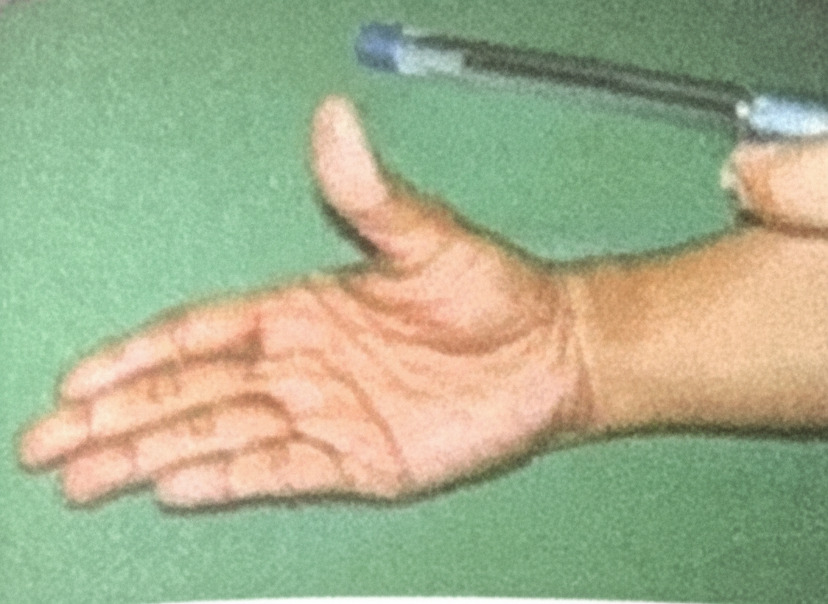
Pen Test is for which nerve
A 45-year-old woman presents to her primary care provider for wrist pain. She reports a 4-month history of gradually worsening pain localized to the radial side of her right wrist. The pain is dull, non-radiating, and intermittent. Her past medical history is notable for rheumatoid arthritis and von Willebrand disease. She does not smoke and drinks alcohol socially. She is active in her neighborhood’s local badminton league. Her temperature is 98.6°F (37°C), blood pressure is 125/75 mmHg, pulse is 80/min, and respirations are 18/min. On exam, she has mild tenderness to palpation in her thenar snuffbox. Nodules are located on the proximal interphalangeal joints of both hands. Ulnar deviation of the hand with her thumb clenched in her palm produces pain. Which of the following muscles in most likely affected in this patient?
A 32-year-old man comes to the physician because of episodic tingling and numbness in his right hand for the past 3 months. His symptoms are worse in the evening. There is no history of trauma. He is employed as a carpenter. He has smoked 1 pack of cigarettes daily for the past 10 years. He drinks a pint of vodka daily. He does not use illicit drugs. His vital signs are within normal limits. Physical examination shows decreased pinch strength in the right hand. Sensations are decreased over the little finger and both the dorsal and palmar surfaces of the medial aspect of the right hand. Which of the following is the most likely site of nerve compression?
A 56-year-old man comes to the emergency department because of pain and swelling in his left leg. He has a history of pancreatic cancer and is currently receiving chemotherapy. Three weeks ago, he had a similar episode in his right arm that resolved without treatment. His temperature is 38.2°C (100.8°F). Palpation of the left leg shows a tender, cord-shaped structure medial to the medial condyle of the femur. The overlying skin is erythematous. Which of the following vessels is most likely affected?
A 64-year-old female with a long-standing history of poorly-controlled diabetes presents with 3 weeks of abnormal walking. She says that lately she has noticed that she keeps dragging the toes of her right foot while walking, and this has led to her stubbing her toes. Upon physical exam, you notice a right unilateral foot drop that is accompanied by decreased sensation in the first dorsal web space. She also walks with a pronounced steppage gait. A deficit in which of the following nerves is likely responsible for this presentation?
A 21-year-old woman is brought to the emergency department following a motor vehicle collision. She has significant pain and weakness in her right arm and hand. Physical examination shows multiple ecchymoses and tenderness in the right upper extremity. She is able to make a fist, but there is marked decrease in grip strength. An x-ray of the right upper extremity shows a midshaft humerus fracture. Which of the following structures is most likely injured?
A 35-year-old woman, gravida 2, para 1, at 40 weeks' gestation, presents to the hospital with contractions spaced 2 minutes apart. Her past medical history is significant for diabetes, which she has controlled with insulin during this pregnancy. Her pregnancy has otherwise been unremarkable. A baby boy is born via a spontaneous vaginal delivery. Physical examination shows he weighs 4.5 kg (9 lb), the pulse is 140/min, the respirations are 40/min, and he has good oxygen saturation on room air. His left arm is pronated and medially rotated. He is unable to move it away from his body. The infant’s right arm functions normally and he is able to move his wrists and all 10 digits. Which of the following nerve roots were most likely damaged during delivery?
A 35-year-old man comes to the physician because of a 3-month history of intermittent right lateral hip pain that radiates to the thigh. Climbing stairs and lying on his right side aggravates the pain. Examination shows tenderness to palpation over the upper lateral part of the right thigh. There is no swelling. When the patient is asked to abduct the right leg against resistance, tenderness is noted. An x-ray of the pelvis shows no abnormalities. Which of the following structures is the most likely source of this patient's pain?
A healthy 28-year-old woman at 30-weeks gestational age, has gained 35lbs since becoming pregnant. She complains of several weeks of bilateral numbness and tingling of her palms, thumbs, index and middle fingers that is worse at night. She also notes weakness gripping objects at the office. Which nerve is most likely affected?
Practice by Chapter
Bones and joints of upper limb
Practice Questions
Muscles and movements of upper limb
Practice Questions
Nerves and blood supply of upper limb
Practice Questions
Clinical correlations of upper limb
Practice Questions
Bones and joints of lower limb
Practice Questions
Muscles and movements of lower limb
Practice Questions
Nerves and blood supply of lower limb
Practice Questions
Clinical correlations of lower limb
Practice Questions
Comparison of upper and lower limb structures
Practice Questions
Want unlimited practice?
Get full access to all questions, explanations, and performance tracking.
Scan to download app