What is the most common site of congenital diaphragmatic hernia?
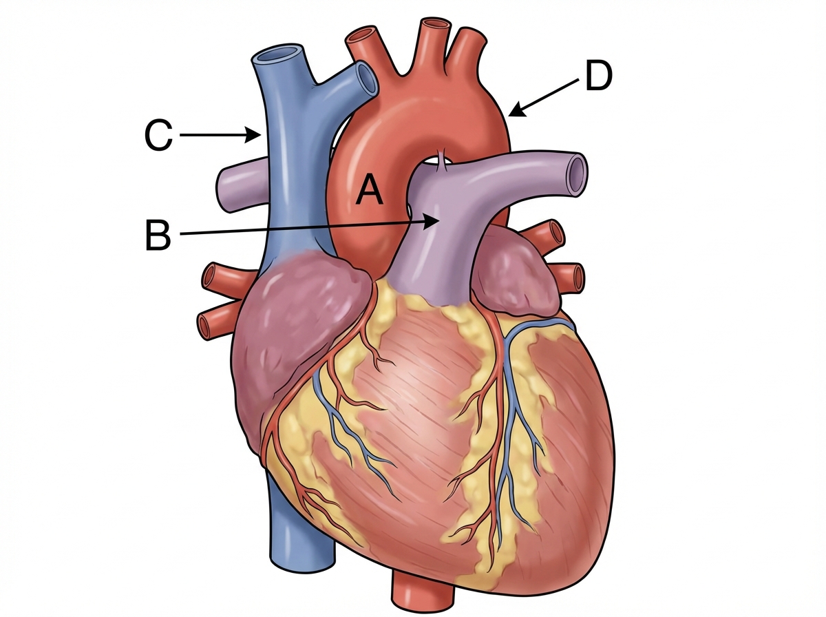
Structure preventing vertical descent of spleen
A 46-year-old male presents in consultation for weight loss surgery. He is 6’0” and weighs 300 pounds. He has tried multiple dietary and exercise regimens but has been unsuccessful in losing weight. The surgeon suggests a sleeve gastrectomy, a procedure that reduces the size of the stomach removing a large portion of the stomach along the middle part of the greater curvature. The surgeon anticipates having to ligate a portion of the arterial supply to this part of the stomach in order to complete the resection. Which of the following vessels gives rise to the vessel that will need to be ligated in order to complete the resection?
A 54-year-old woman comes to the physician because of a 3-month history of upper midthoracic back pain. The pain is severe, dull in quality, and worse during the night. Ten months ago, she underwent a modified radical mastectomy for invasive ductal carcinoma of the right breast. Physical examination shows normal muscle strength. Deep tendon reflexes are 2+ in all extremities. Examination of the back shows tenderness over the thoracic spinous processes. An x-ray of the thoracic spine shows vertebral osteolytic lesions at the levels of T4 and T5. The patient's thoracic lesions are most likely a result of metastatic spread via which of the following structures?
A 35-year-old obese man presents to the office complaining of chronic heartburn and nausea for the past 6 months. These symptoms are relieved when he takes 20 mg of omeprazole twice a day. The patient was prompted to come to the doctor when he recently experienced difficulty breathing and shortness of breath, symptoms which he believes underlies a serious health condition. The patient has no cardiac history but is concerned because his father recently died of a heart attack. Imaging of the patient’s chest and abdomen would most likely reveal which of the following?
A 75-year-old man presents to the clinic for chronic fatigue of 3 months duration. Past medical history is significant for type 2 diabetes and hypertension, both of which are controlled with medications, as well as constipation. He denies any fever, weight loss, pain, or focal neurologic deficits. A complete blood count reveals microcytic anemia, and a stool guaiac test is positive for blood. He is subsequently evaluated with a colonoscopy. The physician notes some “small pouches” in the colon despite poor visualization due to inadequate bowel prep. What is the blood vessel that supplies the area with the above findings?
An 18-year-old man is brought to the emergency department 30 minutes after being stabbed in the chest during a fight. He has no other injuries. His pulse is 120/min, blood pressure is 90/60 mm Hg, and respirations are 22/min. Examination shows a 4-cm deep, straight stab wound in the 4th intercostal space 2 cm medial to the right midclavicular line. The knife most likely passed through which of the following structures?
An 8-year-old boy is brought to the emergency department because of shortness of breath and dry cough for 2 days. His symptoms began after he helped his father clean the basement. He is allergic to shellfish. Respirations are 26/min. Physical examination shows diffuse end-expiratory wheezing and decreased inspiratory-to-expiratory ratio. This patient's symptoms are most likely being caused by inflammation of which of the following structures?
A 24-year-old man presents to the emergency room with a stab wound to the left chest at the sternocostal junction at the 4th intercostal space. The patient is hemodynamically unstable, and the trauma attending is concerned that there is penetrating trauma to the heart. Which cardiovascular structure is most likely to be injured first in this stab wound?
A researcher is investigating the blood supply of the adrenal gland. While performing an autopsy on a patient who died from unrelated causes, he identifies a vessel that supplies oxygenated blood to the inferior aspect of the right adrenal gland. Which of the following vessels most likely gave rise to the vessel in question?
Explanation: ***Posterolateral*** - The **posterolateral** region, specifically the foramen of Bochdalek, is the most common site for congenital diaphragmatic hernia (CDH). - This type of hernia accounts for approximately 80-90% of all CDH cases and usually occurs on the **left side**. *Central tendon* - Hernias through the **central tendon** are extremely rare and are distinct from the more common forms of CDH. - Defects in the central tendon are often associated with **pericardial defects** rather than typical diaphragmatic hernias which allow abdominal contents into the thoracic cavity. *Crural* - Hernias involving the **crura** of the diaphragm are typically **hiatal hernias** (e.g., sliding or paraesophageal), which are different in origin and presentation from CDH. - These are usually acquired and involve the stomach moving into the mediastinum, rather than a congenital defect leading to abdominal viscera migrating into the chest. *Anterolateral* - While congenital diaphragmatic hernias can occur **anterolaterally** through the foramen of Morgagni, these are much less common than posterolateral hernias. - Morgagni hernias account for a small percentage of CDH cases (around 2-5%) and are typically located on the right side, often containing omentum or colon. *Esophageal hiatus* - The **esophageal hiatus** is the normal opening in the diaphragm through which the esophagus passes. - While hiatal hernias can occur at this site, these are typically **acquired hernias** in adults, not congenital diaphragmatic hernias. - Congenital CDH refers to developmental defects in the diaphragm itself, not enlargement of normal openings.
Explanation: ***Phrenocolic ligament*** - The **phrenocolic ligament** is a fold of peritoneum that extends from the left colic (splenic) flexure of the colon to the diaphragm. - It forms a shelf or sling underneath the spleen, providing crucial support and preventing its **vertical descent**. *Ligamentum teres* - The **ligamentum teres hepatis** is the remnant of the obliterated umbilical vein, found in the free margin of the falciform ligament. - It connects the umbilicus to the liver and plays no role in supporting the spleen. *Ligamentum flavum* - The **ligamentum flavum** is a series of elastic ligaments connecting the laminae of adjacent vertebrae in the spinal column. - It is a component of the vertebral column and has no anatomical or functional relationship with the spleen. *Hepatogastric ligament* - The **hepatogastric ligament** is part of the lesser omentum, extending from the liver to the lesser curvature of the stomach. - Its primary function is to contain the **gastric arteries** and connect these organs, not to support the spleen. *Lienorenal ligament* - The **lienorenal ligament** (splenorenal ligament) connects the hilum of the spleen to the anterior surface of the left kidney. - While it provides **lateral support** to the spleen, it does not prevent **vertical descent** as effectively as the phrenocolic ligament.
Explanation: ***Splenic artery*** - A sleeve gastrectomy involves resecting a large portion of the **greater curvature of the stomach**. This portion is primarily supplied by the **short gastric arteries** and the **left gastroepiploic artery**. - The **splenic artery** is the main vessel that gives rise to the **short gastric arteries** and the **left gastroepiploic artery**, which originate from the distal portion of the splenic artery prior to its terminal branches. Therefore, ligation of branches from the splenic artery would be necessary. *Right gastroepiploic artery* - The **right gastroepiploic artery** primarily supplies the distal part of the greater curvature and arises from the **gastroduodenal artery**. - While it contributes to the vascular supply of the greater curvature, the bulk of the vessels needing ligation for a *sleeve gastrectomy* are the short gastrics and left gastroepiploic, which stem from the **splenic artery**. *Left gastric artery* - The **left gastric artery** primarily supplies the lesser curvature of the stomach near the cardia and arises directly from the **celiac trunk**. - Its branches would not be the primary vessels ligated in a procedure focused on the **greater curvature**. *Gastroduodenal artery* - The **gastroduodenal artery** arises from the **common hepatic artery** and typically supplies the pylorus, duodenum, and head of the pancreas, giving rise to the **right gastroepiploic artery**. - It is not the main source of arterial supply to the proximal and middle greater curvature resected during a sleeve gastrectomy. *Right gastric artery* - The **right gastric artery** typically arises from the **common hepatic artery** or **proper hepatic artery** and primarily supplies the lesser curvature of the stomach. - Its role in the robust blood supply to the **greater curvature** is minimal, and its ligation would not be central to a sleeve gastrectomy focused on this region.
Explanation: ***Azygos vein*** - The **vertebral venous plexus** communicates with both the intercostal veins (draining the chest wall and ultimately the breast) and the azygos vein system, providing a direct pathway for **hematogenous spread** to the spine without passing through the portal or caval systems. - The **Batson venous plexus**, a valveless system within the vertebral venous plexus, allows for easy retrograde flow of cancer cells from breast cancer to the thoracic spine due to changes in intra-abdominal or intrathoracic pressure. *Lateral axillary lymph nodes* - While **axillary lymph nodes** are the primary site of lymphatic spread for breast cancer, they would typically lead to spread to other lymphatic structures or secondary hematogenous spread via systemic circulation, not direct metastasis to the spine. - Lymphatic spread to axillary nodes typically causes **lymphadenopathy** in the axilla and can be a prognostic indicator, but it is not the direct pathway for vertebral osteolytic lesions. *Thyrocervical trunk* - The **thyrocervical trunk** is an artery that supplies structures in the neck and shoulder, such as the thyroid gland and scapular muscles. - It is an **arterial structure** and is not a common pathway for the metastatic spread of breast cancer to the thoracic spine. *Thoracic duct* - The **thoracic duct** is the main lymphatic vessel of the body, collecting lymph from the lower body, left arm, and left side of the head and neck, eventually draining into the left subclavian vein. - While breast cancer can spread via lymphatics, the thoracic duct is not the typical or direct route for metastasis to the **thoracic vertebrae**. *Intercostal artery* - **Intercostal arteries** supply blood to the intercostal spaces, chest wall, and pleura. - As an **arterial structure**, it is involved in blood supply to the region but is not a primary pathway for the metastatic spread of cancer cells to the bones.
Explanation: ***Protrusion of fundus of the stomach through the diaphragm into the thoracic cavity*** - The patient's symptoms of chronic heartburn, nausea, and relief with omeprazole are classic for **Gastroesophageal Reflux Disease (GERD)**, which is frequently caused or exacerbated by a **hiatal hernia**. - A hiatal hernia involves the **fundus of the stomach** protruding through the esophageal hiatus of the diaphragm into the chest cavity, leading to reflux and potentially respiratory symptoms due to compression or irritation of surrounding structures. *Cardiomegaly with pulmonary effusion* - While an obese patient could have **cardiac issues**, his symptoms, particularly the response to omeprazole, do not primarily point to cardiomegaly or pulmonary effusion. - This option suggests a **cardiac origin** for shortness of breath, which is less likely given the prominent GI symptoms. *Lung hypoplasia due to a defect in the diaphragm* - **Lung hypoplasia** typically occurs in infancy or childhood due to conditions like congenital diaphragmatic hernia, not in a 35-year-old presenting with chronic heartburn. - It describes **underdevelopment of the lungs**, which wouldn't be the primary finding in an adult with these symptoms. *Widened mediastinum with evidence of esophageal rupture* - A **widened mediastinum** with **esophageal rupture** (Boerhaave syndrome) is an acute, life-threatening condition associated with severe pain, vomiting, and shock, not chronic heartburn and gradual onset shortness of breath. - This condition is typically an **acute surgical emergency**, and the patient's presentation does not fit this high-acuity scenario. *"Hourglass stomach" due to upward displacement of the gastroesophageal junction* - An **"hourglass stomach"** usually refers to a severe stricture or narrowing of the mid-stomach, often due to chronic ulcers or malignancy, not just upward displacement of the gastroesophageal junction. - While it can be associated with GI symptoms, it doesn't directly explain the **fundic protrusion** characteristic of a hiatal hernia, which is more consistent with the chronic reflux.
Explanation: ***Inferior mesenteric artery*** - The patient's **microcytic anemia** and **positive stool guaiac test** indicate chronic gastrointestinal blood loss, highly suggestive of **diverticulosis** presenting as "small pouches" in the colon. - Diverticulosis commonly affects the **descending colon** and **sigmoid colon**, which are primarily supplied by branches of the **inferior mesenteric artery**. *Ileocolic artery* - The ileocolic artery is a branch of the **superior mesenteric artery** and supplies the **ileum**, **cecum**, and **ascending colon**. - Diverticula are less commonly found in these regions compared to the left colon. *Superior mesenteric artery* - The superior mesenteric artery supplies the **midgut derivatives**, including the **small intestine** and the **right half of the large intestine** (up to the distal transverse colon). - While it supplies a large portion of the GI tract, the typical location of diverticulosis (descending and sigmoid colon) is outside its primary distribution. *Middle colic artery* - The middle colic artery is a branch of the **superior mesenteric artery** and supplies the **transverse colon**. - While diverticula can occur in the transverse colon, it is not the most common location, and the inferior mesenteric artery supplies the areas most frequently affected. *Right colic artery* - The right colic artery is a branch of the **superior mesenteric artery** and supplies the **ascending colon**. - Diverticula are less frequently found in the ascending colon compared to the descending and sigmoid colon.
Explanation: ***Pectoral fascia, transversus thoracis muscle, right lung*** * The stab wound is in the **4th intercostal space**, 2 cm medial to the right midclavicular line, placing it over the anterior chest wall. This trajectory would first penetrate the **pectoral fascia**. * Deeper structures in this region include the **transversus thoracis muscle** and, given the depth, the **right lung** as it extends superiorly behind the anterior chest wall. * *Serratus anterior muscle, pleura, inferior vena cava* * The **serratus anterior muscle** is more laterally positioned, typically covering the side of the rib cage. * The **inferior vena cava** is located more medially and posteriorly within the mediastinum, deep to the diaphragm, making it an unlikely target for an anterior 4th intercostal stab. * *External oblique muscle, superior epigastric artery, azygos vein* * The **external oblique muscle** is part of the abdominal wall and would not be penetrated in the 4th intercostal space. * The **superior epigastric artery** is lower, typically extending into the abdominal wall, and the **azygos vein** is in the posterior mediastinum, not in the path of this superficial anterior stab wound. * *Pectoralis minor muscle, dome of the diaphragm, right lobe of the liver* * The **pectoralis minor muscle** is located deep to the pectoralis major, which would be penetrated. However, a stab at the 4th intercostal space would be too high to directly involve the **dome of the diaphragm** or the **right lobe of the liver**, which are typically below the 5th intercostal space, especially in forced expiration. * *Intercostal muscles, internal thoracic artery, right heart* * The **intercostal muscles** would certainly be traversed. * However, the **internal thoracic artery** runs paramedially (about 1-2 cm from the sternum), and getting to the **right heart** would require a more medial and deeper trajectory, potentially causing immediate tamponade or severe hemorrhage.
Explanation: ***Terminal bronchioles*** - The combination of **shortness of breath**, **dry cough**, **diffuse end-expiratory wheezing**, and decreased inspiratory-to-expiratory ratio in an allergic child points strongly to **bronchiolar inflammation** and **bronchoconstriction**, characteristic of asthma. - The **terminal bronchioles** are the primary site of airway obstruction in asthma, where inflammation, mucus plugging, and smooth muscle constriction lead to the classic wheezing sound upon expiration. *Pleural cavity* - Inflammation or fluid in the **pleural cavity** (e.g., pleurisy, pleural effusion) typically causes **sharp pleuritic chest pain** and diminished breath sounds, not diffuse wheezing. - While it can cause shortness of breath, the hallmark signs like wheezing and decreased I:E ratio are not characteristic of pleural involvement. *Respiratory bronchioles* - While respiratory bronchioles are involved in gas exchange and can be affected by some lung diseases, the **terminal bronchioles** are the main site of **bronchoconstriction** and airflow obstruction responsible for the wheezing seen in asthma. - Inflammation primarily affecting respiratory bronchioles might lead to more insidious symptoms or **bronchiolitis obliterans**, not acute, widespread wheezing as described. *Alveoli* - Inflammation of the **alveoli** (e.g., pneumonia, pulmonary edema) leads to impaired gas exchange, often presenting with **crackles**, consolidation, or hypoxemia. - It does not typically cause **diffuse wheezing** or a decreased inspiratory-to-expiratory ratio, which are signs of airway obstruction. *Distal trachea* - Inflammation or narrowing of the **distal trachea** can cause stridor or a monophonic wheeze, but typically affects both inspiration and expiration equally (not predominantly end-expiratory). - It would not typically present with the diffuse, widespread wheezing characteristic of **small airway obstruction** seen in this patient.
Explanation: ***Right ventricle*** - The **right ventricle** lies most anteriorly in the chest, directly behind the sternum and costal cartilages, making it the most likely chamber to be injured in an anterior stab wound. - Its anatomical position makes it vulnerable to penetrating trauma at the sternocostal junction, especially at the **4th intercostal space**. *Left ventricle* - The **left ventricle** is located more posteriorly and to the left of the midline, making it less susceptible to a central anterior stab wound. - While possible, it would typically require a stab wound further to the left or a deeper penetration. *Aorta* - The **aorta** is a very deep, large vessel located posterior to the heart, making it generally protected from isolated anterior stab wounds to the sternocostal junction. - Injury to the aorta typically occurs with more extensive, high-energy trauma or deeper penetration. *Right atrium* - The **right atrium** is located to the right and slightly posterior to the right ventricle, so it is less exposed to a direct anterior stab wound than the right ventricle. - Although it is a relatively anterior structure, its position is slightly less exposed than the right ventricle. *Left atrium* - The **left atrium** is the most posterior chamber of the heart, nestled against the esophagus, and is therefore very well protected from anterior penetrating trauma. - Injury to the left atrium is extremely rare with an anterior stab wound and would suggest a very severe and deep injury.
Explanation: ***Renal artery*** - The **inferior suprarenal artery**, which supplies the inferior part of the adrenal gland, typically arises from the **renal artery**. - The adrenal glands receive a rich blood supply from three main arterial sources: superior, middle, and inferior suprarenal arteries. *Inferior phrenic artery* - The **superior suprarenal arteries** typically arise from the **inferior phrenic arteries** and supply the superior aspect of the adrenal glands. - While critical for adrenal blood supply, they do not typically contribute to the inferior aspect directly. *Abdominal aorta* - The **middle suprarenal artery** usually arises directly from the **abdominal aorta**. - This vessel supplies the central part of the adrenal gland, but not primarily the inferior aspect. *Superior mesenteric artery* - The **superior mesenteric artery** primarily supplies structures of the midgut (e.g., small intestine, ascending colon) and does not typically give rise to vessels supplying the adrenal glands. - It is located inferior to the origin of the renal arteries and the adrenal glands. *Common iliac artery* - The **common iliac arteries** supply the lower limbs and pelvic organs, originating from the abdominal aorta bifurcation. - These arteries are located much too far inferior to supply the adrenal glands, which are retroperitoneal structures in the upper abdomen.
Explanation: ***Gastroduodenal artery*** - The **gastroduodenal artery** courses posterior to the first part of the duodenum and often supplies the **second part of the duodenum** via its branches, the anterior and posterior superior pancreaticoduodenal arteries. - An ulcer in the **posteromedial wall of the second portion of the duodenum** is most likely to erode into the gastroduodenal artery, leading to significant hemorrhage as seen in this patient. *Dorsal pancreatic artery* - The **dorsal pancreatic artery** typically arises from the splenic artery or hepatic artery and supplies the **pancreas**, particularly the neck and body. - It is not a primary blood supply to the **duodenum**, and its erosion due to a duodenal ulcer is highly unlikely. *Left gastroepiploic artery* - The **left gastroepiploic artery** (also known as the left gastro-omental artery) primarily supplies the **greater curvature of the stomach** and the **greater omentum**. - Its location and distribution make it an improbable source of bleeding for an ulcer located in the **duodenum**. *Inferior pancreaticoduodenal artery* - The **inferior pancreaticoduodenal artery** typically arises from the superior mesenteric artery and supplies the **head of the pancreas** and the **third and fourth parts of the duodenum**. - While it supplies the duodenum, it is less likely to be eroded by an ulcer in the **second portion of the duodenum** compared to the gastroduodenal artery. *Greater pancreatic artery* - The **greater pancreatic artery** (also known as the artery of the pancreatic magna) is a branch of the **splenic artery** and supplies the **pancreatic body and tail**. - This artery is exclusively involved in the blood supply of the **pancreas** and is not a direct or likely source of bleeding from a **duodenal ulcer**.
Explanation: **Gastroduodenal artery** - The **gastroduodenal artery (GDA)** runs immediately posterior to the **duodenal bulb** and is the most common artery eroded by posterior duodenal ulcers. - **Bleeding** from a posterior duodenal ulcer can be severe and life-threatening due to the proximity and size of the GDA. *Superior pancreaticoduodenal artery* - The superior pancreaticoduodenal artery branches off the GDA and supplies the head of the pancreas and duodenum. - While it contributes to the duodenal blood supply, it is less commonly directly eroded by a duodenal bulb ulcer compared to its parent artery, the GDA. *Right gastroepiploic artery* - The right gastroepiploic artery (right gastroomental artery) branches from the GDA and runs along the greater curvature of the stomach. - It is unlikely to be affected by an ulcer in the duodenal bulb due to its anatomical location away from the posterior duodenal wall. *Inferior pancreaticoduodenal artery* - The inferior pancreaticoduodenal artery branches from the superior mesenteric artery and supplies the head of the pancreas and duodenum. - It is anatomically located inferior and posterior to the duodenal bulb, making it less vulnerable to direct erosion by an ulcer in the duodenal bulb, which is typically supplied by branches of the GDA. *Dorsal pancreatic artery* - The dorsal pancreatic artery is a branch of the splenic artery or hepatic artery, supplying the body and tail of the pancreas. - It is anatomically situated away from the duodenal bulb and would not be at risk from a duodenal bulb ulcer.
Explanation: ***Rectosigmoid colon*** - The **inferior mesenteric artery (IMA)** supplies the distal third of the transverse colon, descending colon, sigmoid colon, and superior part of the rectum, which includes the **rectosigmoid colon**. - The **rectosigmoid region** is entirely dependent on IMA branches (sigmoid arteries and superior rectal artery) and represents a classic watershed area vulnerable to ischemia. - An occlusion in the IMA would compromise blood flow to these structures, leading to ischemia and symptoms like severe abdominal pain, bloody diarrhea, and peritoneal signs. *Hepatic flexure* - The **hepatic flexure** is primarily supplied by branches of the **superior mesenteric artery (SMA)**, specifically the middle colic artery. - An occlusion in the IMA would generally spare the hepatic flexure, as its blood supply comes from a different major arterial system. *Ascending colon* - The **ascending colon** receives its blood supply from the **superior mesenteric artery (SMA)** via the ileocolic and right colic arteries. - Therefore, an occlusion in the IMA would not directly affect the blood supply to the ascending colon. *Transverse colon* - While the **IMA** supplies the **distal one-third of the transverse colon** via the left colic artery, the term "transverse colon" as an anatomical structure includes both IMA and SMA territories. - The proximal two-thirds are supplied by the **superior mesenteric artery (SMA)** via the middle colic artery, with robust collateral circulation through the marginal artery of Drummond. - The rectosigmoid colon is the more specific and entirely IMA-dependent structure, making it the most likely to be affected. *Lower rectum* - The **lower rectum** receives its blood supply primarily from the **internal iliac arteries** via the middle and inferior rectal arteries. - The IMA supplies the superior part of the rectum, but the lower rectum has a separate and robust blood supply, making it less likely to be affected by an isolated IMA occlusion.
Explanation: ***Right intermediate bronchus*** - Due to the **anatomy of the tracheobronchial tree**, aspirated foreign bodies preferentially enter the **right bronchial tree** because the right main bronchus is wider, shorter, and more vertically oriented than the left. - In the right bronchial tree, foreign bodies most commonly lodge in the **right lower lobe bronchus or right intermediate bronchus region** (the intermediate bronchus is the segment between the right upper lobe takeoff and the middle/lower lobe bifurcation). - Among the options provided, the **right intermediate bronchus** is the most anatomically accurate location, as it represents the pathway through which most aspirated foreign bodies travel in the right lung. - Classic presentation includes sudden onset **cough, shortness of breath, inspiratory wheezing**, and **unilateral diminished breath sounds**. *Left upper lobe bronchus* - The **left main bronchus** is narrower and branches at a more acute angle (40-60°) from the trachea compared to the right (20-30°), making aspiration into the left side significantly less common. - The left upper lobe bronchus branches superiorly and is even less likely to receive aspirated material due to its upward trajectory. *Left lower lobe bronchus* - While the **left lower lobe bronchus** is more vertically oriented than the left upper lobe, the entire left bronchial tree receives aspirated foreign bodies much less frequently than the right side. - If aspiration occurs on the left, this would be the more likely site than the upper lobe, but right-sided aspiration predominates (approximately 60-80% of cases). *Right middle lobe bronchus* - The **right middle lobe bronchus** branches anterolaterally from the intermediate bronchus and takes a horizontal course, making it less likely to receive aspirated foreign bodies compared to the more vertical right lower lobe pathway. - Foreign bodies following gravity tend to bypass this horizontal branch and continue into the lower lobe. *Left main bronchus* - While a foreign body could lodge in the **left main bronchus**, this is much less common than right-sided aspiration due to the **more acute angle** (40-60°) at which the left main bronchus branches from the trachea. - The right main bronchus is the preferential pathway in approximately 60-80% of aspiration cases in young children.
Explanation: ***Descending colon*** - The **descending colon** is located in the left abdominal cavity, specifically in the left upper quadrant and extending into the left lower quadrant, making it highly susceptible to injury from a gunshot wound in the **left abdominal quadrant** just inferior to the left lateral costal border. - Its position aligns directly with the described entry point and bullet trajectory. *Transverse colon* - The **transverse colon** lies more centrally in the upper abdomen, spanning from the right to the left upper quadrants. - While possible to be hit by a left-sided entry wound, the trajectory described as "inferior to the left lateral costal border" makes the descending colon a more direct and likely target. *Ascending colon* - The **ascending colon** is located in the **right abdominal cavity**, specifically in the right upper and lower quadrants. - A wound inferior to the left lateral costal border would be on the opposite side of the abdomen and thus unlikely to penetrate the ascending colon. *Sigmoid colon* - The **sigmoid colon** is located more inferiorly in the **left lower quadrant** and pelvis. - While on the left side, the entry wound described as "inferior to the left lateral costal border" is generally higher than the typical location of the sigmoid colon. *Superior duodenum* - The **superior duodenum** is located in the **right upper quadrant** of the abdomen, anterior to the head of the pancreas. - Its position on the right side makes it highly unlikely to be penetrated by a gunshot wound to the left abdominal quadrant.
Explanation: ***Greater splanchnic nerves to the spinal cord*** - The **greater splanchnic nerves** (T5-T9) carry **visceral afferent fibers** from the gallbladder, transmitting pain to the spinal cord segments corresponding to the upper back (T5-T9). - This explains the **dull, cramping right upper quadrant pain** that **radiates to the upper back**, characteristic of visceral pain from the gallbladder. *Right thoraco-abdominal intercostal nerves* - These nerves primarily innervate the **parietal peritoneum** and abdominal wall, responsible for sharp, localized somatic pain. - While they could be involved in localized pain, they don't typically account for the **referred dull, cramping pain to the back** originating from a visceral organ like the gallbladder. *The phrenic nerve* - The **phrenic nerve** innervates the diaphragm and carries pain from the **diaphragmatic pleura and peritoneum**, often resulting in referred pain to the shoulder tip. - Gallbladder pain can sometimes irritate the diaphragm, but the primary referral to the **upper back** is more characteristic of splanchnic nerve involvement. *Left greater splanchnic nerve* - The **left greater splanchnic nerve** primarily innervates organs on the left side of the upper abdomen, such as the stomach and spleen. - Since the gallbladder is on the **right side**, its afferent pain signals travel via the right greater splanchnic nerves. *The pain endings of the visceral peritoneum* - The **visceral peritoneum** itself is generally insensitive to pain from cutting or burning; it senses stretch and inflammation. - However, the pain signals from the stretched or inflamed gallbladder are transmitted via **visceral afferent fibers within the splanchnic nerves**, not directly by the visceral peritoneum's own pain endings.
Explanation: ***Splenic artery*** - The splenic artery supplies the **fundus** and **greater curvature** of the stomach via the **short gastric arteries** and the **left gastroepiploic artery**. - An embolic occlusion of the splenic artery would lead to **necrosis** in these regions of the stomach as described in the vignette. *Superior mesenteric artery* - The superior mesenteric artery primarily supplies the **midgut** (from the distal duodenum to the proximal two-thirds of the transverse colon) and **pancreas**. - An occlusion would typically cause symptoms related to **small intestinal ischemia** and not primarily the stomach's greater curvature. *Left gastric artery* - The left gastric artery supplies the **lesser curvature** and **cardia** of the stomach. - Its occlusion would affect these areas, not the greater curvature. *Right gastroepiploic artery* - The right gastroepiploic artery, a branch of the **gastroduodenal artery**, supplies the **distal portion of the greater curvature** of the stomach. - While it supplies the greater curvature, the question states necrosis of the "proximal portion," making the splenic artery (via short gastrics/left gastroepiploic) a more direct and proximal supply to that region *Inferior mesenteric artery* - The inferior mesenteric artery supplies the **hindgut** (distal transverse colon to the superior rectum). - Its occlusion would cause symptoms related to **large intestinal ischemia**, distinct from gastric involvement.
Explanation: **Inferior thyroid artery** - The esophageal mass is located just distal to the **upper esophageal sphincter**, which is in the neck, close to the **thyroid gland**. - During surgery for an esophageal tumor in this region, the **inferior thyroid artery**, which supplies the thyroid and adjacent structures, is at the greatest risk of injury due to its proximity. *Bronchial branch of thoracic aorta* - The **bronchial branches** of the thoracic aorta primarily supply the bronchi and lungs. - These vessels are located deeper in the thorax, away from the **upper esophageal sphincter** and the initial surgical field for an upper esophageal tumor. *Left gastric artery* - The **left gastric artery** supplies the stomach and is a branch of the celiac trunk. - This artery is located in the **abdomen**, far from the surgical site involving an esophageal mass near the upper esophageal sphincter. *Left inferior phrenic artery* - The **left inferior phrenic artery** primarily supplies the diaphragm. - This vessel originates from the aorta in the **abdominal region**, which is distant from the upper esophageal sphincter. *Esophageal branch of thoracic aorta* - **Esophageal branches** directly supply the esophagus; however, the question refers to the **thoracic aorta branches**. - Tumors near the **upper esophageal sphincter** are usually accessed via a cervical incision, making thoracic branches less likely to be injured compared to arteries located in the neck.
Explanation: ***Vagus nerve*** - The **esophagus** passes through the diaphragm at the level of the **T10 vertebra**, accompanied by the **anterior and posterior vagal trunks**. Damage to these nerves is a known complication of esophageal surgery. - The vagus nerves provide **parasympathetic innervation** to the gastrointestinal tract, and their close proximity to the esophagus makes them vulnerable during tumor resection. *Azygos vein* - The **azygos vein** typically passes through the diaphragm at the level of **T12** through the **aortic hiatus**, not with the esophagus at T10. - It drains into the superior vena cava and is located more posteriorly in the mediastinum. *Right phrenic nerve* - The **right phrenic nerve** passes through the diaphragm with the **inferior vena cava** at the level of **T8**, innervating the diaphragm. - It is located more anteriorly and laterally to the esophagus, making direct damage during esophageal surgery less likely than the vagus nerves. *Inferior vena cava* - The **inferior vena cava (IVC)** passes through its own opening in the central tendon of the diaphragm at the level of **T8**, not with the esophagus at T10. - Damage to the IVC would result in significant hemorrhage and is typically a separate surgical concern. *Thoracic duct* - The **thoracic duct** passes through the **aortic hiatus** at the level of **T12** along with the aorta, collecting lymph from most of the body. - Its location makes it less likely to be damaged during a standard esophageal resection at T10 compared to the vagus nerves.
Explanation: ***Sternocleidomastoid muscles*** - In advanced **amyotrophic lateral sclerosis (ALS)**, progressive motor neuron degeneration affects both the diaphragm and intercostal muscles - The **paradoxical inward movement of the abdomen** during inspiration indicates severe diaphragmatic weakness or paralysis - The **shallow respiratory movements** and **severe respiratory distress** (respiratory rate 40/min) suggest that both primary inspiratory muscle groups (diaphragm and external intercostals) are significantly compromised - At this stage, **accessory muscles of inspiration**, particularly the **sternocleidomastoid muscles**, become critically important for maintaining ventilation by elevating the sternum and upper ribs - The dramatic improvement when sitting upright (orthopnea relief) supports accessory muscle recruitment, as this position optimizes sternocleidomastoid mechanical advantage - **Clinical pearl:** In neuromuscular respiratory failure, neck muscle recruitment (visible SCM contraction) is a key sign of impending respiratory failure requiring ventilatory support *External intercostal muscles* - The **external intercostal muscles** are normally primary muscles of inspiration that elevate the ribs - However, in advanced ALS with **2 years of progressive disease** and worsening dyspnea over the past month, these muscles would also be significantly weakened by the neurodegenerative process - The **lack of breath sounds in the lower lungs bilaterally** suggests poor chest wall expansion, indicating compromised intercostal function - While they continue to contribute, they are insufficient to maintain adequate ventilation alone at this stage of disease *Internal intercostal muscles* - The **internal intercostal muscles** function primarily in **forced expiration** by depressing the ribs - They do not play a significant role in inspiration *Muscles of anterior abdominal wall* - The **anterior abdominal wall muscles** (rectus abdominis, external/internal obliques, transversus abdominis) are **expiratory muscles** used in forced expiration and coughing - The **paradoxical inward movement** of the abdomen during inspiration is a passive phenomenon resulting from diaphragmatic weakness—the negative intrathoracic pressure pulls the weakened diaphragm upward, which in turn draws the abdominal wall inward - These muscles are not contributing to inspiration in this patient *Trapezius muscle* - The **trapezius** primarily functions in scapular movement and neck stabilization - While it provides some mechanical stability for the shoulder girdle during accessory muscle breathing, it is not directly involved in rib cage elevation - It plays a minor supportive role compared to the sternocleidomastoid in respiratory distress
Explanation: ***Medial pectoral*** - The **medial pectoral nerve** innervates both the pectoralis major and pectoralis minor muscles. - Damage to this nerve during mastectomy can lead to **atrophy of the lower lateral portion of the pectoralis major**, as this area relies heavily on its innervation. *Long thoracic* - The **long thoracic nerve** innervates the **serratus anterior muscle**. - Damage to this nerve would cause **scapular winging**, not atrophy of the pectoralis major. *Intercostobrachial* - The **intercostobrachial nerve** is primarily a **sensory nerve** supplying the skin of the upper medial arm and axilla. - Damage to this nerve would result in **sensory changes** (numbness, pain) in that area, not muscle atrophy. *Lateral intercostal* - The **lateral intercostal nerves** are primarily sensory and motor to the intercostal muscles and overlying skin. - Damage would typically cause **pain or numbness** along the chest wall or trunk, and possibly weakness of intercostal muscles, not pectoralis major atrophy. *Lateral pectoral* - The **lateral pectoral nerve** primarily innervates the **clavicular head** and **upper sternocostal portion** of the pectoralis major. - Damage to this nerve would cause atrophy in the **upper and medial parts** of the pectoralis major, not specifically the lower lateral portion.
Explanation: ***Left atrium*** - The patient's symptoms of **dysphagia (difficulty swallowing)** and **hoarseness** suggest compression of anatomical structures by an enlarged cardiac chamber, which the echocardiogram confirms. - An enlarged **left atrium**, typically due to **mitral stenosis**, can compress the esophagus (leading to dysphagia) and the **recurrent laryngeal nerve** (leading to hoarseness, known as Ortner's syndrome). The **opening snap** at the apex is also highly characteristic of mitral stenosis. *Patent ductus arteriosus* - A **patent ductus arteriosus (PDA)** is a congenital heart defect that typically causes a **continuous murmur** and may lead to pulmonary hypertension or heart failure, but not direct compression of the esophagus or recurrent laryngeal nerve. - The symptoms of PDA are usually present earlier in life, though uncorrected large PDAs can cause symptoms in adulthood, they do not cause dysphagia or hoarseness through direct esophageal compression. *Right ventricle* - An enlarged **right ventricle** usually causes symptoms related to right heart failure like **peripheral edema** or **dyspnea** due to pulmonary hypertension. - It is not anatomically positioned to compress the esophagus or recurrent laryngeal nerve in a way that would cause dysphagia or hoarseness. *Left ventricle* - An enlarged **left ventricle** (e.g., due to hypertension or aortic stenosis) primarily causes symptoms like **dyspnea on exertion** or **angina**. - While a severely dilated left ventricle can displace other structures, it does not typically cause direct esophageal compression leading to dysphagia or recurrent laryngeal nerve compression leading to hoarseness. *Right atrium* - An enlarged **right atrium** might be seen in conditions like tricuspid regurgitation or right heart failure but can manifest as **edema** or **jugular venous distention**. - It is not anatomically positioned to cause dysphagia or hoarseness from esophageal or recurrent laryngeal nerve compression.
Explanation: ***Left ventricle*** - The patient presents with symptoms and ECG findings consistent with **unstable angina** or **non-ST elevation myocardial infarction (NSTEMI)**, indicating myocardial ischemia. - With **75% left main coronary artery stenosis**, there is high risk of progression to **transmural myocardial infarction (STEMI)**, particularly affecting the anterior wall and septum. - Mural thrombi in the left ventricle typically form **3-7 days post-infarction** in areas of **dyskinetic or akinetic myocardium** due to blood stasis, endocardial injury, and hypercoagulability (Virchow's triad). - Left main disease affecting such a large territory makes the **left ventricle the most likely site** for mural thrombus formation. *Left atrium* - Mural thrombi in the left atrium are most commonly associated with **atrial fibrillation** due to blood stasis in the **left atrial appendage**. - This patient's symptoms are characteristic of coronary artery disease affecting the left ventricle, not an atrial arrhythmia. *Aorta* - While thrombi can form in the aorta (e.g., in the setting of **atherosclerosis** or **aneurysms**), they are typically mural thrombi associated with specific vascular pathologies. - The symptoms of **chest pain, ST depression**, and **coronary artery narrowing** point toward a myocardial event, making the left ventricle the most likely site for mural thrombus in this clinical context. *Right atrium* - Thrombi in the right atrium are usually associated with conditions leading to **venous stasis, such as deep vein thrombosis**, **central venous catheters**, or **right-sided heart failure**. - The patient's presentation with exertional chest pain and left main coronary artery narrowing is unrelated to right atrial thrombosis. *Right ventricle* - The right ventricle is **much less commonly** affected by ischemic events leading to mural thrombi compared to the left ventricle, due to its **lower oxygen demand** and **different blood supply** (right coronary artery). - While right ventricular infarction can occur (usually with inferior MI), the **left main coronary artery** supplies the left ventricle, making it the primary concern for mural thrombus formation in this patient.
Explanation: ***His pain is transmitted by right somatic nerve fibers.*** - The **migration of pain from the periumbilical region to the right lower quadrant** and becoming **severe and constant** indicates parietal peritoneal irritation. - **Somatic nerve fibers** innervate the parietal peritoneum and are responsible for transmitting **sharp, localized pain** typically associated with appendicitis in the right lower quadrant. *His pain is transmitted bilaterally by somatic afferent nerve fibers of the abdomen.* - While **visceral pain** from the initial appendiceal inflammation can be perceived bilaterally in the periumbilical region due to **bilateral innervation of visceral organs**, the **localized right lower quadrant pain** signifies involvement of **unilaterally innervated parietal peritoneum**. - The physical exam findings of **rebound tenderness** strongly suggest **localized peritoneal inflammation**, which is transmitted by **unilateral somatic nerves** at the site of inflammation, not bilaterally across the abdomen. *His pain is transmitted by somatic afferent nerve fibers located in the right flank.* - The **right flank** refers to the lateral aspect of the abdomen, while the pain is specifically localized to the **right lower quadrant**. - Although somatic nerves are involved, stating "right flank" is **too broad and imprecise** given the very specific localization of the pain to the right lower quadrant where the inflamed appendix is typically situated. *His pain is transmitted by the pelvic nerves.* - **Pelvic nerves** primarily carry parasympathetic fibers and visceral afferent fibers from pelvic organs, not the somatic pain from the parietal peritoneum in the right lower quadrant. - Pain from **pelvic organs** or **pelvic peritoneum** would be transmitted via these nerves, but the localized pain here is distinctly higher than typical pelvic organ pain. *His pain is mainly transmitted by the right splanchnic nerve.* - **Splanchnic nerves** primarily carry **visceral afferent fibers** responsible for the dull, poorly localized, initial periumbilical pain of appendicitis. - They do not transmit the **sharp, well-localized somatic pain** associated with parietal peritoneal irritation, which is characteristic of the pain migrating to the right lower quadrant.
Explanation: ***Superior mesenteric vein*** - The patient has a **cecal mass** and **liver lesions** consistent with **colon cancer** with **liver metastasis**. Cancer cells from the cecum drain predominantly into the **superior mesenteric vein**, before traveling to the liver via the **portal vein system**. - The **superior mesenteric vein (SMV)** drains blood from the cecum, ascending colon, and transverse colon. Metastatic cells from these regions would use this route to reach the liver. *Right gonadal vein* - The right gonadal vein drains into the **inferior vena cava (IVC)**, bypassing the portal system. - Metastasis to the liver would be less direct via this route, and the **cecum** does not primarily drain into the gonadal veins. *Inferior mesenteric vein* - The **inferior mesenteric vein (IMV)** drains the descending colon, sigmoid colon, and rectum. - While it eventually joins the **splenic vein** and then the **portal vein**, it is not the primary drainage for the cecum. *Inferior rectal vein* - The **inferior rectal vein** drains the lower rectum and anal canal, primarily into the **internal iliac veins** and then the **IVC**, bypassing the portal system. - This route is not relevant for metastasis from a **cecal mass**. *Right renal vein* - The **right renal vein** drains blood from the right kidney into the **inferior vena cava (IVC)**. - This vein is unrelated to the drainage of the gastrointestinal tract and would not be involved in metastasis from a **cecal mass**.
Explanation: ***Gastroduodenal artery*** - A deep ulcer on the **posterior wall of the duodenal bulb** is anatomically very close to the **gastroduodenal artery**. - Erosion into this artery can lead to **life-threatening upper gastrointestinal bleeding**, a severe complication of peptic ulcer disease. *Splenic vein* - The **splenic vein** is located more posteriorly and superiorly, primarily in relation to the pancreas and spleen, making it less likely to be eroded by a duodenal bulb ulcer. - While erosion into major vessels can occur, the gastroduodenal artery is in a much more direct and immediate proximity to the posterior duodenal bulb. *Descending aorta* - The **descending aorta** is a retroperitoneal structure located much more posteriorly and medially, far from the duodenal bulb. - Erosion into the aorta is an extremely rare and catastrophic event, not typically associated with duodenal ulcers. *Pancreatic duct* - The **pancreatic duct** (Wirsung's duct) is located within the pancreas, which lies posterior to the duodenum. While a *deep* ulcer could hypothetically penetrate the pancreas, the primary structure at risk for hemorrhage from a posterior duodenal bulb ulcer is the gastroduodenal artery. - Erosion into the pancreatic duct would likely cause **pancreatitis** or **fistula formation**, rather than acute hemorrhage. *Transverse colon* - The **transverse colon** is located inferior to the duodenum, separated by the greater omentum. - Ulcers would typically erode anteriorly or directly posteriorly, not inferiorly into the transverse colon, which would involve fistula formation rather than arterial erosion.
Explanation: **Phrenic nerve** - The **phrenic nerve** innervates the diaphragm and also carries sensory fibers from the **mediastinal and diaphragmatic pleura**, as well as the **pericardium**. - Irritation of the phrenic nerve, due to its **C3-C5 cervical origin**, can cause **referred pain to the ipsilateral shoulder** or neck. *Thoracic spinal nerves* - These nerves primarily serve the **intercostal muscles** and skin of the chest wall. - While they can transmit pain from the chest wall, they are not typically associated with **referred shoulder pain** from intrathoracic structures. *Vagus nerve* - The **vagus nerve** provides parasympathetic innervation to many thoracic and abdominal organs and carries visceral afferents. - It plays a role in regulating lung function but does not transmit sensory information that would be perceived as **shoulder pain** from diaphragmatic irritation. *Pulmonary plexus* - The **pulmonary plexus** is formed by branches of the vagus and sympathetic nerves, primarily involved in regulating **bronchial and vascular tone** in the lungs. - It does not transmit sensory input that would cause referred pain to the shoulder. *Intercostal nerves* - These nerves run along the ribs and innervate the **intercostal muscles** and skin of the **thoracic wall**. - Pain from these nerves would typically be felt along the **rib cage** or chest wall, not as referred shoulder pain.
Explanation: ***The posterior mediastinum*** - The posterior mediastinum contains the **esophagus**, descending aorta, thoracic duct, azygos and hemiazygos veins, and lymph nodes. Given the esophageal stricture, this is the most likely location. - An esophageal obstruction at **24 cm from the incisors** typically corresponds to the mid-to-distal esophagus, which is located in the posterior mediastinum. *The superior mediastinum* - The superior mediastinum extends from the **thoracic inlet** to the level of the sternal angle (T4/T5 vertebrae). - It contains the **trachea**, great vessels, thymus, and upper esophagus but an obstruction at 24 cm is usually below this region. *The epigastrium* - The epigastrium is an **abdominal region** of the upper abdomen, inferior to the xiphoid process and superior to the umbilicus. - It contains structures like the stomach and pancreas, but not the **thoracic esophagus**. *The anterior mediastinum* - The anterior mediastinum contains the **thymus gland**, lymph nodes, and internal mammary vessels. - The **esophagus is not located** in the anterior mediastinum. *The diaphragm* - The diaphragm is a **musculofibrous septum** that separates the thoracic and abdominal cavities. - While the esophagus passes through an opening in the diaphragm (the **esophageal hiatus**), the stricture itself is within the esophageal tube, not the diaphragm itself, and 24 cm from the incisors is proximal to the hiatus.
Explanation: ***Left gastro-omental artery*** - This artery runs along the **greater curvature** of the stomach, making it the most probable vessel to be seen pulsing in an ulcer located in the **proximal part of the greater curvature**. - Its anatomical location directly underlies this area, making it vulnerable to erosion from a penetrating ulcer. *Left gastric artery* - The **left gastric artery** supplies the **lesser curvature** of the stomach, which is not the location described for the ulcer. - An ulcer on the greater curvature would not typically expose this vessel. *Cystic artery* - The **cystic artery** supplies the **gallbladder** and is located much further away from the stomach, making it an unlikely vessel to be exposed by a gastric ulcer. - It arises from the right hepatic artery and is not in close proximity to the stomach's curvature. *Common hepatic artery* - The **common hepatic artery** is located **posterior** to the stomach and more superiorly, supplying the liver, pylorus, and duodenum through its branches. - It is not directly adjacent to the greater curvature of the stomach in a position that would be exposed by an ulcer there. *Right gastro-omental artery* - The **right gastro-omental artery** also runs along the **greater curvature**, but it is located more **distally** than the left gastro-omental artery. - A pulsing artery in the **proximal part** of the greater curvature makes the left gastro-omental artery a more precise and likely answer.
Explanation: ***Short gastric vein*** - This patient's heavy alcohol use, fatigue, abdominal distention with **ascites** (fluid wave, shifting dullness), and **scleral icterus** are highly suggestive of **decompensated cirrhosis** with **portal hypertension**. - **Portal hypertension** causes blood to back up into the **splenic vein** and its tributaries, including the **short gastric veins**, leading to **gastric varices** which are prone to rupture and bleeding. *Inferior epigastric vein* - The inferior epigastric vein drains into the **external iliac vein** and is part of the systemic venous circulation, not directly impacted by portal hypertension. - While systemic venous pressure can increase in conditions like heart failure, it's not the primary vessel affected by **portal hypertension** due to cirrhosis. *Azygos vein* - The **azygos vein** is a part of the systemic venous system in the chest and typically becomes engorged in conditions causing **superior vena cava obstruction** or severe right-sided heart failure. - While it can indirectly become distended in severe portal hypertension through portosystemic shunts, the **short gastric veins** are more directly and significantly affected by elevated splenic vein pressure. *Gastroduodenal artery* - This is an **artery**, and arterial pressure is distinct from venous pressure; it is typically not directly increased due to cirrhosis and portal hypertension. - Arteries carry oxygenated blood away from the heart, while the issue here is with venous drainage from the portal system. *Splenic artery* - This is an **artery** that supplies the spleen, and its pressure is not directly increased in **portal hypertension**. - While the spleen itself can enlarge due to venous congestion (**splenomegaly**) from portal hypertension, this refers to changes in venous, not arterial, pressure.
Explanation: ***Left upper lobe of the lung*** - The **left upper lobe of the lung** extends to the 4th intercostal space at the midclavicular line, making it the most probable structure to be traversed by a penetrating injury at this location. - The **pleural cavity** and lung tissue are superficially located in this region, making them highly susceptible to injury from a nail gun. *Right atrium of the heart* - The **right atrium** is located predominantly on the right side of the sternum, more centrally, and slightly to the right of the midclavicular line. - An injury at the **left 4th intercostal space at the midclavicular line** would typically be too lateral and superior to directly injure the right atrium. *Inferior vena cava* - The **inferior vena cava (IVC)** enters the right atrium from below, primarily located within the abdomen and passing through the diaphragm at the level of T8. - Its position is far too **inferior and posterior** relative to the 4th intercostal space to be directly injured by this wound. *Left atrium of the heart* - The **left atrium** is the most posterior chamber of the heart and is largely covered by the left ventricle. - Although part of the heart is on the left, an injury at the **4th intercostal space, midclavicular line**, would likely impact the left ventricle or lung tissue before reaching the left atrium, which is located more posteriorly and medially. *Superior vena cava* - The **superior vena cava (SVC)** is located to the right of the midline, formed by the brachiocephalic veins behind the right first costal cartilage. - Its position is too **medial and superior**, on the right side, to be directly injured by a nail penetrating the left 4th intercostal space at the midclavicular line.
Explanation: ***Esophagus*** - The patient's history of recurrent joint pain and fever in childhood, along with the cardiac exam findings of an **opening snap** followed by a **late diastolic rumble (best heard at the fifth intercostal space in the left midclavicular line)**, are classic for **rheumatic mitral stenosis**. - **Mitral stenosis** leads to chronic elevation of **left atrial pressure**, causing **left atrial enlargement**. The **esophagus** lies directly posterior to the left atrium, making it susceptible to compression by an enlarged left atrium. Patients may present with **dysphagia** due to this compression. *Trachea* - The trachea is located anterior to the esophagus and typically superior to the heart at the level of the atria, making it less likely to be directly compressed by an enlarged left atrium. - While significant cardiac enlargement can distort mediastinal structures, direct tracheal compression by an isolated enlarged left atrium is uncommon. *Hemiazygos vein* - The hemiazygos vein is located on the left side of the vertebral column in the posterior mediastinum and is not typically in close proximity to the left atrium in a manner that would lead to compression from left atrial enlargement. - It drains into the azygos vein, which is more medially located, and its compression is not a recognized complication of isolated left atrial enlargement. *Thoracic duct* - The thoracic duct ascends in the posterior mediastinum, largely posterior and to the left of the esophagus. Its course makes it less susceptible to direct compression by an enlarged left atrium. - Compression of the thoracic duct would typically lead to **chylothorax**, which is not associated with mitral stenosis. *Vagus nerve* - The vagus nerves (right and left) descend through the mediastinum in close proximity to the trachea and esophagus, but they are generally less vulnerable to direct compression by an enlarged left atrium compared to the esophagus. - Compression of the left recurrent laryngeal nerve (a branch of the left vagus) can occur in cases of extreme left atrial enlargement (**Ortner's syndrome**), leading to **hoarseness**, but direct compression of the main vagus nerve itself causing broader symptoms is less common than esophageal compression.
Explanation: ***T10*** - The initial **diffuse abdominal pain** in appendicitis is typically referred umbilical pain, which is mediated by the **T10 nerve root** due to the visceral afferent fibers primarily from the midgut. - As the inflammation of the appendix progresses to involve the **parietal peritoneum**, the pain localizes to the right lower quadrant, corresponding to somatic innervation. *T7* - The **T7 nerve root** supplies innervation to the **epigastric region**, above the umbilicus, and is generally associated with conditions affecting the stomach or duodenum. - This dermatome is too high to account for the initial visceral pain of appendicitis, which typically originates closer to the umbilical region. *L1* - The **L1 nerve root** primarily innervates the **inguinal region** and parts of the lower abdomen and hip. - While relevant for conditions like groin hernias, it does not typically mediate the initial diffuse umbilical pain associated with appendicitis. *T4* - The **T4 nerve root** corresponds to the nipple dermatome and is associated with pain in the **chest wall** or upper abdomen, like esophageal pain. - This dermatome is located significantly higher than the umbilical region and is not involved in the visceral pain pathway of appendicitis. *C6* - The **C6 nerve root** provides sensory innervation to the **thumb** and radial forearm, and motor innervation to muscles involved in wrist extension and elbow flexion. - This nerve root is in the cervical spine and has no involvement in abdominal pain.
Explanation: ***Left fifth intercostal space in the midclavicular line*** - The **apex of the heart**, predominantly formed by the **left ventricle**, is typically located in the **fifth intercostal space in the midclavicular line**. - A stab wound in this location could therefore directly penetrate the **left ventricle** and the adjacent **left lung**. *Left fifth intercostal space just lateral to the sternum* - This location is closer to the **sternum** and would be more likely to damage the **right ventricle** or the **sternum** itself, rather than directly injuring the left ventricle and lung in a single penetration. - The **left ventricle** is located more laterally, closer to the midclavicular line, especially at the apex. *Left fifth intercostal space in the midaxillary line* - The **midaxillary line** is too lateral to injure both the **left lung** and the **left ventricle** with a single penetrating stab in cases where the heart is in a normal anatomical position. - While it would certainly penetrate the **left lung**, the heart is not anterior enough to be struck at this lateral position. *Left seventh intercostal space in the midclavicular line* - The **seventh intercostal space** is below the typical location of the **heart's apex** (fifth intercostal space). - A stab wound here would likely hit the **diaphragm** or structures below it (e.g., spleen, stomach) rather than the heart. *Left seventh intercostal space in the midaxillary line* - This location is both too inferior and too lateral to simultaneously penetrate the **left lung** and **left ventricle** in a single stab. - It would most likely injure the **diaphragm**, **spleen**, or the inferior aspects of the **left lung** (pulmonary base).
Explanation: ***Teniae coli*** - The **teniae coli** are three distinct longitudinal bands of smooth muscle that run along the length of the large intestine, converging at the base of the appendix. They serve as reliable anatomical landmarks for locating the appendix during surgery. - Given the patient's symptoms (periumbilical pain migrating to the right lower quadrant, fever, vomiting, and right lower quadrant tenderness), **acute appendicitis** is highly suspected, making the teniae coli crucial for surgical identification of the inflamed appendix. *McBurney's point* - **McBurney's point** is a clinical landmark on the abdominal wall, two-thirds of the way from the umbilicus to the right anterior superior iliac spine, that often corresponds to the base of the appendix. It is used to elicit tenderness during physical examination. - While tenderness at McBurney's point is a strong indicator of appendicitis, it is a **surface landmark** for diagnosis and not an internal anatomical structure that aids the surgeon in _finding_ the appendix during a laparoscopic procedure. *Linea Semilunaris* - The **linea semilunaris** is the curved tendinous intersection found at the lateral border of the rectus abdominis muscle, extending from the costal margin to the pubic tubercle. - It defines the lateral extent of the rectus sheath but has **no direct anatomical relationship** to the appendix or its surgical identification. *Transumbilical plane* - The **transumbilical plane** is an imaginary horizontal plane passing through the umbilicus. It is used in topographical anatomy for abdominal segmentation. - It is a **surface and arbitrary anatomical plane** for regional description, not an internal structure that guides surgical access to or identification of the appendix. *Arcuate line* - The **arcuate line** is a crescent-shaped anatomical landmark located on the posterior wall of the rectus sheath, inferior to the umbilicus, marking the transition where the aponeuroses of the transverse abdominis and internal oblique muscles pass anterior to the rectus abdominis. - This line is relevant to the integrity of the rectus sheath but is **anatomically distant from the appendix** and does not assist in its surgical localization.
Explanation: ***Superior segment of right inferior lobe*** - The patient was found to be lying on his back while eating peanuts, which is a position that predisposes to aspiration into the **superior segment of the right inferior lobe**. - Aspiration during supine positioning typically leads to foreign body entry into the most posterior-inferiorly directed airways. *Posterior segment of right superior lobe* - Aspiration into the **posterior segment of the right superior lobe** is less common in a supine position unless the patient is positioned slightly to the side. - While the right lung is generally more prone to aspiration due to the straighter main bronchus, the specific segment depends on body position. *Inferior segment of right inferior lobe* - The **inferior segment of the right inferior lobe** would be more likely affected if the patient were in an upright position (e.g., sitting or standing) when aspiration occurred. - In a supine position, gravity directs aspirated material more towards the superior segment of the inferior lobe. *Anterior segment of right superior lobe* - Aspiration into the **anterior segment of the right superior lobe** is rare regardless of body position during aspiration. - This segment is anatomically less susceptible to gravitational flow of aspirated material compared to more posterior or inferior segments. *Inferior segment of left inferior lobe* - The **left main bronchus** branches at a sharper angle than the right, making aspiration into the left lung in general less common than into the right lung. - If aspiration were to occur in the left lung, the specific segment would still largely depend on the patient's body position.
Explanation: ***Right marginal artery*** - The right marginal artery typically arises from the **right coronary artery** and supplies the **right ventricle**. - Given the injury location to the **right of the sternum** and the 4th intercostal space, the right ventricle is the most superficial and anterior chamber and thus the most likely to be injured. *Left anterior descending artery* - The left anterior descending artery supplies the **anterior** two-thirds of the **interventricular septum** and the anterior wall of the **left ventricle**. - While located anteriorly, it is generally to the left of the sternum and would be protected by the more anterior right ventricle from an injury to the right of the sternum. *Left coronary artery* - The left coronary artery is a **short main stem** that quickly branches into the left anterior descending and left circumflex arteries. - It is located more superiorly and to the left, making it less likely to be directly injured by a penetrating trauma to the **right of the sternum** at the 4th intercostal space. *Posterior descending artery* - The posterior descending artery supplies the **posterior** wall of both ventricles and the posterior one-third of the **interventricular septum**. - This vessel is located on the posterior aspect of the heart, making it extremely unlikely to be injured by an anterior penetrating trauma. *Left circumflex coronary artery* - The left circumflex coronary artery supplies the **lateral and posterior walls of the left ventricle** and the left atrium. - Its location on the posterior-lateral aspect of the heart makes it much less vulnerable to a penetrating injury coming from the **anterior chest**.
Explanation: ***Thoracic aorta, right bronchial artery*** - The **femoral artery** leads directly into the **aorta**. From the aorta, the catheter can be navigated to the **thoracic aorta**, where the **bronchial arteries** typically originate. - The **bronchial arteries** usually arise directly from the **descending thoracic aorta** (most commonly T5-T6 vertebral level) to supply the lung parenchyma and airways. *Thoracic aorta, right superior epigastric artery, right bronchial artery* - The **superior epigastric artery** is a terminal branch of the **internal thoracic artery**, supplying the anterior abdominal wall, and is not a direct path to the bronchial arteries. - Navigating from the superior epigastric artery to the main bronchial artery without passing through intermediary large vessels would be anatomically incorrect and impractical. *Thoracic aorta, left ventricle, left atrium, pulmonary artery, right bronchial artery* - This path describes the venous and then pulmonary circulation (right heart, lungs), which is incorrect for reaching the **arterial system** of the bronchial arteries. - A catheter inserted via the **femoral artery** remains within the arterial system and would not cross into the pulmonary circulation or the left heart chambers in this manner. *Thoracic aorta, brachiocephalic trunk, right subclavian artery, right internal thoracic artery, right bronchial artery* - This pathway involves ascending from the **thoracic aorta** to the **brachiocephalic trunk** and subsequently into the **right subclavian** and **internal thoracic arteries**, which is a route primarily to the upper limb and chest wall. - While the internal thoracic artery can sometimes have small anastomoses, it is not the primary or direct route for embolizing a bronchial artery, which typically originates directly from the descending thoracic aorta. *Thoracic aorta, right subclavian artery, right internal thoracic artery, right bronchial artery* - Similar to the previous incorrect option, this route involves navigating through the **subclavian** and **internal thoracic arteries**, which is an indirect and unnecessarily complex path to the bronchial arteries. - The **bronchial arteries** are direct branches of the **thoracic aorta**, making this a much more convoluted and less likely route for therapeutic embolization.
Thoracic wall and diaphragm
Practice Questions
Pleura and lungs
Practice Questions
Mediastinum and heart
Practice Questions
Great vessels and lymphatics
Practice Questions
Abdominal wall and inguinal region
Practice Questions
Peritoneum and peritoneal cavity
Practice Questions
GI tract anatomy (esophagus to rectum)
Practice Questions
Liver, gallbladder and biliary tree
Practice Questions
Spleen and pancreas
Practice Questions
Kidneys and suprarenal glands
Practice Questions
Retroperitoneal structures
Practice Questions
Get full access to all questions, explanations, and performance tracking.
Start For Free