Question 11: A 73-year-old man comes to the physician because of worsening bilateral lower extremity pain for the past 8 months. The pain begins after walking one to two blocks and radiates bilaterally down the buttocks with cramping and tingling. He reports that the pain is worse while walking downhill and is relieved by sitting and leaning forward. He has hypertension, hyperlipidemia, and type 2 diabetes mellitus. He had a myocardial infarction at the age of 55 years and an abdominal aortic aneurysm repair at the age of 60 years. He has smoked one pack of cigarettes daily for the past 30 years. He does not drink alcohol or use illicit drugs. His current medications include sitagliptin, metformin, atorvastatin, metoprolol succinate, amlodipine, and hydrochlorothiazide. His temperature is 37.5°C (99.5°F), pulse is 82/min, respirations are 17/min, and blood pressure is 150/87 mm Hg. Examination shows full muscle strength. Sensation is reduced bilaterally in the feet and toes. Straight leg raise is negative. X-ray of the spine shows degenerative changes. Which of the following is the most appropriate next step in diagnosis?
- A. MRI scan of the spine (Correct Answer)
- B. Measurement of the ankle brachial index
- C. Measurement of serum creatine kinase
- D. Polysomnography
- E. Measurement of HLA-B27 antigen
Explanation: ***MRI scan of the spine***
- The patient's symptoms of **neurogenic claudication** (pain worse with walking downhill, relieved by sitting and leaning forward) are highly suggestive of **spinal stenosis**.
- An MRI provides detailed imaging of the spinal canal, nerve roots, and surrounding soft tissues, which is essential for confirming **spinal stenosis** and identifying the precise location and extent of nerve compression.
*Measurement of the ankle brachial index*
- An ABI measures peripheral arterial disease (PAD), which can cause **vascular claudication** (pain worse with uphill walking, relieved by standing still).
- The patient's symptoms (pain worse downhill, relieved by leaning forward) are not typical for **vascular claudication** despite his vascular risk factors.
*Measurement of serum creatine kinase*
- Creatine kinase is an enzyme released from damaged muscle, used to diagnose **myopathies** or significant muscle injury.
- The clinical presentation is more consistent with a **neurological compression** than a primary muscle disorder, and the patient has full muscle strength.
*Polysomnography*
- Polysomnography is a sleep study used to diagnose sleep disorders such as **sleep apnea** or **narcolepsy**.
- It is unrelated to lower extremity pain and claudication symptoms.
*Measurement of HLA-B27 antigen*
- HLA-B27 is a genetic marker associated with **spondyloarthropathies** like ankylosing spondylitis, reactive arthritis, and psoriatic arthritis.
- The patient's age of onset and specific symptoms of neurogenic claudication are not characteristic of inflammatory rheumatologic conditions.
Question 12: 
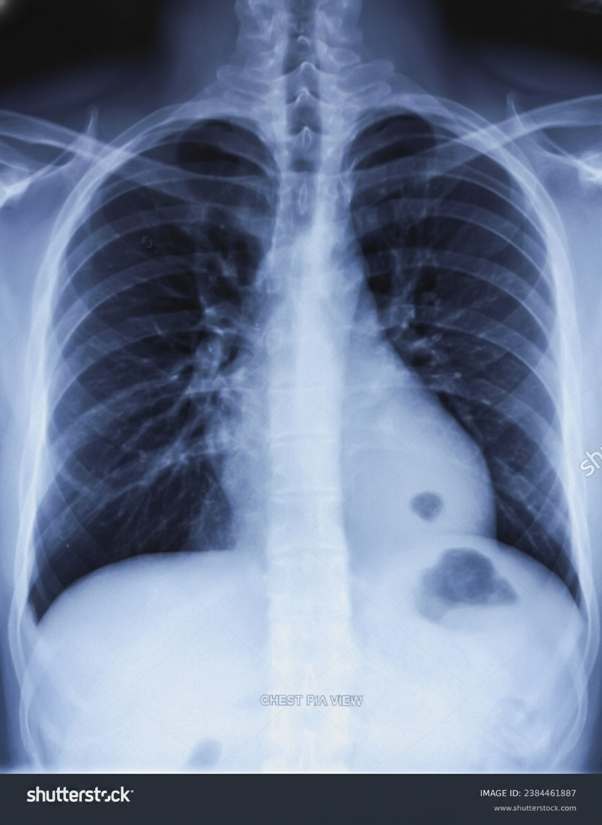
A 45-year-old man presents for a follow-up visit as part of his immigration requirements into the United States. Earlier this week, he was administered the Mantoux tuberculin skin test (TST). Today’s reading, 3 days after being administered the test, he shows an induration of 10 mm. Given his recent immigration from a country with a high prevalence of tuberculosis, he is requested to obtain a radiograph of the chest, which is shown in the image. Which of the following is true regarding this patient’s chest radiograph (CXR)?
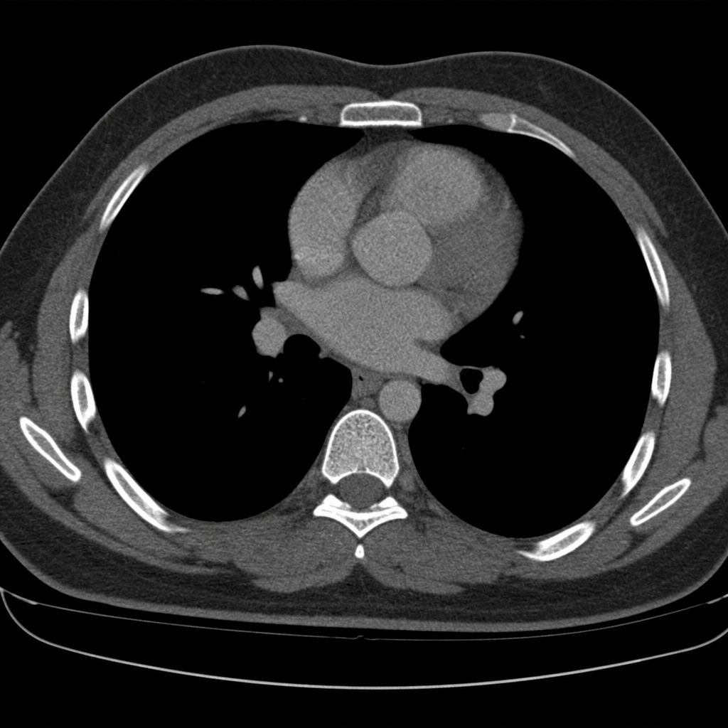
- A. Posterior ribs 9 and 10 are visible only in an expiratory film.
- B. The right lower boundary of the mediastinal silhouette belongs to the right ventricle.
- C. If the spinous process is not in-between the two clavicular heads, the image is repeated. (Correct Answer)
- D. The film is taken in a supine position.
- E. The view is anteroposterior (AP).
Explanation: ***If the spinous process is not in-between the two clavicular heads, the image is repeated.***
- Proper patient positioning is crucial for an accurate chest X-ray; **rotation of the patient** can distort the appearance of the heart and lungs, simulating pathology or obscuring real findings.
- Symmetrical positioning, indicated by the **spinous process being equidistant from the medial ends of the clavicles**, ensures an ideal posteroanterior (PA) view for interpretation.
*Posterior ribs 9 and 10 are visible only in an expiratory film.*
- In a properly **inspired chest X-ray**, at least 9 or 10 posterior ribs (or 5-6 anterior ribs) should be visible above the diaphragm.
- If fewer ribs are visible on a standard PA film, it indicates a **poor inspiratory effort**, which can compress lung fields and mimic pathology.
*The right lower boundary of the mediastinal silhouette belongs to the right ventricle.*
- The **right lower boundary of the mediastinal silhouette** is formed by the right atrium, not the right ventricle.
- The right ventricle forms part of the **anterior cardiac border** and is typically not seen as a distinct border on a standard PA chest X-ray.
*The film is taken in a supine position.*
- The presence of the **gastric bubble visible below the left hemidiaphragm** confirms an upright position, as gas rises in the stomach.
- A supine film would typically result in a **magnified cardiac silhouette** and less prominent gastric air.
*The view is anteroposterior (AP).*
- In an AP view, the **medial ends of the clavicles overlay the lung apices**, and the scapulae are often within the lung fields; this image shows the scapulae largely clear of the lung fields, consistent with a PA view.
- AP films also tend to **magnify the heart shadow** due to the divergent X-ray beam, which is not evident here.
Question 13: A 74-year-old male is brought to the emergency department 1 hour after he fell from the top of the staircase at home. He reports pain in his neck as well as weakness of his upper extremities. He is alert and immobilized in a cervical collar. He has hypertension treated with hydrochlorothiazide. His pulse is 90/min and regular, respirations are 18/min, and blood pressure is 140/70 mmHg. Examination shows bruising and midline cervical tenderness. Neurologic examination shows diminished strength and sensation to pain and temperature in the upper extremities, particularly in the hands. Upper extremity deep tendon reflexes are absent. Strength, sensation, and reflexes in the lower extremities are intact. Anal sensation and tone are present. Babinski's sign is absent bilaterally. Which of the following is most likely to confirm the cause of this patient's neurologic examination findings?
- A. CT angiography of the neck
- B. MRI of the cervical spine without contrast (Correct Answer)
- C. X-ray of the cervical spine
- D. CT of the cervical spine with contrast
- E. Cervical myelography
Explanation: ***MRI of the cervical spine without contrast***
- This patient presents with symptoms consistent with **central cord syndrome**, characterized by greater weakness in the upper extremities than the lower extremities, often following a hyperextension injury in older adults with pre-existing cervical spondylosis. **MRI is the gold standard for visualizing soft tissue injuries**, including spinal cord compression, edema, or hemorrhage, which are typical causes of central cord syndrome.
- Given the primary concern for spinal cord injury and the detailed neurological deficits indicating specific cord involvement, **MRI** offers the best resolution for evaluating the extent of cord damage, disc herniation, ligamentous injury, and pre-existing degenerative changes.
*CT angiography of the neck*
- **CT angiography** primarily evaluates the **vascular structures** of the neck (e.g., carotid and vertebral arteries) for dissection, stenosis, or occlusion.
- While vascular injury could occur in trauma, the patient's neurological findings (disproportionate upper extremity weakness, pain and temperature sensation loss) point more directly to **spinal cord pathology** rather than isolated vascular compromise as the primary cause.
*X-ray of the cervical spine*
- **X-rays** are useful for initial screening of **bony fractures** and significant dislocations but offer limited information about soft tissues, such as the spinal cord, ligaments, or intervertebral discs.
- They cannot adequately visualize the spinal cord damage responsible for the patient's specific neurological deficits, making it insufficient for confirming the cause of central cord syndrome.
*CT of the cervical spine with contrast*
- **CT scans** excel at visualizing **bony structures** and acute fractures, but even with contrast, they provide less detail of the **spinal cord parenchyma** and soft tissue ligaments compared to MRI.
- **Contrast** is typically used to highlight vascular structures, inflammatory processes, or tumors, which are not the primary diagnostic concerns suggested by this patient's acute post-traumatic presentation of central cord syndrome.
*Cervical myelography*
- **Myelography** involves injecting contrast into the subarachnoid space, followed by X-ray or CT imaging, to outline the spinal cord and nerve roots.
- While it can identify **spinal cord compression**, it is an **invasive procedure** with risks (e.g., headache, seizures) and has largely been replaced by the non-invasive and superior soft tissue imaging capabilities of MRI, especially in acute trauma.