
Female reproductive organs — MCQs

A 26-year-old woman presents to her primary care physician for 5 days of increasing pelvic pain. She says that the pain has been present for the last 2 months; however, it has become increasingly severe recently. She also says that the pain has been accompanied by unusually heavy menstrual periods in the last few months. Physical exam reveals a mass in the right adnexa, and ultrasonography reveals a 9 cm right ovarian mass. If this mass is surgically removed, which of the following structures must be diligently protected?
During a surgical procedure to repair an abdominal aortic aneurysm, the surgeon must be careful to avoid injury to which of the following arterial structures that originates near the level of the renal vessels?
A 28-year-old woman presents to an outpatient clinic for a routine gynecologic examination. She is concerned about some swelling on the right side of her vagina. She senses that the right side is larger than the left and complains that sometimes that area itches and there is a dull ache. She denies any recent travel or history of trauma. She mentions that she is sexually active in a monogamous relationship with her husband; they use condoms inconsistently. On physical examination her vital signs are normal. Examination of the pelvic area reveals a soft, non-tender, mobile mass that measures approximately 2 cm in the greatest dimension at the 8 o’clock position on the right side of the vulva, just below the vaginal wall. Which of the following is the most likely diagnosis?
A 24-year-old woman presents to her primary care doctor with a lesion on her labia. She first noticed the lesion 2 days ago. It is not painful. She denies vaginal discharge or dysuria. She has no past medical history and takes no medications. She has had 4 sexual partners in the past 8 months and uses the pull-out method as contraception. She drinks 12-16 alcoholic beverages per week and is a law student. Her temperature is 97.8°F (36.6°C), blood pressure is 121/81 mmHg, pulse is 70/min, and respirations are 16/min. On exam, she has an indurated non-tender ulcer on the left labia majora. There is no appreciable inguinal lymphadenopathy. Multiple tests are ordered and pending. This patient's condition is most likely caused by a pathogen with which of the following characteristics on microscopic examination?
A 22-year-old woman presents to the gynecologist for evaluation of amenorrhea and dyspareunia. The patient states that she recently got married and has been worried about getting pregnant. The patient states that she has never had a period and that sex has always been painful. On examination, the patient is Tanner stage 5 with no obvious developmental abnormalities. The vaginal exam is limited with no identified vaginal canal. What is the most likely cause of this patient’s symptoms?
A 57-year-old woman comes to the physician because of several years of recurrent pelvic pain and constipation. She has increased fecal urgency and a sensation of incomplete evacuation following defecation. She has had no problems associated with urination. Her last menstrual period was 6 years ago. She has had three uncomplicated vaginal deliveries. Physical examination shows normal external genitalia. Speculum examination of the vagina and the cervix shows bulging of the posterior vaginal wall during Valsalva maneuver. Weakness of which of the following structures is the most likely cause of this patient's symptoms?
A 22-year-old Caucasian male is stabbed in his left flank, injuring his left kidney. As the surgeon undertakes operative repair, she reviews relevant renal anatomy. All of the following are correct regarding the left kidney EXCEPT?
A 60-year-old post-menopausal female presents to her gynecologist with vaginal bleeding. Her last period was over 10 years ago. Dilation and curettage reveals endometrial carcinoma so she is scheduled to undergo a total abdominal hysterectomy and bilateral salpingo-oophorectomy. During surgery, the gynecologist visualizes paired fibrous structures arising from the cervix and attaching to the lateral pelvic walls at the level of the ischial spines. Which of the following vessels is found within each of the paired visualized structure?
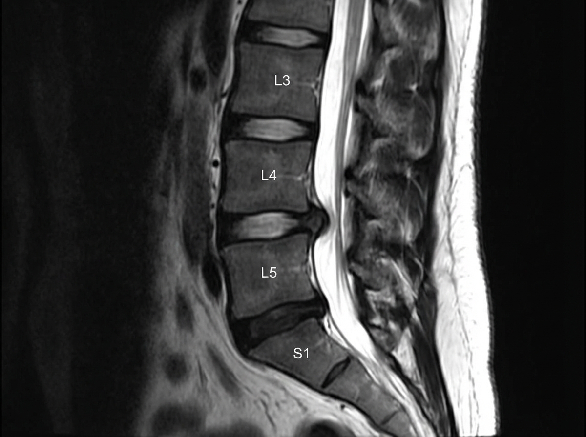
A slipped disc at the level shown in the image would most likely involve which nerve root?
An 11-year-old girl is brought in to her pediatrician by her parents due to developmental concerns. The patient developed normally throughout childhood, but she has not yet menstruated and has noticed that her voice is getting deeper. The patient has no other health issues. On exam, her temperature is 98.6°F (37.0°C), blood pressure is 110/68 mmHg, pulse is 74/min, and respirations are 12/min. The patient is noted to have Tanner stage I breasts and Tanner stage II pubic hair. On pelvic exam, the patient is noted to have a blind vagina with slight clitoromegaly as well as two palpable testes. Through laboratory workup, the patient is found to have 5-alpha-reductase deficiency. Which of the following anatomic structures are correctly matched homologues between male and female genitalia?
Want unlimited practice?
Get full access to all questions, explanations, and performance tracking.
Scan to download app