
Pelvis/Perineum — MCQs

On this page
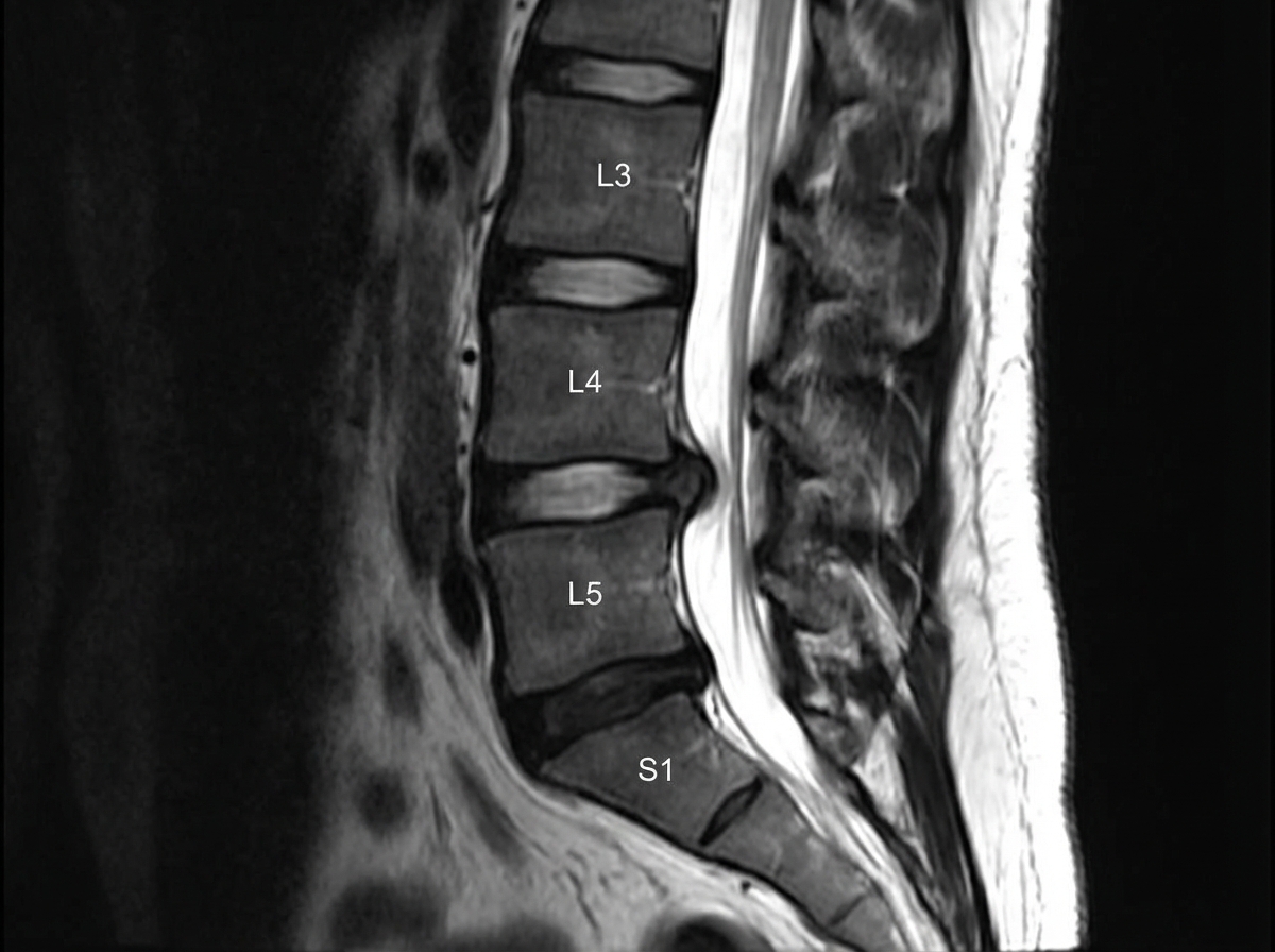
A slipped disc at the level shown in the image would most likely involve which nerve root?
An 11-year-old girl is brought in to her pediatrician by her parents due to developmental concerns. The patient developed normally throughout childhood, but she has not yet menstruated and has noticed that her voice is getting deeper. The patient has no other health issues. On exam, her temperature is 98.6°F (37.0°C), blood pressure is 110/68 mmHg, pulse is 74/min, and respirations are 12/min. The patient is noted to have Tanner stage I breasts and Tanner stage II pubic hair. On pelvic exam, the patient is noted to have a blind vagina with slight clitoromegaly as well as two palpable testes. Through laboratory workup, the patient is found to have 5-alpha-reductase deficiency. Which of the following anatomic structures are correctly matched homologues between male and female genitalia?
A 47-year-old woman comes to the physician because of involuntary leakage of urine for the past 4 months, which she has experienced when bicycling to work and when laughing. She has not had any dysuria or urinary urgency. She has 4 children that were all delivered vaginally. She is otherwise healthy and takes no medications. The muscles most likely affected by this patient's condition receive efferent innervation from which of the following structures?
A 56-year-old man undergoes a cystoscopy for the evaluation of macroscopic hematuria. During the procedure, an opening covered with a mucosal flap is visualized at the base of the trigone. Which of the following best describes this structure?
A 45-year-old man is brought to the emergency department following a motor vehicle collision. He reports right hip pain and numbness along the right thigh. Physical examination shows decreased sensation to light touch over a small area of the proximal medial thigh. X-rays of the pelvis show a displaced pelvic ring fracture. Further evaluation of this patient is most likely to show which of the following findings?
A 59-year-old woman presents to the emergency room with severe low back pain. She reports pain radiating down her left leg into her left foot. She also reports intermittent severe lower back spasms. The pain started after lifting multiple heavy boxes at her work as a grocery store clerk. She denies bowel or bladder dysfunction. Her past medical history is notable for osteoporosis and endometrial cancer. She underwent a hysterectomy 20 years earlier. She takes alendronate. Her temperature is 99°F (37.2°C), blood pressure is 135/85 mmHg, pulse is 85/min, and respirations are 22/min. Her BMI is 21 kg/m^2. On exam, she is unable to bend over due to pain. Her movements are slowed to prevent exacerbating her muscle spasms. A straight leg raise elicits severe radiating pain into her left lower extremity. The patient reports that the pain is worst along the posterior thigh and posterolateral leg into the fourth and fifth toes. Palpation along the lumbar vertebral spines demonstrates mild tenderness. Patellar reflexes are 2+ bilaterally. The Achilles reflex is decreased on the left. Which nerve root is most likely affected in this patient?
A 68-year-old man presents to his primary care physician complaining of a bulge in his scrotum that has enlarged over the past several months. He is found to have a right-sided inguinal hernia and undergoes elective hernia repair. At his first follow-up visit, he complains of a tingling sensation on his scrotum. Which of the following nerve roots communicates with the injured tissues?
A previously healthy 20-year-old man comes to the physician because of a 6-month history of a painless mass in his left groin that has been gradually increasing in size. Physical examination shows a 3x3-cm oval, non-tender left inguinal mass and a fluctuant, painless left scrotal swelling that increase in size with coughing. Which of the following is the most likely cause of this patient's symptoms?
A 29-year-old man presents to his primary care provider complaining of testicular pain. He reports a four-day history of dull chronic pain in his left testicle that is worse with standing. His past medical history is notable for asthma and major depressive disorder. He takes inhaled albuterol as needed and sertraline. He is sexually active with a single female partner and always uses barrier protection. His temperature is 99.2°F (37.3°C), blood pressure is 125/75 mmHg, pulse is 85/min, and respirations are 17/min. Physical examination reveals a non-tender twisted mass along the left spermatic cord that disappears when the patient lies supine. This patient’s condition most likely stems from decreased laminar flow at which of the following vascular junctions?
A 59-year-old truck driver presents to the emergency department after returning from his usual week-long trucking trip with excruciating pain around his anus. The patient admits to drinking beer when not working and notes that his meals usually consist of fast food. He has no allergies, takes no medications, and his vital signs are normal. On examination, he was found to have a tender lump on the right side of his anus that measures 1 cm in diameter. The lump is bluish and surrounded by edema. It is visible without the aid of an anoscope. It is soft and tender with palpation. The rest of the man’s history and physical examination are unremarkable. Which vein drains the vessels responsible for the formation of this lump?
Practice by Chapter
Pelvic girdle and ligaments
Practice Questions
Pelvic floor muscles
Practice Questions
Male reproductive organs
Practice Questions
Female reproductive organs
Practice Questions
Urinary bladder and urethra
Practice Questions
Rectum and anal canal
Practice Questions
Perineum and ischioanal fossa
Practice Questions
Pudendal nerve and internal pudendal vessels
Practice Questions
Pelvic vasculature and lymphatics
Practice Questions
Clinical correlations in pelvis/perineum
Practice Questions
Want unlimited practice?
Get full access to all questions, explanations, and performance tracking.
Scan to download app