
Skin histology — MCQs

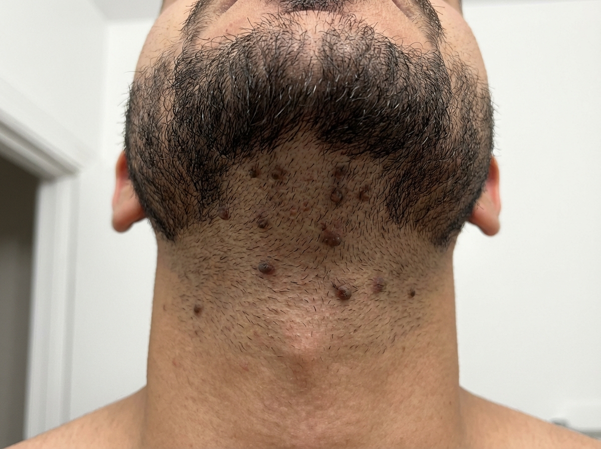
A 21-year-old man comes to the physician because of painful, firm, dark bumps on his neck and jawline. He has no history of serious illness and takes no medications. His brother had a similar rash. A photograph of the rash is shown. Which of the following is the most likely underlying mechanism of this patient's condition?
A 65-year-old man presents with a slowly growing, hyperkeratotic lesion on his right temple. The lesion has been present for approximately 8 months. He has a history of significant sun exposure. Examination reveals a 1.5 cm scaly, erythematous plaque with adherent scale. Biopsy shows atypical keratinocytes extending from the epidermis into the dermis. Which of the following is the most likely diagnosis?
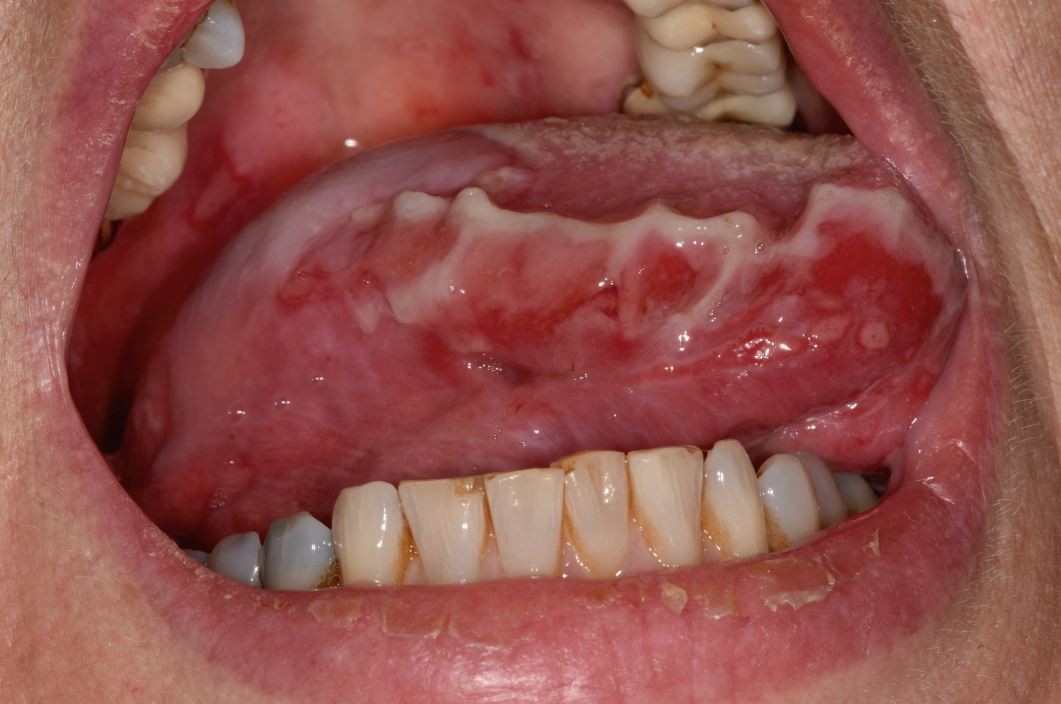
A 46-year-old woman presents to your office with oral lesions as shown in Image A. On examination, you find that her back has flaccid bullae that spread when you apply lateral pressure with your fingertips. This patient most likely has autoantibodies directed against which of the following?
A 15-year-old boy comes to the physician because of skin changes on his face, chest, and back over the past year. Treatment with over-the-counter benzoyl peroxide has been ineffective. Physical examination shows numerous open comedones, inflammatory papules, and pustules on his face, chest, and back. Which of the following is the most likely underlying mechanism of this patient’s skin condition?
A 37-year-old man presents to the clinic because of painful, severe blistering over his buttocks for the past week. About a year ago, he noticed a similar outbreak on his inner thighs, but it receded within a few days on its own. Physical examination shows the blisters are tense, and rubbing the affected skin does not result in ‘popping’ of the blisters. A biopsy shows the entire epidermis lifting away from the basal lamina with extensive inflammatory infiltrates abundant with eosinophils. Immunofluorescence shows a linear pattern of immune complex deposits. Which of the following cellular structures, if defective, is most likely involved in the formation of these blisters?
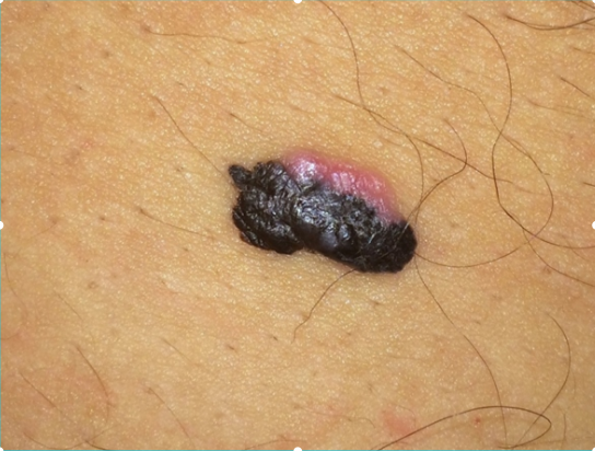
A 59-year-old man comes to the physician for evaluation of a progressively enlarging, 8-mm skin lesion on the right shoulder that developed 1 month ago. The patient has a light-skinned complexion and has had several dysplastic nevi removed in the past. A photograph of the lesion is shown. The lesion is most likely derived from cells that are also the embryological origin of which of the following tumors?
A 65-year-old man comes to the physician because he is worried about a mole on his right forearm. He has had the mole for several years, but it has grown in size in the past 3 months. Physical examination shows a hyperpigmented plaque with irregular borders and small area of ulceration. Histopathologic analysis of a full-thickness excisional biopsy confirms the diagnosis of malignant melanoma. Invasion of which of the following layers of skin carries the highest risk of mortality for this patient?
Research is being conducted on embryoblasts. The exact date of fertilization is unknown. There is the presence of a cytotrophoblast and syncytiotrophoblast, marking the time when implantation into the uterus would normally occur. Within the embryoblast, columnar and cuboidal cells are separated by a membrane. Which of these cell layers begins to line the yolk sac cavity?
A 52-year-old woman sees you in your office with a complaint of new-onset headaches over the past few weeks. On exam, you find a 2 x 2 cm dark, irregularly shaped, pigmented lesion on her back. She is concerned because her father recently passed away from skin cancer. What tissue type most directly gives rise to the lesion this patient is experiencing?
A 32-year-old woman presents with amenorrhea and galactorrhea. MRI shows a pituitary adenoma. Histological examination of the surgical specimen shows cells arranged in cords and nests with sinusoidal capillaries. Special staining reveals three distinct cell types: chromophobes (50%), acidophils (40%), and basophils (10%). Immunohistochemistry shows the tumor cells staining strongly for prolactin. Evaluate the relationship between normal pituitary architecture and tumor development to determine which cell type most likely gave rise to this neoplasm.
Want unlimited practice?
Get full access to all questions, explanations, and performance tracking.
Scan to download app