
Organ-specific histology — MCQs

On this page
A 23-year-old woman presents to her primary care physician with 3 days of fatigue and back pain after she started a drug for malaria prophylaxis. She says that her urine has also been darker over the same time period. Her past medical history is significant for allergies as well as a broken elbow that was treated in a cast 10 years ago. She does not take any medications, does not smoke, and drinks socially. Peripheral blood smear reveals both red blood cells with dark intracellular inclusions as well as abnormally shaped red blood cells. The immune cells responsible for the shape of these red blood cells are located in which of the following places?
A 31-year-old woman presents to the emergency room with high-grade fever and abdominal pain for the past 2 days. She also complains of malaise and has vomited several times since last night. The past medical history is benign. The vital signs include: temperature 40.0°C (104.0°F), pulse 120/min, respiratory rate 28/min, and blood pressure 120/89 mm Hg. On physical examination, severe costovertebral angle tenderness is noted. She is admitted to the medical floor and blood is drawn. The laboratory testing reveals leukocytosis with predominant neutrophilia and increased C-reactive protein and ferritin levels. She is suspected to have a retroperitoneal organ infection. Which of the following best describes the involved organ?
A 38-year-old man comes to the clinic complaining of recurrent abdominal pain for the past 2 months. He reports a gnawing, dull pain at the epigastric region that improves with oral ingestion. He has been taking calcium carbonate for the past few weeks; he claims that “it used to help a lot but it’s losing its effects now.” Laboratory testing demonstrated increased gastrin levels after the administration of secretin. A push endoscopy visualized several ulcers at the duodenum and proximal jejunum. What characteristics distinguish the jejunum from the duodenum?
A scientist is studying the anatomy and function of bone growth. He is able to create a cell line of osteocytes with a mutation that prevents the osteocytes from exchanging nutrients and waste products within neighboring lamellae. This mutation most likely affected which of the following cell structures?
An investigator is studying the immune response and the spleen in a mouse model infected with Escherichia coli. Which of the following anatomical sites in the spleen is important for the secondary maturation and affinity maturation of B cells that will ultimately target Escherichia coli?
A 58-year-old woman with refractory gastrointestinal complaints undergoes a bowel biopsy. On histology, the pathologist observes that submucosal glands of Brunner are present in the specimen. Which portion of the bowel was most likely biopsied?
A 35-year-old woman presents to a pre-operative evaluation clinic prior to an elective cholecystectomy. She has a 5 pack-year smoking history. The anesthesiologist highly recommends to discontinue smoking for at least 8 weeks prior to the procedure for which she is compliant. What is the most likely histology of her upper respiratory tract's epithelial lining at the time of her surgery?
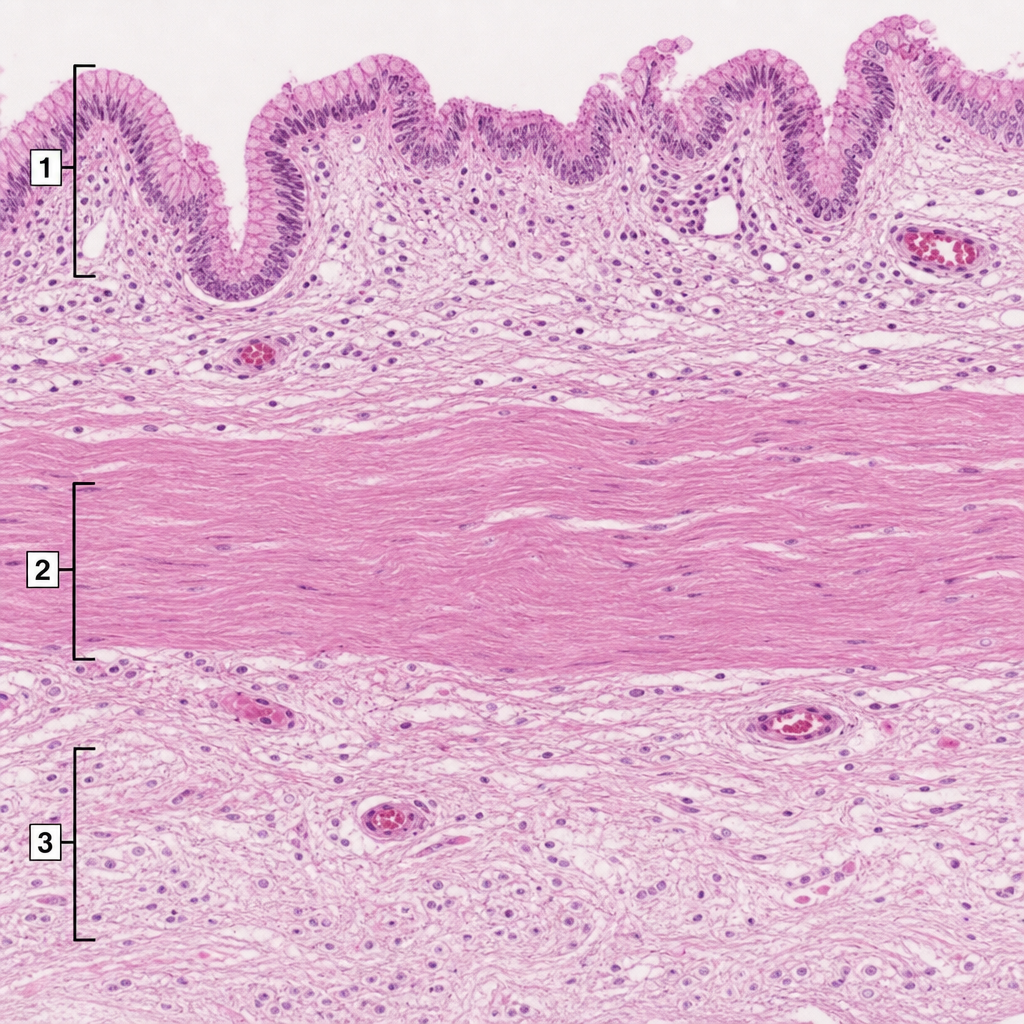
A 36-year-old man undergoes surgical intervention due to a right upper quadrant stab wound. His gallbladder was found to be lacerated and is removed. It is sent for histological evaluation. The pathologist examines the slide shown in the exhibit and identifies several structures numbered the image. Which of the following statements is correct?
A 62-year-old man with prostate cancer comes to the physician because of low back pain for 2 weeks and a 4.5-kg (10-lb) weight loss. Physical examination shows localized tenderness over the lumbar spine. An x-ray of the lumbar spine shows several osteoblastic lesions at the level of L2 and L4 vertebrae. Microscopic examination of a bone biopsy specimen from the L4 vertebra shows irregular bone trabeculae and star-shaped cells with long, cytoplasmic processes located deep within the lacunae. Exchange of nutrients and waste products between these cells most likely occurs through which of the following structures?
Practice by Chapter
Cardiovascular histology
Practice Questions
Respiratory system histology
Practice Questions
GI tract histology
Practice Questions
Liver and gallbladder histology
Practice Questions
Pancreas and salivary gland histology
Practice Questions
Urinary system histology
Practice Questions
Male reproductive histology
Practice Questions
Female reproductive histology
Practice Questions
Endocrine gland histology
Practice Questions
Skin histology
Practice Questions
Lymphoid tissue histology
Practice Questions
Central and peripheral nervous system histology
Practice Questions
Want unlimited practice?
Get full access to all questions, explanations, and performance tracking.
Scan to download app