
Neuroanatomy — MCQs

On this page
A 27-year-old woman comes to the clinic for blisters on both hands. The patient has a past medical history of asthma, eczema, and a car accident 2 years ago where she sustained a concussion. She also reports frequent transient episodes of blurred vision that clear with artificial tears. When asked about her blisters, the patient claims she was baking yesterday and forgot to take the pan out with oven gloves. Physical examination demonstrates weeping blisters bilaterally concentrated along the palmar surfaces of both hands and decreased pinprick sensation along the arms bilaterally. What is the most likely explanation of this patient’s symptoms?
A 60-year-old man comes to the physician because his wife has noticed that his left eye looks smaller than his right. He has had worsening left shoulder and arm pain for 3 months. He has smoked two packs of cigarettes daily for 35 years. Examination shows left-sided ptosis. The pupils are unequal but reactive to light; when measured in dim light, the left pupil is 3 mm and the right pupil is 5 mm. Which of the following is the most likely cause of this patient's ophthalmologic symptoms?
A 60-year-old African American woman presents to her ophthalmologist with blurry vision. She reports a 2-month history of decreased vision primarily affecting her right eye. Her past medical history is notable for type 1 diabetes and hypertension. She takes insulin and enalapril. She has a 40-pack-year smoking history and drinks a glass of wine at dinner each night. Her family history is notable for glaucoma in her mother and severe diabetes complicated by nephropathy and retinopathy in her father. Her temperature is 99°F (37.2°C), blood pressure is 134/82 mmHg, pulse is 88/min, and respirations are 18/min. On exam, she is well-appearing and in no acute distress. The physician asks the patient to look forward and shines a penlight first in one eye, then the other, alternating quickly to observe the pupillary response to the light. When the light is shined in the right eye, both pupils partially constrict. When the light is shined in the left eye, both pupils constrict further. When the light is moved back to the right eye, both eyes dilate slightly to a partially constricted state. Where is the most likely site of this patient’s lesion?
A 32-year-old previously healthy female presents to her primary care physician with double vision. She first noted the double vision yesterday and saw no improvement this morning. She does not think it is worsening. She has not had any changes in her normal routine though she recalls one episode of right arm weakness 2 months ago. She did not seek treatment and the weakness subsided after several days. She does not have a history of head trauma. She denies headache, fever, chills, nausea, vomiting, paresthesias, extremity pain, or weakness. On exam she has right adduction palsy on leftward gaze. She has no focal weakness. Which of the following additional physical exam findings is associated with the lesion responsible for her ocular findings?
A 78-year-old right-handed male is brought in by ambulance after being found down in his home. After being aroused, the patient has difficulty answering questions and appears to be frustrated by his inability to communicate. He is able to speak his name and a few other words but his speech is not fluent. Subsequent neurologic exam finds that the patient is able to comprehend both one and two step instructions; however, he is unable to repeat phrases despite being able to understand them. He also has difficulty writing despite retaining fine motor control. CT reveals an acute stroke to his left hemisphere. Damage to which of the following sets of structures would be most likely to result in this pattern of deficits?
A 23-year-old man presents to the emergency room following a stab wound to the back. He was in a bar when he got into an argument with another man who proceeded to stab him slightly right of the midline of his back. He is otherwise healthy and does not take any medications. He has one previous admission to the hospital for a stab wound to the leg from another bar fight 2 years ago. His temperature is 99°F (37.2°C), blood pressure is 115/80 mmHg, pulse is 100/min, and pulse oximetry is 99% on room air. Cardiopulmonary and abdominal exams are unremarkable; however, he has an abnormal neurologic exam. If this wound entered his spinal cord but did not cross the midline, which of the following would most likely be seen in this patient?
A 63-year-old man is brought to the emergency department by his wife because she is concerned he is having another stroke. The patient says he woke up with right-sided facial weakness and drooping. Past medical history is significant for a recent case of shingles treated with acyclovir, and a stroke, diagnosed 10 years ago, from which he recovered with no residual functional deficits. On physical examination, there is weakness and drooping of the entire right side of the face. Sensation is intact. The remainder of the physical examination is unremarkable. Which of the following additional findings would also most likely be seen in this patient?
A 25-year-old previously healthy woman presents to her PCP reporting cessation of menses for the past 6 months. Previously, her period occurred regularly, every 30 days. She also complains of decreased peripheral vision, most noticeably when she is driving her car. She denies any recent sexual activity and a pregnancy test is negative. Upon further work-up, what physical exam finding is most likely to be identified?
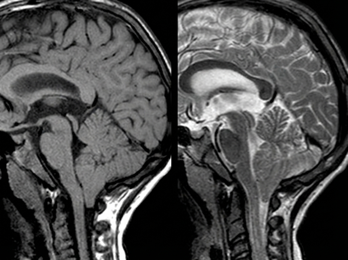
A 4-year-old boy is brought to the physician because of a progressive headache and neck pain for 2 weeks. During this period, he has had multiple episodes of dizziness and tingling sensations in his arms and hands. A year ago, he underwent closed reduction of a dislocated shoulder that he suffered after a fall. He underwent surgical removal of a sac-like protuberance on his lower back, soon after being born. His temperature is 36.7°C (98°F), pulse is 80/min, and blood pressure is 100/80 mm Hg. His neck is supple. Neurological examination shows sensorineural hearing loss bilaterally and normal gross motor function. Fundoscopy reveals bilateral optic disk swelling. An MRI of the brain is shown. Which of the following is the most likely cause of this patient's symptoms?
A 71-year-old woman presents to the emergency department with a headache for the past 30 minutes. She says that this is the worst headache of her life and that it came on suddenly after she hit her head. She says that she has also been experiencing visual problems with double vision when she looks to the left or the right. Visual examination reveals that her right eye cannot move right past the midline and her left eye cannot move left past the midline. Which of the following is most likely responsible for this patient's visual defects?
Practice by Chapter
Cerebral cortex and lobes
Practice Questions
Basal ganglia
Practice Questions
Thalamus and hypothalamus
Practice Questions
Limbic system
Practice Questions
Cerebellum
Practice Questions
Spinal cord organization
Practice Questions
CSF production and circulation
Practice Questions
Meninges and blood-brain barrier
Practice Questions
Sensory pathways
Practice Questions
Motor pathways
Practice Questions
Functional neuroanatomy of vision
Practice Questions
Functional neuroanatomy of hearing and balance
Practice Questions
Want unlimited practice?
Get full access to all questions, explanations, and performance tracking.
Scan to download app