
Anatomical variants in imaging — MCQs

A 75-year-old man presents to the clinic for chronic fatigue of 3 months duration. Past medical history is significant for type 2 diabetes and hypertension, both of which are controlled with medications, as well as constipation. He denies any fever, weight loss, pain, or focal neurologic deficits. A complete blood count reveals microcytic anemia, and a stool guaiac test is positive for blood. He is subsequently evaluated with a colonoscopy. The physician notes some “small pouches” in the colon despite poor visualization due to inadequate bowel prep. What is the blood vessel that supplies the area with the above findings?
A 56-year-old man presents to the emergency room after being in a motor vehicle accident. He was driving on an icy road when his car swerved off the road and ran head on into a tree. He complains of severe pain in his right lower extremity. He denies loss of consciousness during the accident. His past medical history is notable for poorly controlled hypertension, hyperlipidemia, and major depressive disorder. He takes enalapril, atorvastatin, and sertraline. His temperature is 99.1°F (37.3°C), blood pressure is 155/85 mmHg, pulse is 110/min, and respirations are 20/min. On exam, he is alert and fully oriented. He is unable to move his right leg due to pain. Sensation is intact to light touch in the sural, saphenous, tibial, deep peroneal, and superficial peroneal distributions. His leg appears adducted, flexed, and internally rotated. An anteroposterior radiograph of his pelvis would most likely demonstrate which of the following findings?
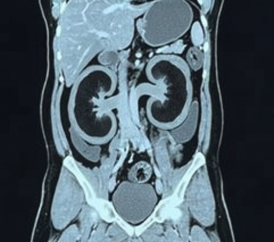
An 11-year-old girl is brought to the office by her mother due to complaint of intermittent and severe periumbilical pain for 1 day. She does not have any significant past medical history. She provides a history of a recent school trip to the suburbs. On physical examination, there is a mild tenderness around the umbilicus without any distension or discharge. There is no rebound tenderness. Bowel sounds are normal. An abdominal imaging shows enlarged mesenteric lymph nodes, and she is diagnosed with mesenteric lymphadenitis. However, incidentally, a mass of tissue was seen joining the inferior pole of both kidneys as shown in the image. Which of the following best describes this renal anomaly?
A newborn of a mother with poor antenatal care is found to have a larger than normal head circumference with bulging fontanelles. Physical examination reveals a predominant downward gaze with marked eyelid retraction and convergence-retraction nystagmus. Ultrasound examination showed dilated lateral ventricles and a dilated third ventricle. Further imaging studies reveal a solid mass in the pineal region. Which of the following is the most likely underlying pathophysiological mechanism responsible for the neurological signs in this patient?
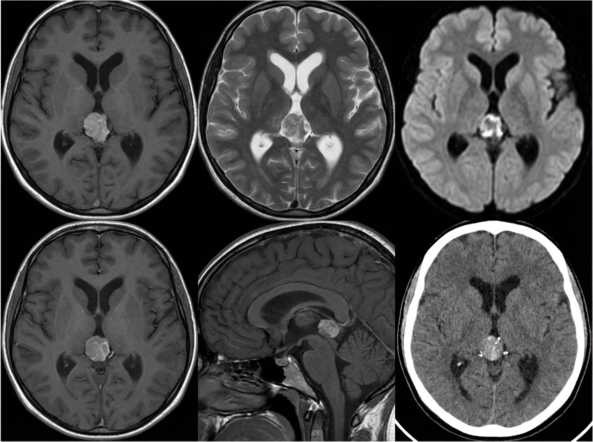
A 10-year-old girl is brought to the physician by her parents due to 2 months of a progressively worsening headache. The headaches were initially infrequent and her parents attributed them to stress from a recent move. However, over the last week the headaches have gotten significantly worse and she had one episode of vomiting this morning when she woke up. Her medical history is remarkable for a hospitalization during infancy for bacterial meningitis. On physical exam, the patient has difficulty looking up. The lower portion of her pupil is covered by the lower eyelid and there is sclera visible below the upper eyelid. A magnetic resonance imaging (MRI) of the brain is shown. Which of the following is the most likely diagnosis?
A 72-year-old man presents to his physician’s office with complaints of a cough and painful breathing for the last 2 months. He says that he has also observed a 5 kg (11 lb) weight loss during the past month. He is relatively healthy but the sudden change in his health worries him. Another problem that he has been facing is the swelling of his face and arms at unusual times of the day. He says that the swelling is more prominent when he is supine. He has also lately been experiencing difficulty with his vision. He consumes alcohol occasionally and quit smoking last year following a 25-year history of smoking. On examination, the patient is noted to have distended veins in the chest and arms. His jugular veins are distended. Physical examination shows ptosis of the right eye and miosis of the right pupil. His lungs are clear to auscultation. He is sent for an X-ray for further evaluation of his condition. Which of the following is the most likely site for the detection of the nodule on CT scan?
A 2-year-old child is brought to the emergency department with rapid breathing and a severe cyanotic appearance of his lips, fingers, and toes. He is known to have occasional episodes of mild cyanosis, especially when he is extremely agitated. This is the worst episode of this child’s life, according to his parents. He was born with an APGAR score of 8 via a normal vaginal delivery. His development is considered delayed compared to children of his age. History is significant for frequent squatting after strenuous activity. On auscultation, there is evidence of a systolic ejection murmur at the left sternal border. On examination, his oxygen saturation is 71%, blood pressure is 81/64 mm Hg, respirations are 42/min, pulse is 129/min, and temperature is 36.7°C (98.0°F). Which of the following will most likely be seen on chest x-ray (CXR)?
A 65-year-old man presents to the emergency department with abdominal pain and a pulsatile abdominal mass. Further examination of the mass shows that it is an abdominal aortic aneurysm. A computed tomography scan with contrast reveals an incidental finding of a horseshoe kidney, and the surgeon is informed of this finding prior to operating on the aneurysm. Which of the following may complicate the surgical approach in this patient?
A 23-year-old woman presents to the emergency department with an acute exacerbation of her 3-month history of low back and right leg pain. She says she has had similar symptoms in the past, but this time the pain was so excruciating, it took her breath away. She describes the pain as severe, shock-like, and localized to her lower back and radiating straight down the back of her right thigh and to her calf, stopping at the ankle. Her pain is worse in the morning, and, sometimes, the pain wakes her up at night with severe buttock and posterior thigh pain but walking actually makes the pain subside somewhat. The patient reports no smoking history or alcohol or drug use. She has been working casually as a waitress and does find bending over tables a strain. She is afebrile, and her vital signs are within normal limits. On physical examination, her left straight leg raise test is severely limited and reproduces her buttock pain at 20° of hip flexion. Pain is worsened by the addition of ankle dorsiflexion. The sensation is intact. Her L4 and L5 reflexes are normal, but her S1 reflex is absent on the right side. A CT of the lumbar spine shows an L5–S1 disc protrusion with right S1 nerve root compression. Which of the following muscle-nerve complexes is involved in producing an S1 reflex?
A 27-year-old female ultramarathon runner presents to the physician with complaints of persistent knee pain. She describes the pain to be located in the anterior area of her knee and is most aggravated when she performs steep descents down mountains, though the pain is present with running on flat roads, walking up and down stairs, and squatting. Which of the following would most likely be an additional finding in this patient’s physical examination?
Want unlimited practice?
Get full access to all questions, explanations, and performance tracking.
Scan to download app