
Abdominal wall development — MCQs

A 34-year-old woman comes to the emergency department because of a 2-hour history of abdominal pain, nausea, and vomiting that began an hour after she finished lunch. Examination shows abdominal guarding and rigidity; bowel sounds are reduced. Magnetic resonance cholangiopancreatography shows the dorsal pancreatic duct draining into the minor papilla and a separate smaller duct draining into the major papilla. The spleen is located anterior to the left kidney. A disruption of which of the following embryological processes is the most likely cause of this patient's imaging findings?
During a surgical procedure to repair an abdominal aortic aneurysm, the surgeon must be careful to avoid injury to which of the following arterial structures that originates near the level of the renal vessels?
A 52-year-old woman sees you in your office with a complaint of new-onset headaches over the past few weeks. On exam, you find a 2 x 2 cm dark, irregularly shaped, pigmented lesion on her back. She is concerned because her father recently passed away from skin cancer. What tissue type most directly gives rise to the lesion this patient is experiencing?
A child is in the nursery one day after birth. A nurse notices a urine-like discharge being expressed through the umbilical stump. What two structures in the embryo are connected by the structure that failed to obliterate during the embryologic development of this child?
A 23-year-old woman, gravida 2, para 1, at 26 weeks gestation comes to the physician for a routine prenatal visit. Physical examination shows a uterus consistent in size with a 26-week gestation. Fetal ultrasonography shows a male fetus with a thick band constricting the right lower arm; the limb distal to the constrictive band cannot be visualized. The most likely condition is an example of which of the following embryological abnormalities?
A 4700-g (10.3-lb) male newborn is delivered at 37 weeks' gestation to a 30-year-old woman, gravida 2, para 1. Apgar scores are 7 and 8 at 1 and 5 minutes, respectively. The newborn appears pale. Temperature is 37°C (98.6°F), pulse is 180/min, and blood pressure is 90/60 mm Hg. Examination in the delivery room shows midfacial hypoplasia, infraorbital creases, and a large tongue. The right side of the body is larger than the left. Abdominal examination shows that the abdominal viscera protrudes through the abdominal wall at the umbilicus; the viscera are covered by the amniotic membrane and the peritoneum. The liver is palpated 2–3 cm below the right costal margin. Fingerstick blood glucose concentration is 60 mg/dL. Ultrasonography of the abdomen shows enlarged kidneys bilaterally. In addition to surgical closure of the abdominal wall, which of the following is the most appropriate next step in management?
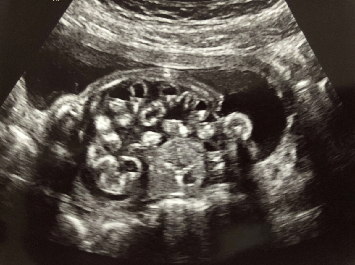
A 19-year-old woman, gravida 1, para 0, at 21 weeks’ gestation comes to the physician for a follow-up prenatal visit. At her previous appointment, her serum α-fetoprotein concentration was elevated. She had smoked 1 pack of cigarettes daily for 3 years but quit at 6 weeks' gestation. Examination shows a uterus consistent in size with a 21-week gestation. Ultrasonography shows fetal viscera suspended freely into the amniotic cavity. Which of the following is the most likely diagnosis?
During development, a fetus is found to have incomplete fusion of the neural tube. Which of the following structures would most likely be affected by this developmental defect?
A newborn boy born vaginally to a healthy 37-year-old G3P1 from a pregnancy complicated by hydramnios fails to pass meconium after 24 hours of life. The vital signs are within normal limits for his age. The abdomen is distended, the anus is patent, and the rectal examination reveals pale mucous with non-pigmented meconium. Based on a barium enema, the boy is diagnosed with sigmoid colonic atresia. Disruption of which structure during fetal development could lead to this anomaly?
A male newborn is born at 37 weeks' gestation after spontaneous vaginal delivery. The mother had no prenatal care. Physical examination shows a urethral opening on the dorsal aspect of the penis, 4 mm proximal to the glans. There is a 3-cm defect in the midline abdominal wall superior to the pubic symphysis with exposure of moist, erythematous mucosa. Which of the following is the most likely underlying cause of this patient's findings?
Want unlimited practice?
Get full access to all questions, explanations, and performance tracking.
Scan to download app