
Cardiac cross-sections — MCQs

A 58-year-old female presents to her primary care physician with complaints of chest pain and palpitations. A thorough past medical history reveals a diagnosis of rheumatic fever during childhood. Echocardiography is conducted and shows enlargement of the left atrium and narrowing of the mitral valve opening. Which of the following should the physician expect to hear on cardiac auscultation?
A previously healthy 19-year-old man is brought to the emergency department by his girlfriend after briefly losing consciousness. He passed out while moving furniture into her apartment. She said that he was unresponsive for a minute but regained consciousness and was not confused. The patient did not have any chest pain, palpitations, or difficulty breathing before or after the episode. He has had episodes of dizziness when exercising at the gym. His blood pressure is 125/75 mm Hg while supine and 120/70 mm Hg while standing. Pulse is 70/min while supine and 75/min while standing. On examination, there is a grade 3/6 systolic murmur at the left lower sternal border and a systolic murmur at the apex, both of which disappear with passive leg elevation. Which of the following is the most likely cause?
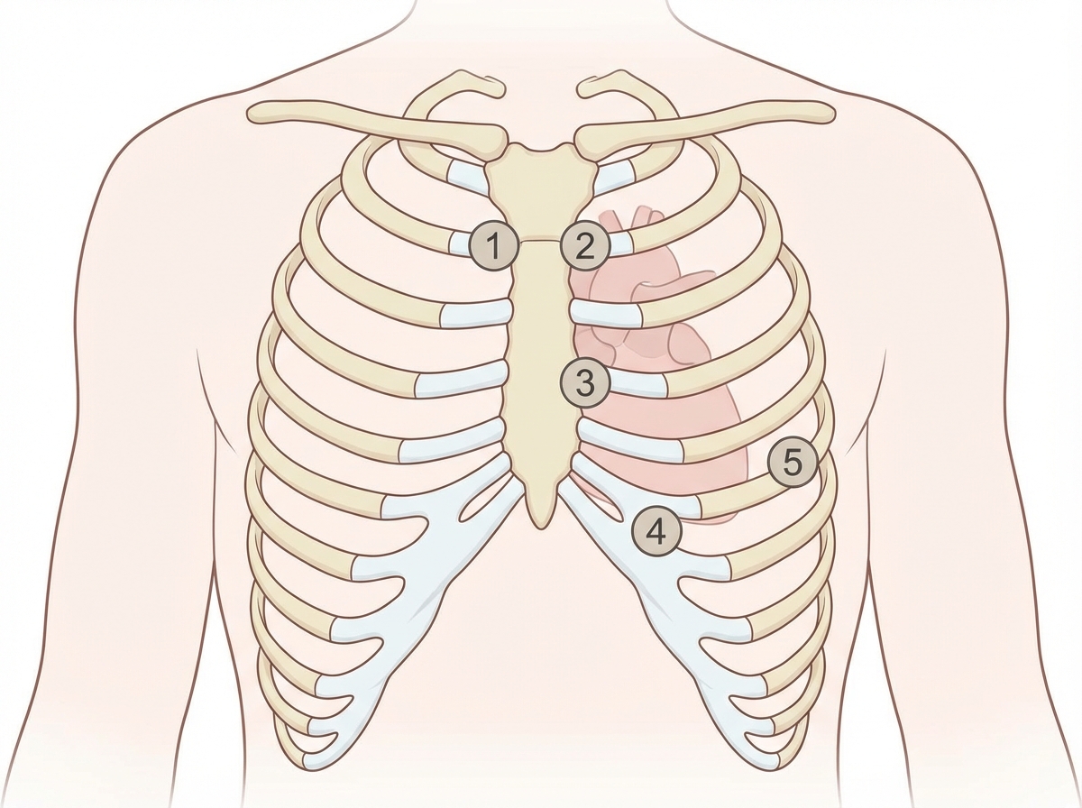
A 27-year-old woman, who recently immigrated from Bangladesh, presents to her primary care physician to discuss birth control. During a review of her past medical history, she reports that as a child she had a recurrent sore throat and fever followed by swollen and aching hip and knee joints. These symptoms returned every season and were never treated but went away on their own only to return with the next typhoon season. When asked about any current complaints, the patient says that she sometimes has shortness of breath and palpitations that do not last long. A physical exam is performed. In which of the auscultation sites will a murmur most likely be heard in this patient?
A 67-year-old man with type 2 diabetes mellitus comes to the emergency department because of lightheadedness over the past 2 hours. He reports that he has had similar episodes of lightheadedness and palpitations over the past 3 days. His only medication is metformin. His pulse is 110/min and irregularly irregular. An ECG shows a variable R-R interval and absence of P waves. The patient undergoes transesophageal echocardiography. During the procedure, the tip of the ultrasound probe is angled posteriorly within the esophagus. This view is most helpful for evaluating which of the following conditions?
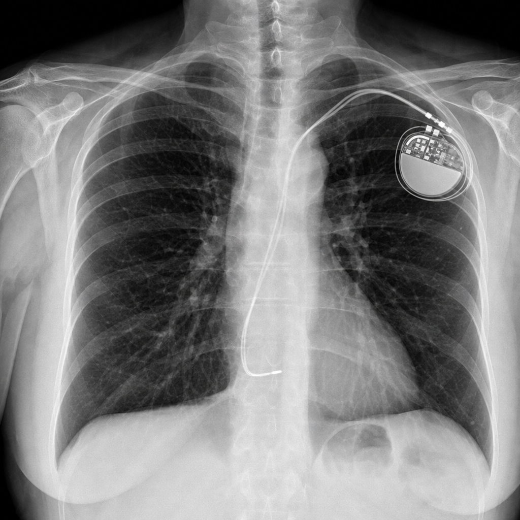
A 67-year-old woman comes to the emergency department because of a 4-month history of fatigue, shortness of breath with exertion, and dizziness. She has a history of atrial fibrillation and had a single-chamber pacemaker placed five years ago after an episode of syncope. Her pulse is 66/min and blood pressure is 98/66 mm Hg. An x-ray of the chest is shown. The x-ray confirms termination of the pacemaker lead in which of the following structures?
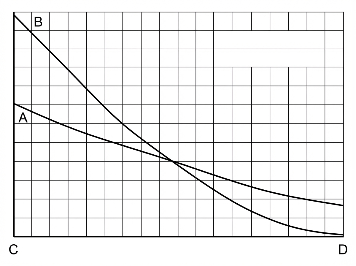
A young researcher is responsible for graphing laboratory data involving pulmonary blood flow and ventilation pattern obtained from a healthy volunteer who was standing in an upright position. After plotting the following graph, the researcher realizes he forgot to label the curves and the x-axis (which represents the position in the lung). Which of the following is the appropriate label for each point on the graph?
A 70-year-old woman presents with substernal chest pain. She says that the symptoms began 2 hours ago and have not improved. She describes the pain as severe, episodic, and worse with exertion. She reports that she has had multiple similar episodes that have worsened and increased in frequency over the previous 4 months. Past medical history is significant for diabetes and hypertension, both managed medically. The vital signs include temperature 37.0°C (98.6°F), blood pressure 150/100 mm Hg, pulse 80/min, and respiratory rate 15/min. Her serum total cholesterol is 280 mg/dL and high-density lipoprotein (HDL) is 30 mg/dL. The electrocardiogram (ECG) shows ST-segment depression on multiple chest leads. Coronary angiography reveals 75% narrowing of her left main coronary artery. In which of the following anatomical locations is a mural thrombus most likely to form in this patient?
A 23-year-old man is brought to the emergency department by a coworker for an injury sustained at work. He works in construction and accidentally shot himself in the chest with a nail gun. Physical examination shows a bleeding wound in the left hemithorax at the level of the 4th intercostal space at the midclavicular line. Which of the following structures is most likely injured in this patient?
A 55-year-old man visits the clinic with his wife. He has had difficulty swallowing solid foods for the past 2 months. His wife adds that his voice is getting hoarse but they thought it was due to his recent flu. His medical history is significant for type 2 diabetes mellitus for which he is on metformin. He suffered from many childhood diseases due to lack of medical care and poverty. His blood pressure is 125/87 mm Hg, pulse 95/min, respiratory rate 14/min, and temperature 37.1°C (98.7°F). On examination, an opening snap is heard over the cardiac apex. An echocardiogram shows an enlarged cardiac chamber pressing into his esophagus. Changes in which of the following structures is most likely responsible for this patient’s symptoms?
A 71-year-old man undergoes CT angiography for suspected mesenteric ischemia. Axial sections at the L1 level show a dissection flap in the superior mesenteric artery with the true lumen severely narrowed. The false lumen extends into a vessel that crosses anterior to the left renal vein. Coronal reconstructions show this vessel arising from the anterolateral aspect of the aorta at L2. The patient has left flank pain and hematuria in addition to abdominal pain. Synthesize the cross-sectional and vascular anatomy to determine the additional vessel involved.
Want unlimited practice?
Get full access to all questions, explanations, and performance tracking.
Scan to download app