
Vascular Surgery — MCQs

On this page
Which treatment is considered more effective for the management of varicose veins in terms of long-term outcomes and recurrence rates?
What is the standard treatment for a patient diagnosed with a small, unruptured abdominal aortic aneurysm?
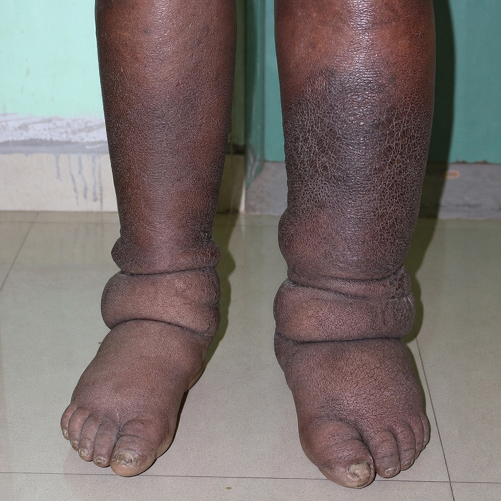
Identify the condition shown in the provided image.
What is the appropriate management strategy for an asymptomatic abdominal aortic aneurysm (AAA)?
CEAP score indicates-
Percutaneous chemical lumbar sympathectomy is practised using -
Which of the following lumbar vertebrae is typically spared in a lumbar sympathectomy?
Which of the following statements is MOST accurate regarding the use of grafts in vascular surgery?
Which of the following is the MOST IMPORTANT initial management approach for venous ulcers of the lower limb?
What is the Ankle-Brachial Pressure Index (ABPI) value that indicates imminent risk of necrosis?
Practice by Chapter
Atherosclerotic Disease
Practice Questions
Aortic Aneurysms
Practice Questions
Peripheral Arterial Disease
Practice Questions
Carotid Artery Disease
Practice Questions
Venous Thromboembolism
Practice Questions
Chronic Venous Insufficiency
Practice Questions
Mesenteric Vascular Disease
Practice Questions
Vascular Trauma
Practice Questions
Vascular Access for Hemodialysis
Practice Questions
Endovascular Techniques
Practice Questions
Diabetic Foot Vascular Disease
Practice Questions
Vasculitis
Practice Questions
Want unlimited practice?
Get full access to all questions, explanations, and performance tracking.
Scan to download app