
Vascular Surgery — MCQs

On this page
True about carotid body tumor is all except:
The ideal synthetic material used for femoropopliteal bypass when autologous vein is unavailable is:
A patient is on follow-up for recurrent abdominal pain. USG reveals an aortic aneurysm of 40 mm. What should be the next immediate step?
Falsely elevated ankle brachial index is used for evaluation of?
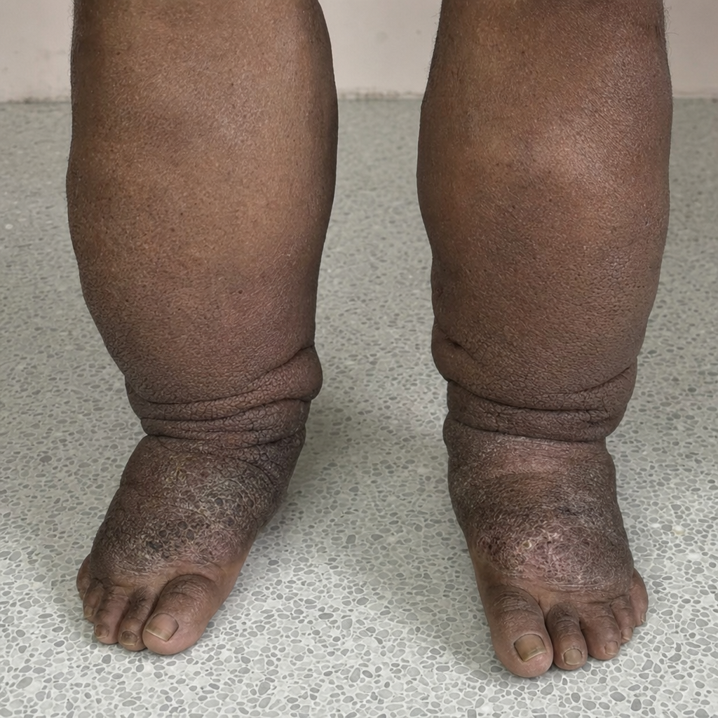
What is the most probable cause of this condition?
Primary vascular access of choice in chronic kidney disease is:
In acute limb ischemia, which finding indicates irreversible damage?
Which of the following is the most common cause of acute mesenteric ischemia?
Which of the following is a true statement about varicose veins?
Which of the following is not true regarding the surgical treatment of varicose veins?
Practice by Chapter
Atherosclerotic Disease
Practice Questions
Aortic Aneurysms
Practice Questions
Peripheral Arterial Disease
Practice Questions
Carotid Artery Disease
Practice Questions
Venous Thromboembolism
Practice Questions
Chronic Venous Insufficiency
Practice Questions
Mesenteric Vascular Disease
Practice Questions
Vascular Trauma
Practice Questions
Vascular Access for Hemodialysis
Practice Questions
Endovascular Techniques
Practice Questions
Diabetic Foot Vascular Disease
Practice Questions
Vasculitis
Practice Questions
Want unlimited practice?
Get full access to all questions, explanations, and performance tracking.
Scan to download app