
Vascular Surgery — MCQs

On this page
Which of the following is NOT a feature of mycotic aneurysm?
A 68-year-old man is admitted to the coronary care unit with an acute myocardial infarction. His postinfarction course is marked by congestive heart failure and intermittent hypotension. On the fourth day in hospital, he develops severe midabdominal pain. On physical examination, blood pressure is 90/60 mm Hg and pulse is 110 beats per minute and regular; the abdomen is soft with mild generalized tenderness and distention. Bowel sounds are hypoactive; stool Hematest is positive. Which of the following is the most appropriate next step in this patient's management?
According to the Spetzler-Martin grading scale, what is the score assigned to an arteriovenous malformation with a 5 cm nidus?
In an emergency, which artery can be safely ligated?
Trendelenburg's test is done for the detection of:
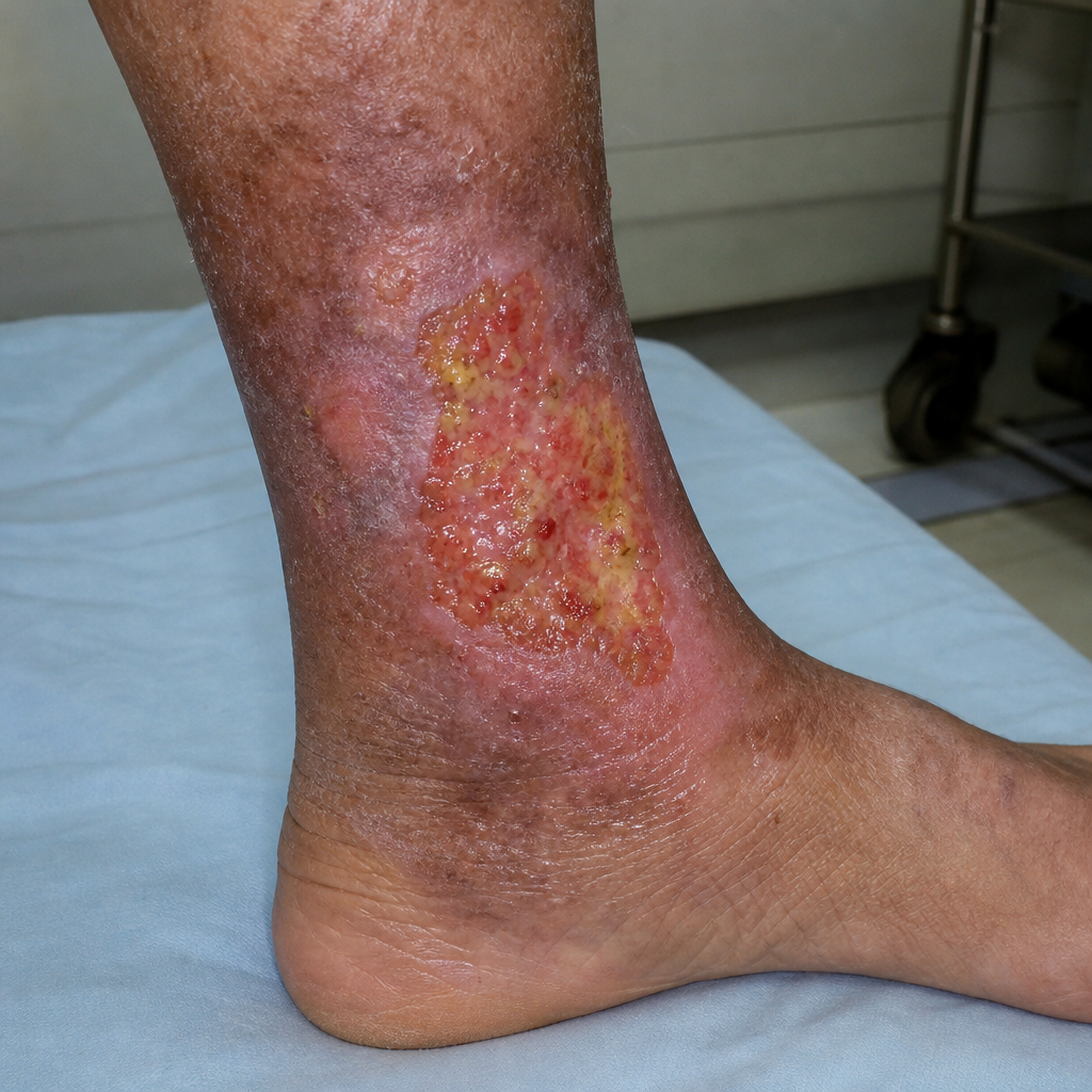
In the given image, which vein is mainly affected?
Identify the type of ulcer? 
A patient with dilated tortuous veins of the leg presented to the OPD and is diagnosed with varicose vein of grade C4a. What is the best preferred treatment?
A patient presents with calf pain while walking a certain distance. The pain is severe enough that he must stop and rest before continuing. According to Boyd's grading, which of the following grades best describes this condition?
Which is the least commonly used graft for coronary artery bypass graft (CABG)?
Practice by Chapter
Atherosclerotic Disease
Practice Questions
Aortic Aneurysms
Practice Questions
Peripheral Arterial Disease
Practice Questions
Carotid Artery Disease
Practice Questions
Venous Thromboembolism
Practice Questions
Chronic Venous Insufficiency
Practice Questions
Mesenteric Vascular Disease
Practice Questions
Vascular Trauma
Practice Questions
Vascular Access for Hemodialysis
Practice Questions
Endovascular Techniques
Practice Questions
Diabetic Foot Vascular Disease
Practice Questions
Vasculitis
Practice Questions
Want unlimited practice?
Get full access to all questions, explanations, and performance tracking.
Scan to download app