
Vascular Surgery — MCQs

On this page
Surgery for varicose veins is contraindicated by even stripping in?
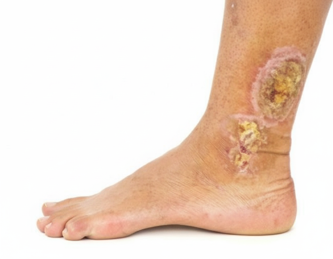
Which of the following treatments would you recommend for this patient?
Sympathectomy is indicated in all of the following conditions except?
Burger disease is:
All of the following are clinical features of thromboangiitis obliterans except?
Perforators are not present at which location?
Fogarty's catheter is used for?
In Deep Venous Thrombosis (DVT), which of the following findings is typically NOT seen?
A 59-year-old woman, who had a left femoral venous thrombosis during a pregnancy 30 years ago and whose left greater saphenous vein had been stripped at age 21, presents with a large, non-healing ulceration over the medial left calf. The ulcer has continuously progressed despite bed rest, elevation, and the use of a support stocking. Descending phlebography of the left leg demonstrates a patent deep venous system with free flow of dye from the groin to the foot. The first profunda femoris valve is competent. What is the most appropriate management?
What is the most common cause of lymphedema?
Practice by Chapter
Atherosclerotic Disease
Practice Questions
Aortic Aneurysms
Practice Questions
Peripheral Arterial Disease
Practice Questions
Carotid Artery Disease
Practice Questions
Venous Thromboembolism
Practice Questions
Chronic Venous Insufficiency
Practice Questions
Mesenteric Vascular Disease
Practice Questions
Vascular Trauma
Practice Questions
Vascular Access for Hemodialysis
Practice Questions
Endovascular Techniques
Practice Questions
Diabetic Foot Vascular Disease
Practice Questions
Vasculitis
Practice Questions
Want unlimited practice?
Get full access to all questions, explanations, and performance tracking.
Scan to download app