
Vascular Surgery — MCQs

On this page
An arteriovenous fistula can be safely ligated if which of the following tests is positive?
An 85-year-old gentleman complains of leg pain while lying down, which lessens when he hangs the foot out of bed. What is the most likely cause?
Which of the following is NOT indicated for an arterial leg ulcer?
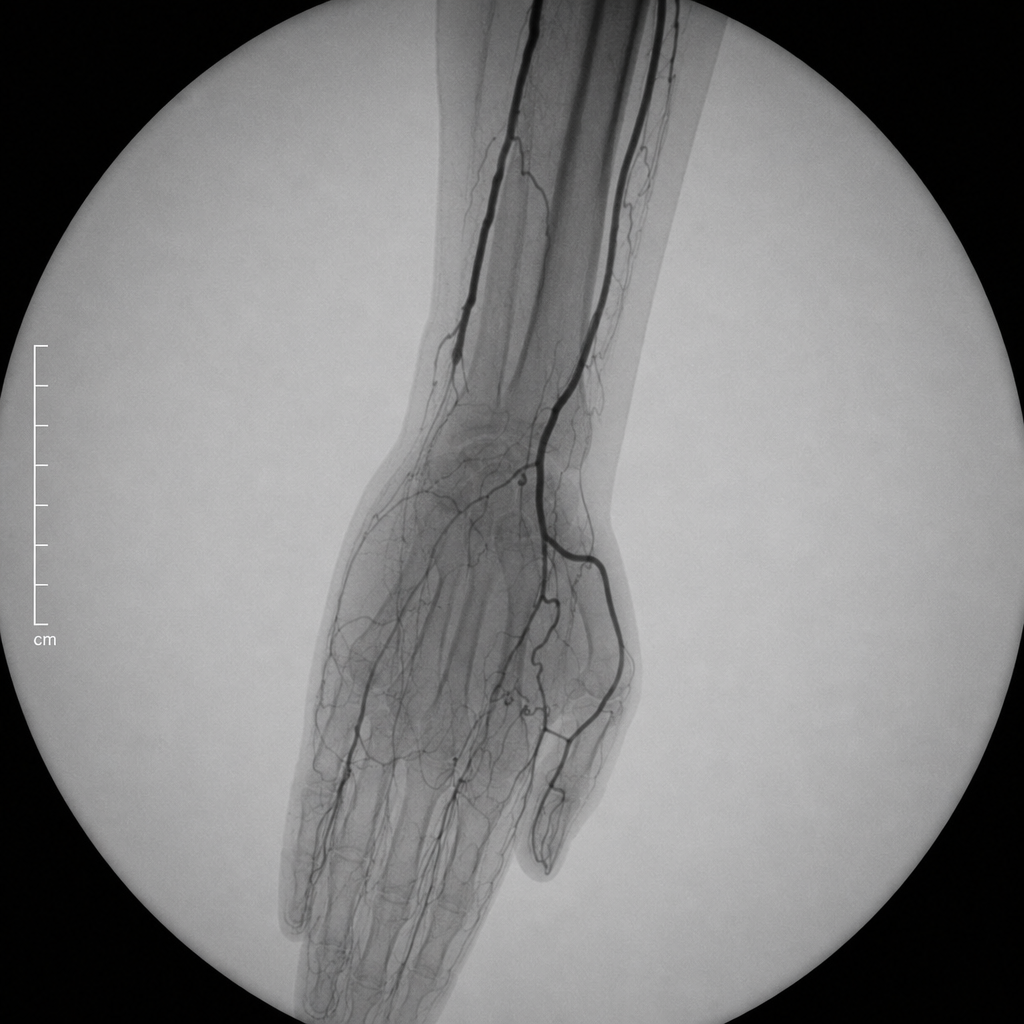
What accounts for this patient's hand pain?
Bilateral pulseless disease in upper limbs is caused by which of the following conditions?
Buerger's disease usually affects all of the following except:
Which of the following does NOT cause lymphoedema?
Lymphoedema is due to all except?
A 55-year-old man suffers from an acute myocardial infarction after occlusion of the left anterior descending coronary artery. The patient undergoes coronary bypass surgery 3 days later. Which of the following is the most frequent cause of saphenous vein graft failure several years following coronary bypass surgery?
Milroy's disease is characterized by which of the following?
Practice by Chapter
Atherosclerotic Disease
Practice Questions
Aortic Aneurysms
Practice Questions
Peripheral Arterial Disease
Practice Questions
Carotid Artery Disease
Practice Questions
Venous Thromboembolism
Practice Questions
Chronic Venous Insufficiency
Practice Questions
Mesenteric Vascular Disease
Practice Questions
Vascular Trauma
Practice Questions
Vascular Access for Hemodialysis
Practice Questions
Endovascular Techniques
Practice Questions
Diabetic Foot Vascular Disease
Practice Questions
Vasculitis
Practice Questions
Want unlimited practice?
Get full access to all questions, explanations, and performance tracking.
Scan to download app