
Vascular Surgery — MCQs

On this page
What is the most commonly used graft for the repair of an aortic coarctation?
Which of the following antibiotics is the drug of choice in the treatment of lymphedema?
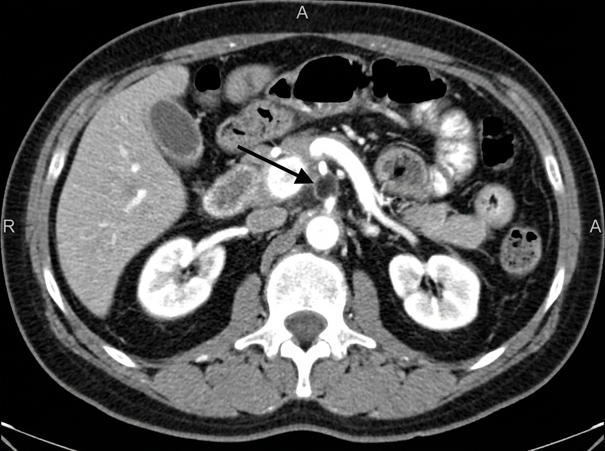
A patient presents to the emergency department with abdominal pain. What is the MOST significant diagnosis?
What is the most common source of pulmonary embolism?
Which of the following is NOT a clinical test used to assess perforator vein incompetence?
Ankle brachial pressure index is more than 1.3 in all except?
What is the initial therapy for documented deep venous thrombosis in a post-operative patient?
All of the following are seen in deep vein thrombosis except?
Grade 4 clinically in varicose veins is characterised by all except?
A patient presented with pulsating varicose veins of the lower limb. On examination, he was having cutaneous hemangioma involving the face with enlarged toes. What is the most probable diagnosis?
Practice by Chapter
Atherosclerotic Disease
Practice Questions
Aortic Aneurysms
Practice Questions
Peripheral Arterial Disease
Practice Questions
Carotid Artery Disease
Practice Questions
Venous Thromboembolism
Practice Questions
Chronic Venous Insufficiency
Practice Questions
Mesenteric Vascular Disease
Practice Questions
Vascular Trauma
Practice Questions
Vascular Access for Hemodialysis
Practice Questions
Endovascular Techniques
Practice Questions
Diabetic Foot Vascular Disease
Practice Questions
Vasculitis
Practice Questions
Want unlimited practice?
Get full access to all questions, explanations, and performance tracking.
Scan to download app