
Vascular Surgery — MCQs

On this page
Which of the following types of dissecting aneurysm does NOT involve the ascending aorta?
Stemmer's sign and buffalo hump are features of which condition?
What is the best graft material for aortic dissection?
A 67-year-old man with a history of hypertension, hyperlipidemia, and tobacco use has been diagnosed with an infra-renal aortic aneurysm of size 3 x 3.5 cm. Which management strategy is best suited for this patient?
Unna boot is used for the treatment of which condition?
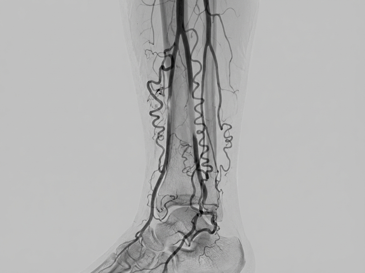
The angiogram depicted below is most typical of a patient whose history includes:
Which of the following statements regarding carotid body tumors is correct?
A patient presents with occasional pain over her middle finger. The surgeon identifies a black spot. Pressing around the black spot does not elicit pain, while pain is sensed on pressing the spot itself. What is the most likely diagnosis?
What is the most common vessel affected in acute limb ischemia?
All of the following are true about varicose veins except:
Practice by Chapter
Atherosclerotic Disease
Practice Questions
Aortic Aneurysms
Practice Questions
Peripheral Arterial Disease
Practice Questions
Carotid Artery Disease
Practice Questions
Venous Thromboembolism
Practice Questions
Chronic Venous Insufficiency
Practice Questions
Mesenteric Vascular Disease
Practice Questions
Vascular Trauma
Practice Questions
Vascular Access for Hemodialysis
Practice Questions
Endovascular Techniques
Practice Questions
Diabetic Foot Vascular Disease
Practice Questions
Vasculitis
Practice Questions
Want unlimited practice?
Get full access to all questions, explanations, and performance tracking.
Scan to download app