The cytogenetics of chromophilic renal cell carcinoma is characterized by:
Management of RCC less than 4 cm in size:
A patient has a cerebellar mass, renal tumor, and a family history of similar conditions. Which of the following mutations is most likely present in the family?
Most common type of renal carcinoma is:
What is the best investigation for diagnosis and staging of renal cell carcinoma with thrombus extending into the IVC?
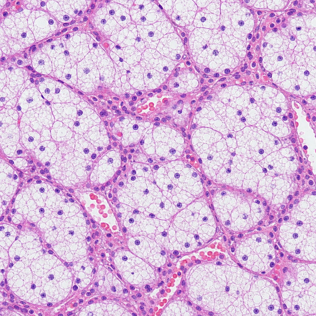
Treatment of Renal cell carcinoma of less than 4 cm will be-
A patient presented with renal cell carcinoma invading the IVC and the renal vein. Which of the following statements is false?
Which of the following statements about hypernephroma is true?
All are true about carcinoma penis except which of the following?
In a patient with stage T1 (tumor invading lamina propria) transitional cell carcinoma of the urinary bladder, the recommended treatment is
Explanation: ***Trisomy 7/17*** - Chromophilic renal cell carcinoma is notably characterized by **trisomy of chromosomes 7 and 17**, which is frequently observed in these tumors [1]. - This genetic alteration is associated with **proliferative changes** in renal cells leading to tumor development. *Loss 3p* - Loss of the **3p chromosome** is more commonly associated with **clear cell renal cell carcinoma**, not chromophilic types [1]. - It involves the **VHL gene**, which is not a primary characteristic of chromophilic renal cell carcinoma. *Mutant VHL gene* - Mutations in the **VHL gene** are primarily linked with **clear cell renal cell carcinoma** and hereditary conditions like **Von Hippel-Lindau syndrome**. - Chromophilic renal cell carcinoma typically does not involve these mutations as a defining feature. *Loss of 5q3* - Loss of **5q3** is not a recognized hallmark for chromophilic renal cell carcinoma; it lacks significance in this context. - This genetic aberration is often unrelated to the development or progression of chromophilic tumors. **References:** [1] Kumar V, Abbas AK, et al.. Robbins and Cotran Pathologic Basis of Disease. 9th ed. The Kidney, pp. 957-959.
Explanation: ***Correct: Partial nephrectomy*** - For **renal cell carcinoma (RCC) less than 4 cm (T1a)**, partial nephrectomy is the **gold standard** as it offers equivalent oncological outcomes to radical nephrectomy while preserving renal function. - This approach minimizes the risk of **chronic kidney disease** and its associated complications without compromising cancer control for appropriately selected smaller tumors. - **Nephron-sparing surgery** is now the preferred approach per EAU and AUA guidelines for small renal masses. *Incorrect: Surgery followed by chemotherapy* - While surgery is the primary treatment, **adjuvant chemotherapy** is generally **not effective** for localized RCC and is not routinely recommended for small tumors. - Systemic therapies are typically reserved for **advanced or metastatic RCC**, or in specific clinical trials. *Incorrect: Radical nephrectomy* - This involves removing the entire kidney, which is typically reserved for **larger tumors (T1b and above)**, centrally located tumors, or those with significant renal parenchymal involvement. - For tumors under 4 cm, radical nephrectomy leads to **unnecessary loss of renal function** compared to partial nephrectomy. *Incorrect: Chemotherapy* - **RCC is notoriously chemoresistant**, meaning traditional chemotherapy drugs have very limited efficacy in treating this cancer. - Chemotherapy alone is **not a primary treatment modality** for localized RCC due to its poor response rates in this cancer type.
Explanation: ***VHL*** - **Von Hippel-Lindau (VHL) disease** is an inherited disorder characterized by the development of tumors and cysts in various parts of the body, including **hemangioblastomas** in the cerebellum and retina, **renal cell carcinomas**, and pheochromocytomas [1]. - The combination of a **cerebellar mass**, renal tumor, and a family history strongly points to VHL disease, which is caused by a germline mutation in the **VHL tumor suppressor gene** [1]. *Neurofibromatosis (NF1)* - **Neurofibromatosis type 1 (NF1)** typically presents with multiple neurofibromas, **café-au-lait spots**, optic pathway gliomas, and Lisch nodules in the iris. - While NF1 can cause tumors, the specific combination of a cerebellar mass and renal tumor is not typical of NF1, and the characteristic skin findings are not mentioned. *Tuberous Sclerosis Complex (TSC)* - **Tuberous Sclerosis Complex (TSC)** is characterized by the growth of benign tumors in the brain (e.g., **subependymal giant cell astrocytomas**), kidneys (e.g., **angiomyolipomas**), heart, lungs, and skin (e.g., facial angiofibromas) [2]. - While TSC can involve brain and kidney tumors, the typical brain tumors are different (astrocytomas vs. hemangioblastomas), and hemangioblastomas are not a common feature of TSC [2]. *Li-Fraumeni syndrome* - **Li-Fraumeni syndrome** is a rare inherited cancer predisposition syndrome characterized by a high risk of developing various cancers, including **sarcomas**, breast cancer, brain tumors (often astrocytomas or medulloblastomas), and adrenocortical carcinoma. - While brain tumors are part of Li-Fraumeni syndrome, renal cell carcinoma is not a primary feature, and the classic cerebellar hemangioblastoma is not typical for this syndrome. **References:** [1] Cross SS. Underwood's Pathology: A Clinical Approach. 6th ed. Common Clinical Manifestations Of Central And Peripheral Nervous System Disease, pp. 724-727. [2] Kumar V, Abbas AK, et al.. Robbins and Cotran Pathologic Basis of Disease. 9th ed. The Central Nervous System, pp. 1318-1319.
Explanation: ***Clear cell type*** - The **clear cell type** is the most common subtype of renal carcinoma, constituting about **70-80%** of cases [3]. - It is typically associated with **von Hippel-Lindau syndrome** and presents with clear or "foamy" cells due to lipid accumulation [3]. *Chromophobe type* - This type comprises about **5-10%** of renal cell carcinomas and usually has a better prognosis [1]. - Characterized by **pale cells with distinct cell borders** and lacks the common features of clear cell carcinoma [1]. *Tubular type* - The tubular variant is less common and does not represent a major subtype of renal cell carcinoma. - It is often confused with other variants but lacks the distinct characteristics of the clear cell type. *Papillary type* - The papillary type accounts for about **10-15%** of renal carcinomas and is characterized by papillary structures [2]. - This type generally has a distinct chromosomal mutation profile compared to the clear cell type [2]. **References:** [1] Kumar V, Abbas AK, et al.. Robbins and Cotran Pathologic Basis of Disease. 9th ed. The Kidney, p. 959. [2] Kumar V, Abbas AK, et al.. Robbins and Cotran Pathologic Basis of Disease. 9th ed. The Kidney, pp. 958-959. [3] Kumar V, Abbas AK, et al.. Robbins and Cotran Pathologic Basis of Disease. 9th ed. The Kidney, pp. 959-961.
Explanation: ***CT scan*** - **CT scan** with contrast is the gold standard for diagnosing renal cell carcinoma and evaluating the extent of tumor thrombus into the **IVC**. - It provides detailed anatomical information on the tumor, staging, and involvement of adjacent structures. *Angiography* - **Angiography** is an invasive procedure primarily used for mapping the vascular supply of the tumor preoperatively or for embolization, not as a primary diagnostic tool. - It carries risks associated with contrast agents and catheterization and provides less comprehensive detail on tumor extension compared to CT. *Colour doppler imaging* - While useful for detecting blood flow and confirming the presence of a thrombus, **color Doppler imaging** (ultrasound) has limitations in accurately assessing the cranial extent of an IVC thrombus. - Its diagnostic accuracy is highly operator-dependent and less reliable for deep structures like the IVC compared to CT. *IVP* - **Intravenous Pyelogram (IVP)** assesses the urinary tract's structure and function but has limited utility in detecting soft tissue masses like renal cell carcinoma or IVC thrombus. - It involves radiation exposure and contrast material, and has largely been replaced by more advanced imaging techniques like CT and MRI for renal masses.
Explanation: **Partial nephrectomy** - For **renal cell carcinoma** (RCC) less than 4 cm, partial nephrectomy is the preferred treatment as it offers comparable oncologic outcomes to radical nephrectomy while preserving renal function. - This approach minimizes the risk of **chronic kidney disease** and its associated complications, which is particularly important given that RCC often occurs in older patients or those with comorbidities. *Radical nephrectomy* - While effective for larger tumors or those with complex involvement, **radical nephrectomy** involves the complete removal of the affected kidney. - For tumors less than 4 cm, it is generally considered an **overtreatment** that unnecessarily sacrifices healthy kidney tissue, increasing the risk of **renal insufficiency**. *Radical nephrectomy + postoperative radiotherapy* - **Postoperative radiotherapy** for RCC does not significantly improve outcomes and is associated with considerable morbidity. - Its use is typically limited to rare cases of positive surgical margins or palliative care, and it is **not indicated** for small, localized tumors. *Radical nephrectomy + chemotherapy* - **Renal cell carcinoma** is historically known to be highly resistant to conventional chemotherapy. - Therefore, **systemic chemotherapy** is generally not used as a primary treatment for localized RCC and has no proven benefit in conjunction with radical nephrectomy for small tumors.
Explanation: ***Correct: IVC involvement indicates inoperability*** - This statement is **FALSE** and therefore the correct answer to this question - IVC involvement does NOT render RCC inoperable - Surgical resection with **radical nephrectomy and tumor thrombectomy** (cavotomy) can be performed successfully - **Even IVC tumor thrombus extending to the right atrium** can be surgically managed by experienced teams - Level of tumor thrombus determines surgical approach but not operability *Incorrect: Preop biopsy is not necessary* - This statement is **TRUE** (not the answer we're looking for) - Pre-operative biopsy typically not required when imaging is diagnostic for RCC - Cross-sectional imaging (CT/MRI) with characteristic features is usually sufficient - Biopsy risks include **tumor seeding** along the tract and potential for non-diagnostic sampling - Biopsy reserved for atypical presentations or when diagnosis unclear *Incorrect: Pre op radiotherapy is not essential* - This statement is **TRUE** (not the answer we're looking for) - RCC is **radioresistant** and responds poorly to radiation therapy - Pre-operative radiotherapy is NOT part of standard management for localized RCC - Surgery (radical nephrectomy) remains the primary curative treatment - Radiation may have palliative role in metastatic disease (bone mets, brain mets) *Incorrect: Chest x-ray should be done to rule out pulmonary metastasis* - This statement is **TRUE** (not the answer we're looking for) - **Lungs are the most common site** of RCC metastases - Chest imaging is essential component of pre-operative staging - **CT chest is preferred** over plain X-ray for better sensitivity in detecting pulmonary metastases - Complete staging includes CT chest, abdomen, and pelvis
Explanation: ***Radiosensitive*** - Hypernephroma, or renal cell carcinoma, is typically resistant to **radiation therapy**, making this statement false. - It is generally treated with **surgery** and targeted therapies rather than radiation. *Usually adenocarcinoma* - Hypernephroma is indeed a type of **adenocarcinoma**, as it originates from the renal tubular epithelium [1]. - It is the most common form of **kidney cancer**, supporting this as a true statement. *May present with rapidly developing varicocele* - Rapidly developing **varicocele** can occur due to **renal vein obstruction** associated with renal tumors [2], so this statement is true. - Varicocele is a recognized clinical feature in renal cell carcinoma due to its anatomical relationships. *Arise from cortex usually from pre existing adenoma* - Hypernephroma does arise from the **renal cortex**, often from pre-existing renal adenomas or other lesions. - This establishes its origin, making the statement accurate. **References:** [1] Kumar V, Abbas AK, et al.. Robbins and Cotran Pathologic Basis of Disease. 9th ed. The Kidney, pp. 959-961. [2] Cross SS. Underwood's Pathology: A Clinical Approach. 6th ed. Common Clinical Problems From Diseases Of The Urinary And Male Genital Tracts, pp. 492-493.
Explanation: ***Spreads by blood borne metastasis*** - Carcinoma penis typically spreads initially via the **lymphatic system** to inguinal lymph nodes. - **Hematogenous spread** is a late event and generally rare, with the most common sites being the lung, liver, and bone. *Leads to erosion of artery* - Local advancement of penile carcinoma can lead to **erosion of penile arteries**, which can cause significant morbidity including bleeding and functional compromise. - This local tissue destruction is a characteristic feature of advanced, uncontrolled penile cancer. *Slowly progressive* - Carcinoma penis is generally a **slowly progressive** malignancy, allowing for early detection and intervention if patients seek medical attention promptly. - The slow growth rate contributes to the fact that many patients present with localized or regionally advanced disease before distant metastases occur. *Most common type is squamous cell carcinoma* - Approximately 95% of penile cancers are **squamous cell carcinomas (SCCs)**, arising from the epithelial cells of the glans or foreskin. - Other rare types include melanoma, basal cell carcinoma, and sarcomas, but SCC vastly predominates.
Explanation: ***Trans urethral resection of bladder tumour (TURBT)*** - For **stage 1 (confined to mucosa and submucosa) transitional cell carcinoma** of the urinary bladder, TURBT is the primary and often curative treatment. - This procedure allows for both diagnostic staging and complete resection of visible tumors. *Radiation therapy* - **Radiation therapy** is generally reserved for more advanced stages or for patients who are not surgical candidates. - It is often used in combination with chemotherapy or as a palliative measure, not typically as monotherapy for early-stage disease. *Systemic chemotherapy* - **Systemic chemotherapy** is typically indicated for advanced, metastatic bladder cancer or as neoadjuvant/adjuvant therapy in muscle-invasive disease. - It is not the primary treatment for early-stage, non-muscle invasive bladder cancer. *Radical cystectomy* - **Radical cystectomy** is a major surgical procedure involving removal of the entire bladder and surrounding structures. - It is reserved for **muscle-invasive bladder cancer** or recurrent high-grade non-muscle invasive bladder cancer that has failed conservative treatments.
Get full access to all questions, explanations, and performance tracking.
Start For Free