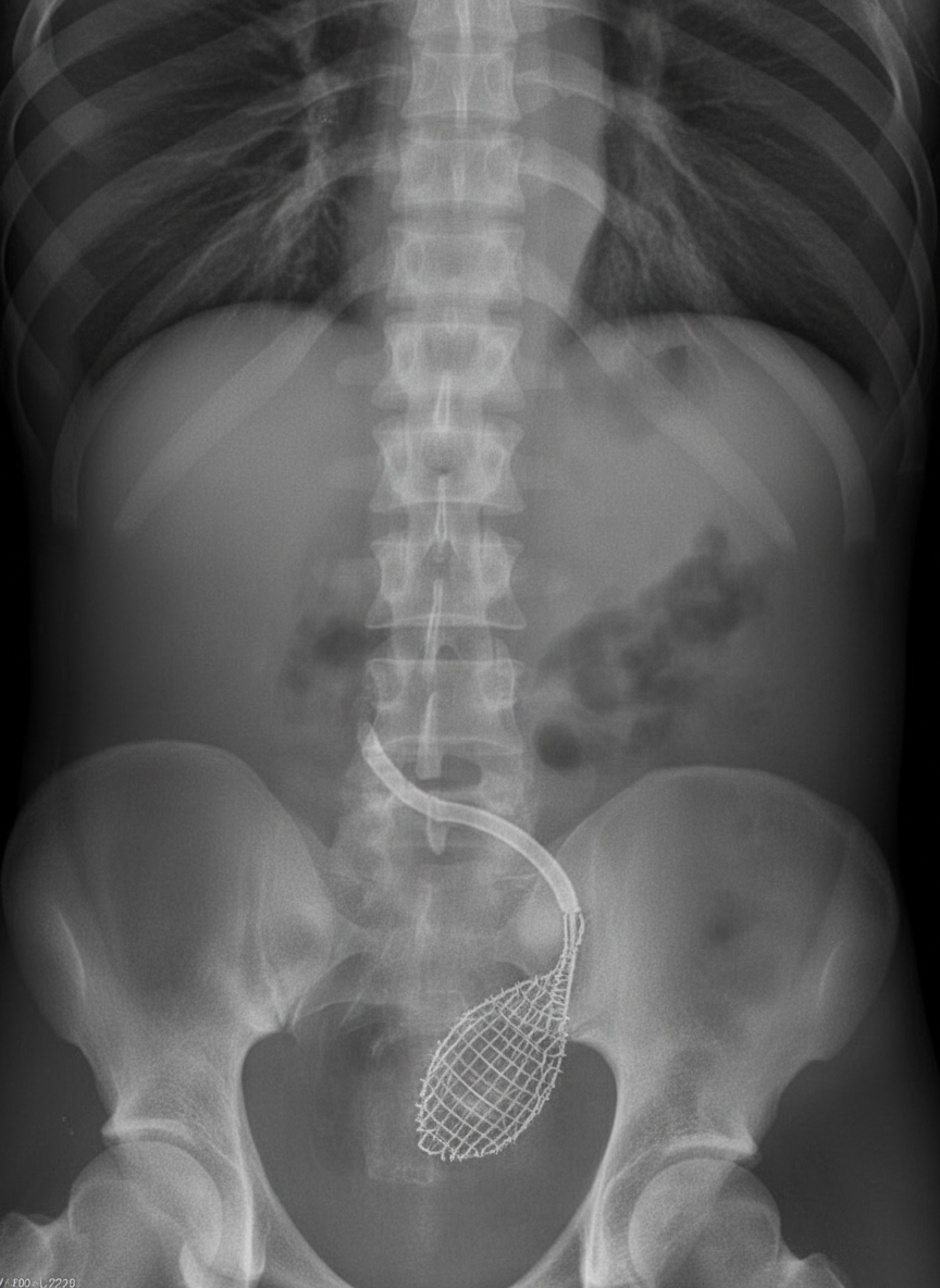
What procedure is shown in the ureter?

All of the following are seen in cystitis EXCEPT:
A patient was brought to the hospital eight hours after an injury. A few drops of blood are noted at the external urethral meatus. He has not passed urine and his bladder is palpable per abdomen. What is the probable diagnosis?
Epidermoid carcinoma of the renal pelvis is usually associated with:
Undescended testis can lead to all of the following complications except:
A 32-year-old man with a pelvic fracture presents with urinary retention and blood at the external meatus. A retrograde urethrogram shows prostatomembranous disruption. What is the most appropriate immediate treatment?
Urinary retention in a child is most commonly caused by which of the following conditions?
All of the following are true about bulbar urethral rupture, except:
A 25-year-old married male presents with infertility. He underwent retroperitoneal lymph node dissection at age 15 for embryonal carcinoma of the right testis. Semen analysis shows a quantity of 0.5 ml, no sperm, and no fructose. Testis biopsy shows normal spermatogenesis. What is the best treatment?
Which is the most common site for ectopic testis?
Explanation: ***Dormia Basket*** - A **Dormia basket** is a wire mesh device used for **stone retrieval** from the ureter, appearing as a characteristic **basket-like radiopaque structure** on fluoroscopy. - The basket is deployed beyond the stone, then **withdrawn** to capture and extract the calculus under direct visualization. *Stenting* - **Ureteric stents** appear as **straight radiopaque tubes** extending from the renal pelvis to the bladder, not basket-shaped structures. - Used for **drainage** and **relief of obstruction** rather than stone extraction procedures. *Dilatation* - **Ureteric dilatation** involves **balloon catheters** that appear as **smooth, elongated structures** when inflated, not wire mesh patterns. - Used to **widen strictures** or **prepare the ureter** for other procedures, not for stone retrieval. *Cannulation* - **Ureteric cannulation** involves **thin catheters** appearing as **fine radiopaque lines** within the ureter. - Used for **contrast injection** during **retrograde pyelography** or **guidewire placement**, not stone extraction.
Explanation: **Explanation:** The correct answer is **Fever**. In clinical urology, it is crucial to differentiate between **Lower Urinary Tract Infections (UTIs)** and **Upper UTIs**. 1. **Why Fever is the correct answer:** Cystitis is a localized inflammation of the bladder mucosa (Lower UTI). Because it is a superficial mucosal infection without systemic involvement or tissue invasion, it typically **does not present with fever**. The presence of fever, chills, or flank pain in a patient with urinary symptoms strongly suggests **Acute Pyelonephritis** (Upper UTI) or systemic involvement like prostatitis. 2. **Analysis of incorrect options:** * **Dysuria:** This is the most common symptom of cystitis, caused by the irritation of the urethral and bladder neck mucosa during voiding. * **Nocturia & Frequency:** Inflammation reduces the functional capacity and compliance of the bladder, leading to an increased urge to void both day and night. * **Hematuria:** "Hemorrhagic cystitis" is common in bacterial infections where the friable, inflamed bladder mucosa bleeds easily. **NEET-PG High-Yield Pearls:** * **Triad of Cystitis:** Frequency, Urgency, and Dysuria. * **Most Common Organism:** *E. coli* (80-85% of cases). * **Gold Standard Diagnosis:** Urine culture (Significant bacteriuria is traditionally defined as $\geq 10^5$ CFU/mL). * **Sterile Pyuria:** If a patient has symptoms of cystitis but negative routine cultures, consider *Chlamydia trachomatis*, *Neisseria gonorrhoeae*, or Renal Tuberculosis. * **Treatment:** Uncomplicated cystitis is typically treated with Nitrofurantoin, Fosfomycin, or TMP-SMX.
Explanation: ### **Explanation** The clinical presentation of **blood at the external urethral meatus** following trauma is the hallmark sign of a **urethral injury**. #### **Why Option A is Correct** The triad of **blood at the meatus**, **inability to void**, and a **palpable/distended bladder** strongly indicates a disruption of the urethra. In this case, the palpable bladder confirms that the kidneys are producing urine and the bladder is intact, but the urine cannot be evacuated due to a mechanical obstruction (the urethral tear). This is most commonly seen in posterior urethral injuries (associated with pelvic fractures) or anterior urethral injuries (straddle injuries). #### **Why Other Options are Incorrect** * **Option B (Rupture bladder):** In a bladder rupture (especially intraperitoneal), the bladder is typically **not palpable** because urine leaks into the peritoneal cavity or perivesical space. Furthermore, blood at the meatus is rare in isolated bladder injuries. * **Option C (Extravasation in retroperitoneum):** While posterior urethral injuries can lead to extravasation, the primary diagnosis remains the injury itself. Moreover, extravasation in the retroperitoneum is more characteristic of extraperitoneal bladder rupture, where a palpable bladder would not be the primary finding. * **Option D (Anuria):** Anuria refers to a failure of the kidneys to produce urine. The fact that the **bladder is palpable** proves that urine is being produced and stored, ruling out hypovolemic anuria. --- ### **Clinical Pearls for NEET-PG** * **Gold Standard Investigation:** Retrograde Urethrogram (RUG) is the initial investigation of choice for suspected urethral injury. **Never** attempt catheterization before an RUG if blood is seen at the meatus. * **Posterior Urethral Injury:** Most common site is the membranous urethra; usually associated with pelvic fractures. Look for a "high-riding prostate" on DRE. * **Anterior Urethral Injury:** Most common site is the bulbar urethra; usually due to a "straddle injury" (falling astride). * **Management:** If the patient cannot void and the bladder is full, a **Suprapubic Cystostomy (SPC)** is the preferred method to divert urine.
Explanation: **Explanation:** Epidermoid carcinoma (Squamous Cell Carcinoma/SCC) of the renal pelvis is a rare but aggressive malignancy. The primary underlying mechanism is **chronic irritation**, which leads to squamous metaplasia of the urothelium, eventually progressing to dysplasia and carcinoma. * **Why Pelvic Calculus is Correct:** Long-standing **renal calculi** (specifically staghorn or pelvic stones) are the most common cause of chronic mechanical irritation and infection in the renal pelvis. Approximately 75–100% of patients with SCC of the renal pelvis have a history of chronic nephrolithiasis. * **Why Incorrect Options are Wrong:** * **Multiple Papillomas:** These are associated with **Transitional Cell Carcinoma (TCC)**, the most common type of renal pelvic tumor. TCC is often multifocal and associated with field defects. * **Tuberculosis of the Kidney:** While TB causes chronic inflammation, it typically leads to strictures, "putty kidney," or autonephrectomy rather than SCC. * **Filariasis:** This usually affects the lymphatic system, leading to chyluria or hydrocele, but is not a recognized risk factor for renal pelvic malignancy. **NEET-PG High-Yield Pearls:** 1. **Most common tumor** of the renal pelvis: **Transitional Cell Carcinoma (TCC)**. 2. **Most common risk factor** for SCC of the renal pelvis: **Chronic Calculi** (Stones). 3. **Schistosomiasis (S. haematobium)** is a major risk factor for SCC of the **Urinary Bladder**, not typically the renal pelvis. 4. SCC of the renal pelvis usually presents at an advanced stage and has a much poorer prognosis compared to TCC.
Explanation: **Explanation:** The correct answer is **Hypertrophy of testes**. In cases of cryptorchidism (undescended testis), the affected testis is typically **atrophic** (small and soft) rather than hypertrophied. This is due to the higher intra-abdominal temperature, which leads to the degeneration of germinal epithelium and peritubular fibrosis. While the contralateral descended testis may undergo compensatory hypertrophy, the undescended testis itself does not. **Analysis of Incorrect Options:** * **Torsion:** Undescended testes are more prone to torsion because they often lack the normal posterior attachment to the scrotum (gubernaculum abnormality) and are frequently associated with a patent processus vaginalis, allowing for increased mobility. * **Sterility:** The higher temperature of the inguinal canal or abdomen inhibits spermatogenesis. While Leydig cells (testosterone production) are relatively resistant, the Sertoli cells and germ cells are damaged, leading to infertility, especially in bilateral cases. * **Carcinoma:** There is a 10–40 times higher risk of germ cell tumors (most commonly **Seminoma**) in undescended testes. Orchiopexy does not eliminate the risk of malignancy but makes the testis accessible for clinical examination and screening. **High-Yield Clinical Pearls for NEET-PG:** * **Most common site:** Inguinal canal. * **Most common complication:** Inguinal hernia (associated with patent processus vaginalis in 90% of cases). * **Most common tumor:** Seminoma (overall); however, if the testis remains intra-abdominal, the risk of Seminoma is significantly higher. * **Timing of Surgery:** Orchiopexy is ideally performed between **6 to 12 months** of age to preserve fertility potential. * **Investigation of choice:** Ultrasonography (initial); Diagnostic Laparoscopy (Gold Standard for impalpable testes).
Explanation: ### Explanation **Correct Answer: C. Suprapubic cystostomy** The clinical triad of **pelvic fracture**, **urinary retention**, and **blood at the urethral meatus** is a classic presentation of a **Posterior Urethral Injury (PUI)**, specifically at the prostatomembranous junction. In the acute setting of a suspected urethral injury, the primary goal is to provide urinary drainage while avoiding further trauma to the injured area. **Suprapubic cystostomy (SPC)** is the gold standard immediate treatment because it diverts urine away from the site of disruption, prevents extravasation into the pelvic space, and allows the pelvic hematoma to resolve without the risk of converting a partial tear into a complete transection. #### Why other options are incorrect: * **A. Urethral catheterization:** This is strictly contraindicated. Blind insertion of a Foley catheter can convert a partial urethral tear into a complete disruption and introduce infection into the pelvic hematoma. * **B. Exploration and repair:** Immediate primary repair is avoided in posterior urethral injuries because the patient is often hemodynamically unstable from the pelvic fracture, and early surgery in a pelvic hematoma increases the risk of massive hemorrhage, impotence, and incontinence. * **C. Perineal urethrostomy:** This is a definitive surgical procedure for complex strictures and is not indicated in the emergency management of acute trauma. #### NEET-PG High-Yield Pearls: * **Gold Standard Investigation:** Retrograde Urethrogram (RUG) is the investigation of choice for suspected urethral injury. * **Posterior vs. Anterior:** Posterior injuries (membranous) are usually associated with pelvic fractures; Anterior injuries (bulbar) are usually due to "straddle" trauma. * **Physical Exam Sign:** A "high-riding prostate" on Digital Rectal Examination (DRE) suggests posterior urethral disruption. * **Definitive Management:** Most posterior injuries are managed with delayed repair (Urethroplasty) 3–6 months after the initial injury.
Explanation: **Explanation:** **Posterior Urethral Valve (PUV)** is the most common cause of lower urinary tract obstruction and urinary retention in **male infants and children**. It is a congenital condition where abnormal mucosal folds (valves) in the prostatic urethra act as a one-way flap, obstructing the outflow of urine. This leads to a classic clinical triad: a palpable distended bladder, a poor/dribbling urinary stream, and bilateral hydroureteronephrosis. **Analysis of Options:** * **Meatal stenosis with ulceration (Option A):** While it can cause painful micturition (dysuria) and a narrow stream, it rarely leads to complete urinary retention. It is usually an acquired condition following circumcision. * **Urethral stricture (Option C):** This is more common in adults (due to trauma or STIs). In children, it is rare and usually follows instrumentation or trauma, rather than being the primary congenital cause of retention. * **Epispadias (Option D):** This is a congenital malformation where the urethra opens on the dorsal aspect of the penis. It typically presents with **urinary incontinence** (due to sphincter defects) rather than retention. **Clinical Pearls for NEET-PG:** * **Gold Standard Investigation:** Voiding Cystourethrogram (VCUG) is the investigation of choice; it classically shows a dilated posterior urethra with a "keyhole" appearance. * **Initial Management:** Catheterization (using a small feeding tube) to decompress the bladder. * **Definitive Treatment:** Endoscopic primary valve ablation (fulguration). * **Antenatal Clue:** Bilateral hydronephrosis and oligohydramnios on maternal ultrasound.
Explanation: ### Explanation The correct answer is **B. Floating prostate on per rectal examination.** #### 1. Why Option B is the Correct Answer (The "Except") A **floating prostate** is a classic clinical sign of **posterior urethral injury** (specifically membranous urethral rupture associated with pelvic fractures). In posterior injuries, the puboprostatic ligaments are torn, allowing the prostate to be displaced superiorly by a pelvic hematoma. In contrast, **bulbar urethral rupture** is an **anterior urethral injury**, typically caused by a "straddle injury" (falling astride a firm object). Because the injury occurs distal to the urogenital diaphragm, the prostate remains in its normal anatomical position and is palpable on a digital rectal exam (DRE). #### 2. Analysis of Incorrect Options * **A & C (Perineal hematoma and Collection of urine):** In bulbar injuries, the rupture occurs below the perineal membrane. If **Buck’s fascia** is torn, urine and blood extravasate into the superficial perineal pouch. This leads to a characteristic **"butterfly-shaped"** hematoma/swelling in the perineum and scrotum. * **D (Bleeding per urethra):** This is the **most common clinical sign** of any urethral injury (both anterior and posterior). Blood at the external meatus indicates a breach in the urethral mucosa. #### 3. Clinical Pearls for NEET-PG * **Mechanism:** Straddle injury = Bulbar (Anterior); Pelvic fracture = Membranous (Posterior). * **Extravasation Limits:** In bulbar injuries with a torn Buck’s fascia, urine can spread to the scrotum, penis, and abdominal wall (deep to Scarpa’s fascia), but **not into the thighs** (due to the attachment of Colles' fascia to the fascia lata). * **Gold Standard Investigation:** Retrograde Urethrogram (RUG). **Never** attempt catheterization before ruling out urethral injury if blood is present at the meatus. * **Management:** Most acute bulbar ruptures are managed initially with a Suprapubic Cystostomy (SPC).
Explanation: ### Explanation **Clinical Analysis:** The patient presents with **obstructive azoospermia** secondary to a history of **Retroperitoneal Lymph Node Dissection (RPLND)**. RPLND often results in damage to the sympathetic nerves (hypogastric plexus), leading to **anejaculation** or **retrograde ejaculation**. The key findings are: 1. **Low semen volume (0.5 ml) and absent fructose:** Suggests a failure of seminal vesicle emission or a physical obstruction/denervation. 2. **Normal spermatogenesis on biopsy:** Confirms that the "factory" (testis) is functioning perfectly, but the "delivery system" is compromised. **Why Option C is Correct:** Since the patient is producing healthy sperm but cannot ejaculate them, the most effective treatment for infertility is **Sperm Retrieval** (such as Micro-TESE or TESA) followed by **Intracytoplasmic Sperm Injection (ICSI)**. This bypasses the transport defect entirely by extracting sperm directly from the source and injecting it into the ovum. **Why Other Options are Incorrect:** * **Option A:** Donor sperm is unnecessary because the patient has documented normal spermatogenesis; his own genetic material can be used. * **Option B:** Penile prostheses are used for erectile dysfunction (impotence), not for infertility or ejaculatory disorders. The patient's issue is emission/ejaculation, not the ability to achieve an erection. **Clinical Pearls for NEET-PG:** * **RPLND Complication:** The most common long-term morbidity of traditional RPLND is **loss of ejaculation** due to injury to the post-ganglionic sympathetic fibers (T12-L2). * **Fructose in Semen:** Fructose is produced by the seminal vesicles. Its absence in azoospermia typically indicates **ejaculatory duct obstruction** or **congenital bilateral absence of the vas deferens (CBAVD)**. * **Nerve-Sparing RPLND:** Modern surgical techniques aim to preserve the hypogastric plexus to maintain normal ejaculation.
Explanation: **Explanation:** The distinction between an **undescended testis (cryptorchidism)** and an **ectopic testis** is a frequent high-yield topic in NEET-PG. An ectopic testis is one that has deviated from the normal path of descent after passing through the external inguinal ring. **Why Inguinal Canal (Superficial Inguinal Pouch) is correct:** The most common site for an ectopic testis is the **Superficial Inguinal Pouch (of Denis Browne)**. This pouch lies superficial to the external oblique aponeurosis. While the question lists "Inguinal canal," in clinical practice and standard surgical textbooks (like Bailey & Love), the superficial inguinal pouch is considered the most frequent site because the testis is diverted here by abnormal fascial attachments (Lockwood’s tails of the gubernaculum) after exiting the canal. **Analysis of Incorrect Options:** * **A. Iliac fossa & C. Abdomen:** These are common sites for **undescended testes** (cryptorchidism), where the testis fails to progress along the normal path. They are rare for ectopic testes, as ectopia occurs *after* the testis has traversed the inguinal canal. * **D. Lumbar:** This is the embryological site of origin for the testes. A testis remaining here is a form of high abdominal arrest (undescended), not ectopia. **Clinical Pearls for NEET-PG:** 1. **Path of Descent:** The most common site for an **undescended** testis is the **Inguinal Canal**. The most common site for an **ectopic** testis is the **Superficial Inguinal Pouch**. 2. **Other Ectopic Sites:** Perineum (2nd most common), femoral canal, base of the penis, and the contralateral scrotal sac (transverse testicular ectopia). 3. **Key Difference:** An undescended testis can often be milked down into the scrotum (though it retracts), whereas an ectopic testis **cannot** be pushed into the scrotum because it is outside the normal anatomical path. 4. **Management:** The treatment of choice for both is **Orchidopexy**, ideally performed between 6 to 12 months of age to preserve fertility and allow for early detection of malignancy.
Urological Anatomy
Practice Questions
Hematuria Evaluation
Practice Questions
Urinary Calculi
Practice Questions
Benign Prostatic Hyperplasia
Practice Questions
Prostate Cancer
Practice Questions
Bladder Cancer
Practice Questions
Renal Cell Carcinoma
Practice Questions
Testicular Tumors
Practice Questions
Urinary Tract Infections
Practice Questions
Urinary Incontinence
Practice Questions
Genitourinary Trauma
Practice Questions
Pediatric Urology Basics
Practice Questions
Get full access to all questions, explanations, and performance tracking.
Start For Free