
Urology — MCQs

On this page
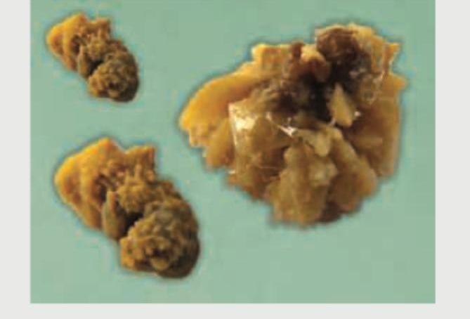
What is the composition of this stone?
A 60-year-old male presents with a poor stream of urine, a post-void residual urine volume of 400 mL, bilateral hydronephrosis, and a prostate weighing 70 g. His urea is 120 mg/dL and creatinine is 3.5 mg/dL. What is the ideal next immediate step?
What is the treatment of choice for ureteric colic?
Which of the following substances is not used as an irrigant during transurethral resection of the prostate?
What is the most common long-term complication causing mortality after urinary diversion?
A 'bag of worms' appearance in the scrotum is typically associated with which condition?
Subcapsular nephrectomy is indicated in which of the following conditions?
Which of the following symptoms does not typically improve following a Transurethral Resection of the Prostate (TURP)?
What is the most common testicular tumor of childhood?
A 9-year-old boy presented with abdominal pain and recurrent UTI. IVP revealed duplication of the left ureter. What is the most likely site of ectopic opening?
Practice by Chapter
Urological Anatomy
Practice Questions
Hematuria Evaluation
Practice Questions
Urinary Calculi
Practice Questions
Benign Prostatic Hyperplasia
Practice Questions
Prostate Cancer
Practice Questions
Bladder Cancer
Practice Questions
Renal Cell Carcinoma
Practice Questions
Testicular Tumors
Practice Questions
Urinary Tract Infections
Practice Questions
Urinary Incontinence
Practice Questions
Genitourinary Trauma
Practice Questions
Pediatric Urology Basics
Practice Questions
Want unlimited practice?
Get full access to all questions, explanations, and performance tracking.
Scan to download app