
Urology — MCQs

On this page
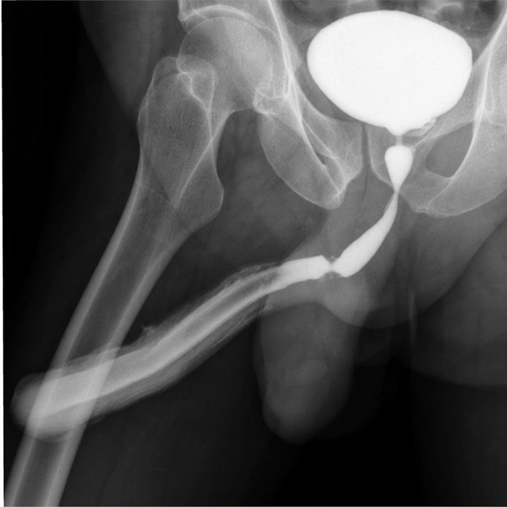
A 50-year-old male presented to the ER with complaints of inability to pass urine after a recent accident. On examination, he has a scrotal hematoma, blood at the urethral meatus, and a high-riding prostate. A retrograde urethrogram has been performed. What should be the next step in the management of the patient?
Which of the following statements is true regarding the management of a patient with hematuria diagnosed to have Stage II Transitional Cell Carcinoma of the bladder?
Urinary catheterization is indicated in cases of acute retention of urine, except in which of the following conditions?
All of the following can be seen in hypernephroma, except:
Which of the following statements about testicular seminoma is FALSE?
What is the most important prognostic factor in a case of penile carcinoma?
Marion's disease is due to which of the following?
All of the following are complications of an undescended testis EXCEPT?
All of the following are indications of percutaneous nephrostomy, EXCEPT:
What scoring system is used to grade the symptoms of Benign Prostatic Hyperplasia (BPH)?
Practice by Chapter
Urological Anatomy
Practice Questions
Hematuria Evaluation
Practice Questions
Urinary Calculi
Practice Questions
Benign Prostatic Hyperplasia
Practice Questions
Prostate Cancer
Practice Questions
Bladder Cancer
Practice Questions
Renal Cell Carcinoma
Practice Questions
Testicular Tumors
Practice Questions
Urinary Tract Infections
Practice Questions
Urinary Incontinence
Practice Questions
Genitourinary Trauma
Practice Questions
Pediatric Urology Basics
Practice Questions
Want unlimited practice?
Get full access to all questions, explanations, and performance tracking.
Scan to download app