
Urology — MCQs

On this page
Testis tumor is associated with secondary hydrocele in what percentage of cases?
A 40-year-old male presents with right loin pain radiating to the right iliac fossa. Investigations including USG abdomen and NCCT KUB reveal a renal stone measuring 8mm. What is the most probable location of this stone?
What is the most common complication seen with prostatitis?
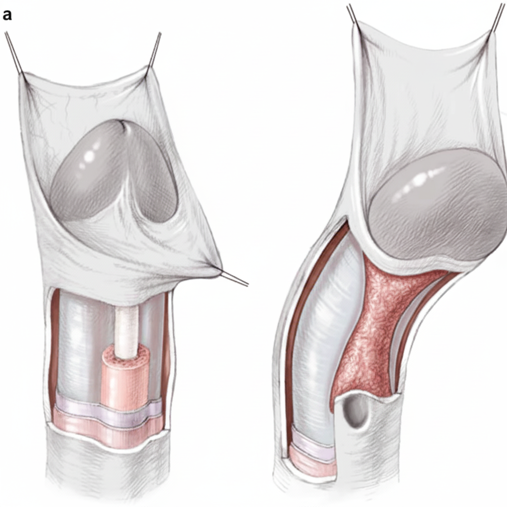
What is the investigation of choice for posterior urethral stricture?
What is the treatment of choice for Stage I seminoma of the testis?
Spermatocoeles are most commonly found at which anatomical location?
A 35-year-old male presents with a non-tender scrotal swelling that is separate from the testes. On transillumination, a "Chinese lantern" pattern is observed. What is the most likely diagnosis?
A 78-year-old male with a known history of prostate cancer presents with multiple painful vertebral metastases. What is the ideal management plan?
Which of the following statements regarding testicular tumors is false?
Which of the following statements is NOT true about this condition?
Practice by Chapter
Urological Anatomy
Practice Questions
Hematuria Evaluation
Practice Questions
Urinary Calculi
Practice Questions
Benign Prostatic Hyperplasia
Practice Questions
Prostate Cancer
Practice Questions
Bladder Cancer
Practice Questions
Renal Cell Carcinoma
Practice Questions
Testicular Tumors
Practice Questions
Urinary Tract Infections
Practice Questions
Urinary Incontinence
Practice Questions
Genitourinary Trauma
Practice Questions
Pediatric Urology Basics
Practice Questions
Want unlimited practice?
Get full access to all questions, explanations, and performance tracking.
Scan to download app