
Urology — MCQs

On this page
Prostate Specific Antigen (PSA) is specific to which of the following conditions?
A 13-year-old boy presents with acute onset right scrotal pain. The pain is not relieved on elevation of the scrotum, and he has no fever or dysuria. The testis is enlarged and tender. His routine urinary examination is normal, and there is no history of trauma. Which of the following is the most appropriate management?
All are features of Fournier's gangrene except?
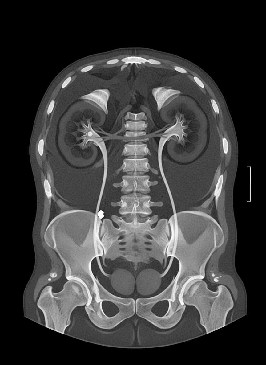
A 40-year-old male presented with severe pain in the left upper abdomen radiating to the groin. Urine routine examination shows 6-8 pus cells and 15-20 RBCs. A CT scan was performed. What is the most likely diagnosis?
Which of the following are indications for percutaneous nephrostomy?
A 60-year-old male presents with a poor stream of urine and a post-void residual volume of 400ml. He has bilateral hydronephrosis and his prostate weighs 70g. His urea is 120 mg/dL and creatinine is 3.5 mg/dL. What is the ideal next immediate step?
A young man presents with primary infertility, and semen analysis reveals low volume, fructose-negative ejaculate with azoospermia. Which of the following is the most useful imaging modality to evaluate the cause of his infertility?
Which of the following is a complication of total parenteral nutrition?
A 30-year-old patient presents with scrotal swelling. Transillumination is positive, and a large hydrocoele is suspected. What is the preferred treatment?
Which statement about Horseshoe Kidney is false?
Practice by Chapter
Urological Anatomy
Practice Questions
Hematuria Evaluation
Practice Questions
Urinary Calculi
Practice Questions
Benign Prostatic Hyperplasia
Practice Questions
Prostate Cancer
Practice Questions
Bladder Cancer
Practice Questions
Renal Cell Carcinoma
Practice Questions
Testicular Tumors
Practice Questions
Urinary Tract Infections
Practice Questions
Urinary Incontinence
Practice Questions
Genitourinary Trauma
Practice Questions
Pediatric Urology Basics
Practice Questions
Want unlimited practice?
Get full access to all questions, explanations, and performance tracking.
Scan to download app