
Urology — MCQs

On this page
All of the following are true about prostate-specific antigen except?
A 19-year-old male college student presents with acute pain and swelling of the scrotum. Physical examination reveals an exquisitely tender, swollen right testis with an absent cremasteric reflex and no swelling in the inguinal area. A urine dipstick is negative for red and white blood cells. What is the most appropriate next step in management?
A patient with a suspected pelvic fracture presents with urethral bleeding and inability to pass urine. What is the immediate management step?
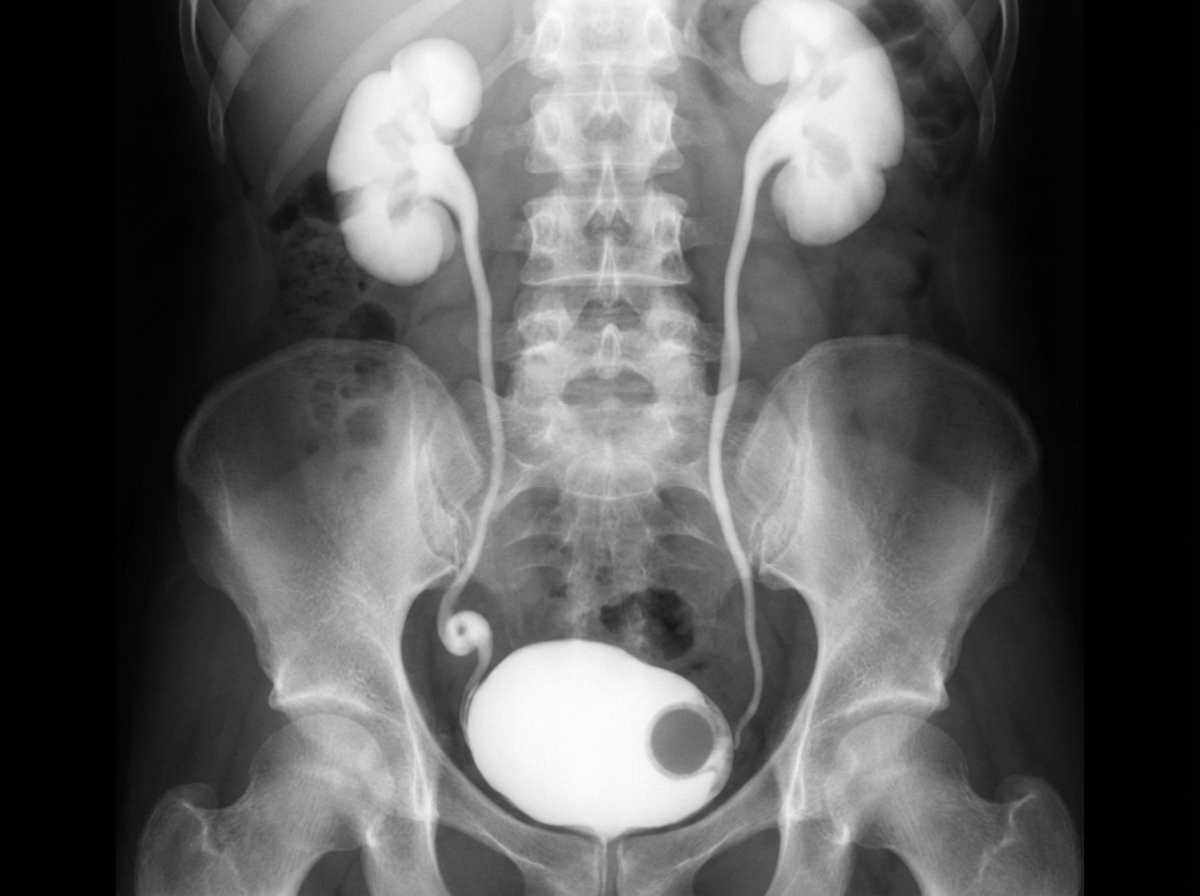
A 10-year-old girl was evaluated for recurrent UTI and dysuria. An IVP finding is shown. What is the preferred treatment option for this case?
Which of the following is an alkaline type of renal calculi?
A patient presents with urethral bleeding and perineal swelling following a straddle injury, indicative of a ruptured bulbar urethra. Which of the following statements regarding his management is FALSE?
Painless hematuria is seen in all of the following conditions except?
Interstitial cystitis is also known as?
What is the commonest cause of death in carcinoma of the penis?
A 28-year-old male presents with lower abdominal pain associated with groin pain and nausea. Elevation of the testis relieves his pain. What is your diagnosis?
Practice by Chapter
Urological Anatomy
Practice Questions
Hematuria Evaluation
Practice Questions
Urinary Calculi
Practice Questions
Benign Prostatic Hyperplasia
Practice Questions
Prostate Cancer
Practice Questions
Bladder Cancer
Practice Questions
Renal Cell Carcinoma
Practice Questions
Testicular Tumors
Practice Questions
Urinary Tract Infections
Practice Questions
Urinary Incontinence
Practice Questions
Genitourinary Trauma
Practice Questions
Pediatric Urology Basics
Practice Questions
Want unlimited practice?
Get full access to all questions, explanations, and performance tracking.
Scan to download app