
Trauma — MCQs

On this page
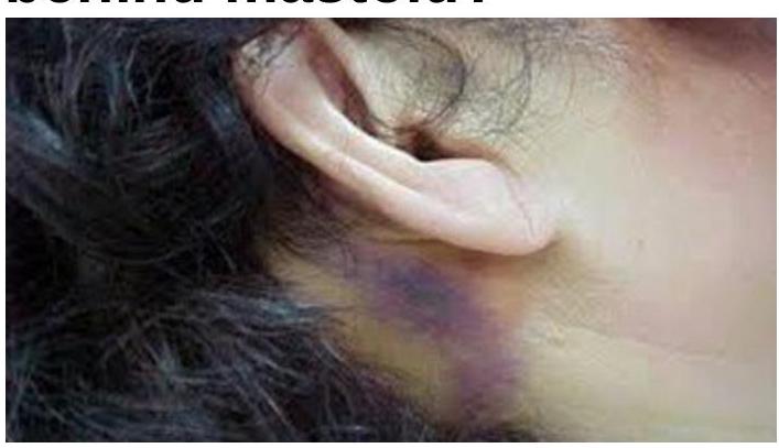
A patient with a basilar skull fracture may present with Battle's sign. What does Battle's sign indicate?
A patient with a basilar skull fracture presents with Battle's sign and hearing loss. Which type of fracture is most likely?
A patient presents with raccoon eyes following a head injury. What is the clinical significance of raccoon eyes and Battle's sign in the context of head trauma?
A 45-year-old male presents with severe abdominal pain following a high-speed car accident. On examination, there is tenderness in the right upper quadrant and epigastrium with guarding. What is the most likely organ to be injured?
What is the best initial management for a burn patient with a suspected inhalation injury?
What does a bluish-purple discoloration behind the mastoid indicate?
A patient presents to the emergency department following blunt abdominal trauma with severe abdominal pain, a pulse of 112 beats per minute, and a systolic blood pressure of 80 mmHg. Based on ATLS principles, what is the immediate next step in management?
Which of the following statements about the management of haematomas is NOT correct?
Estimate volume of Ringer lactate in first 8 hours for a 50 kg male with 40% TBSA second-degree burns?
A patient after a road traffic accident presented with tension pneumothorax. What is the first line of management?
Practice by Chapter
Initial Assessment of Trauma Patient
Practice Questions
Advanced Trauma Life Support (ATLS) Principles
Practice Questions
Chest Trauma
Practice Questions
Abdominal Trauma
Practice Questions
Head Trauma
Practice Questions
Spinal Trauma
Practice Questions
Extremity Trauma
Practice Questions
Vascular Trauma
Practice Questions
Genitourinary Trauma
Practice Questions
Burns Management
Practice Questions
Mass Casualty Management
Practice Questions
Damage Control Surgery
Practice Questions
Want unlimited practice?
Get full access to all questions, explanations, and performance tracking.
Scan to download app