
Trauma — MCQs

On this page
Guerin's sign is associated with which of the following findings?
A patient presents following a motor vehicle accident. The patient moans intermittently, opens eyes only to pain, exhibits no movement in the right limb but moves the left limb in response to pain, and both legs are held in extension. What is the Glasgow Coma Scale (GCS) score?
What is the initial method to prevent infection in burn patients?
All of the following are true about pulmonary contusion except:
A 25-year-old patient presents with upper abdominal pain following a road traffic injury. Their blood pressure is 130/80 mmHg and pulse is 92/min with good volume. A CECT scan reveals findings suggestive of intra-abdominal injury. What is the next line of management?
What is the most effective method for reducing intracranial pressure (ICP)?
All are true about tension pneumothorax except?
Raccoon's eye is a feature of which of the following?
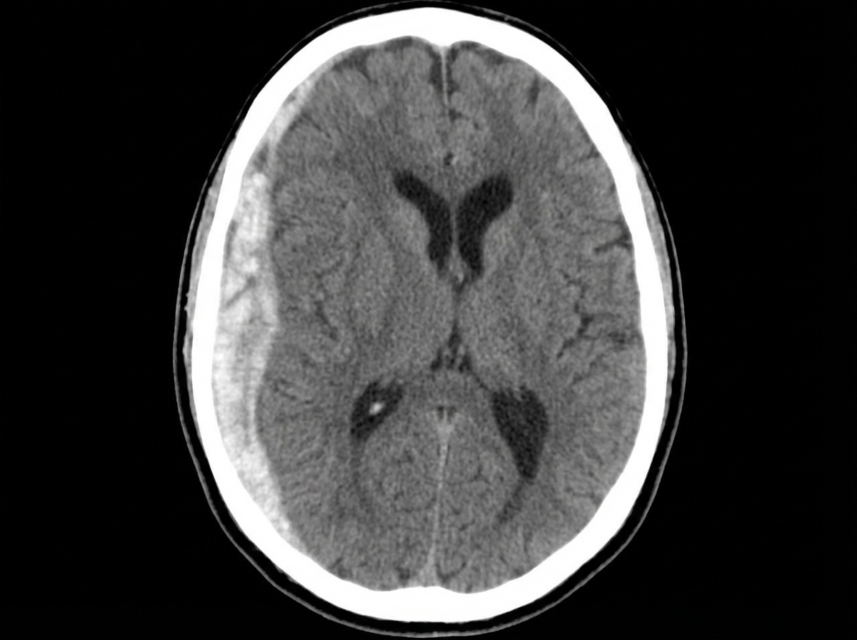
Following a sudden impact in an accident, a 34-year-old race car driver becomes unconscious and is admitted to the hospital. A CT scan reveals a right space-occupying lesion. What is the most likely diagnosis?
A 65-year-old male weighing 50 kg was admitted with 80% burn. According to the Parkland method, how much fluid should be infused in the first 8 hours?
Practice by Chapter
Initial Assessment of Trauma Patient
Practice Questions
Advanced Trauma Life Support (ATLS) Principles
Practice Questions
Chest Trauma
Practice Questions
Abdominal Trauma
Practice Questions
Head Trauma
Practice Questions
Spinal Trauma
Practice Questions
Extremity Trauma
Practice Questions
Vascular Trauma
Practice Questions
Genitourinary Trauma
Practice Questions
Burns Management
Practice Questions
Mass Casualty Management
Practice Questions
Damage Control Surgery
Practice Questions
Want unlimited practice?
Get full access to all questions, explanations, and performance tracking.
Scan to download app