
Preoperative and Postoperative Care — MCQs

On this page
Four days after undergoing subtotal gastrectomy for stomach cancer, a 58-year-old woman complains of right leg and thigh pain, swelling, redness, and tenderness on examination. Deep vein thrombosis is suspected. What is the initial test to establish the diagnosis?
What is true regarding a subphrenic abscess?
A significant increase in energy expenditure, approximately 100% over normal or two times greater than normal, is typically observed in patients with which of the following conditions?
A postoperative patient presents with peritonitis and massive contamination due to a duodenal leak. What is the management of choice?
What is the most important consideration in a patient with borderline pulmonary function undergoing lung resection?
A 65-year-old man undergoes a low anterior resection for rectal cancer. On the fifth day in hospital, his physical examination shows a temperature of 39°C (102°F), blood pressure of 150/90 mm Hg, pulse of 110 beats per minute and regular, and respiratory rate of 28 breaths per minute. A computed tomography (CT) scan of the abdomen reveals an abscess in the pelvis. Which of the following most accurately describes his present condition?
A patient with meningioma and an inflammatory edematous lesion is scheduled for surgery. Which of the following represents a mistake in the junior resident's pre-operative notes?
What is the standard of care regarding vaccination in patients who have undergone splenectomy, excluding which of the following?
The surgeon should be particularly concerned about which coagulation function in patients receiving anti-inflammatory or analgesic medications?
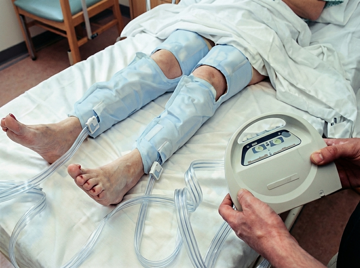
What is the purpose of the device shown below?
Practice by Chapter
Preoperative Risk Assessment
Practice Questions
Perioperative Management of Comorbidities
Practice Questions
Preparation of Patient for Surgery
Practice Questions
Informed Consent Process
Practice Questions
Post-Anesthesia Care
Practice Questions
Pain Management
Practice Questions
Wound Care and Dressings
Practice Questions
Drain Management
Practice Questions
Postoperative Complications Detection
Practice Questions
Early Ambulation and Rehabilitation
Practice Questions
Enhanced Recovery After Surgery (ERAS) Protocols
Practice Questions
Discharge Planning and Follow-up
Practice Questions
Want unlimited practice?
Get full access to all questions, explanations, and performance tracking.
Scan to download app