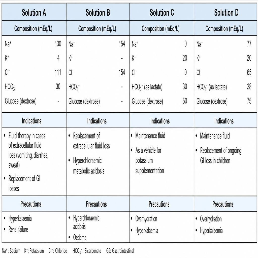
A 70-year-old woman has a small-bowel fistula with an output of 1.5 L/d. Replacement of daily losses should be handled using which of the following fluid solutions based on its composition in mEq/L?
Pyrexia due to wound infection commonly occurs after which postoperative day?
What is the first sign of burst abdomen?
Which of the following is NOT true about the principal hormonal responses to surgical stress?
A 59-year-old man is planning to undergo coronary artery bypass surgery. He has osteoarthritis and consumes nonsteroidal anti-inflammatory drugs (NSAIDs) for the pain. Which of the following is the most appropriate treatment prior to surgery to minimize his risk of bleeding from his NSAID use?
A 65-year-old man is being evaluated for possible right pneumonectomy for bronchogenic carcinoma. Which value is most suggestive of postoperative problems?
A burst abdomen following laparotomy typically occurs after how many days?
A 65-year-old man has an enterocutaneous fistula originating in the jejunum secondary to inflammatory bowel disease. Which of the following would be the most appropriate fluid for replacement of his enteric losses?
Hypocalcemia in the immediate post-operative period following excision of a parathyroid adenoma is due to what?
What is considered the best type of dressing for a surgical wound?
Explanation: The management of small-bowel fistula (SBF) requires meticulous fluid and electrolyte replacement. A high-output SBF (>500 mL/day) leads to significant losses of water, sodium, potassium, and bicarbonate. ### **Explanation of the Correct Answer** **Solution A** is the correct choice because its composition closely mimics the electrolyte profile of **small-bowel succus**. The average electrolyte concentration of small bowel fluid is approximately: * **Sodium (Na⁺):** 100–140 mEq/L * **Potassium (K⁺):** 5–15 mEq/L * **Chloride (Cl⁻):** 90–120 mEq/L * **Bicarbonate (HCO₃⁻):** 20–40 mEq/L Replacement therapy must match these losses to prevent dehydration and metabolic acidosis. **Lactated Ringer’s (LR)** is often the initial fluid of choice as its electrolyte profile (Na⁺ 130, K⁺ 4, Cl⁻ 109, Lactate 28) most closely approximates these values compared to other standard crystalloids. ### **Analysis of Incorrect Options** * **Solution B (Gastric fluid profile):** High in H⁺ and Cl⁻ but low in Na⁺. Replacing SBF with this would lead to hyponatremia and fail to correct the base deficit. * **Solution C (Colonic fluid profile):** Higher in K⁺ (up to 30 mEq/L) and HCO₃⁻ but lower in Na⁺. * **Solution D (Pure Normal Saline):** While it provides Na⁺ (154 mEq/L), it lacks K⁺ and HCO₃⁻, and the high Cl⁻ content can induce hyperchloremic metabolic acidosis. ### **NEET-PG High-Yield Pearls** * **Fistula Classification:** High output is >500 mL/24h; Low output is <200 mL/24h. * **Metabolic Consequence:** Small bowel and pancreatic fistulae typically cause **Normal Anion Gap Metabolic Acidosis (NAGMA)** due to bicarbonate loss. * **Management Priority:** 1. Stabilization/Resuscitation, 2. Sepsis control, 3. Nutritional support (TPN is often required), 4. Definitive surgery (usually delayed 6–12 weeks). * **Drug of Choice:** Octreotide can be used to reduce fistula output but does not increase the spontaneous closure rate.
Explanation: Postoperative fever is a common clinical scenario, and the timing of the fever is the most important clue to its etiology. This is often remembered by the classic mnemonic **"The 5 W’s"** (Wind, Water, Walking, Wound, Wonder drugs). ### **Explanation of the Correct Answer** **Option B (Fifth postoperative day)** is correct because surgical site infections (SSIs) typically require time for bacterial proliferation and the subsequent inflammatory response to manifest systemically. While superficial infections can appear earlier, the classic peak for pyrexia due to a wound infection is between the **5th and 7th postoperative days**. ### **Analysis of Incorrect Options** * **Option D (Second postoperative day):** Fever within the first 48 hours is most commonly due to **atelectasis** (Wind) or the normal metabolic response to surgical trauma. * **Option A (Third postoperative day):** Fever on day 3 is classically associated with **Urinary Tract Infections** (Water), especially in catheterized patients. * **Option C (Seventh postoperative day):** While wound infections can occur here, day 7 and beyond are more frequently associated with **Deep Vein Thrombosis/PE** (Walking) or deep-seated abscesses. ### **NEET-PG High-Yield Pearls** * **The 5 W’s Timeline:** 1. **Wind (Day 1-2):** Atelectasis, Pneumonia. 2. **Water (Day 3):** UTI. 3. **Walking (Day 5):** DVT/Thrombophlebitis. 4. **Wound (Day 5-7):** Surgical Site Infection (SSI). 5. **Wonder drugs (Anytime):** Drug-induced fever or IV line infections. * **Exception:** If a wound infection causes fever within the first 24 hours, suspect **Clostridium perfringens** or **Group A Streptococcus** (Necrotizing fasciitis). * **Immediate Post-op Fever:** Usually due to pre-existing infection, transfusion reaction, or malignant hyperthermia.
Explanation: **Explanation:** **Burst Abdomen (Abdominal Wound Dehiscence)** refers to the partial or complete separation of the layers of a surgical wound before complete healing has occurred. **1. Why Serosanguinous Drainage is Correct:** The hallmark clinical sign of an impending burst abdomen is the sudden discharge of **serosanguinous (pinkish) fluid** from the wound, often described as a "salmon-colored" discharge. This occurs because the deeper layers (fascia) have separated, allowing peritoneal fluid mixed with a small amount of blood to seep through the skin incision. This typically occurs between the **6th and 10th postoperative days**. **2. Analysis of Incorrect Options:** * **A. Bleeding at the site:** While minor oozing can occur, frank bleeding is more characteristic of immediate postoperative complications like reactionary hemorrhage or a slipped ligature, rather than fascial dehiscence. * **C. Pain at the site:** Interestingly, burst abdomen is often **painless**. Patients frequently report a sensation of "something giving way" after a bout of coughing or straining, but acute pain is rarely the presenting sign. **3. NEET-PG High-Yield Pearls:** * **The "Pink Discharge" Sign:** If a question mentions "serosanguinous soakage of dressings" on day 7-8, the diagnosis is always burst abdomen. * **Risk Factors:** Increased intra-abdominal pressure (coughing, vomiting, ascites), malnutrition (hypoalbuminemia), anemia, malignancy, and poor surgical technique (e.g., sutures placed too close to the edge). * **Management:** * **Immediate:** Cover the wound with sterile saline-soaked gauze and provide abdominal binders. * **Definitive:** Urgent surgical re-closure (tension-free) using non-absorbable sutures (e.g., Prolene) often via **interrupted "internal" or "mass" closure.**
Explanation: The metabolic response to surgical stress is characterized by a "neuroendocrine reflex" aimed at mobilizing energy substrates and maintaining cardiovascular stability. ### **Why Option D is Correct** In response to surgical trauma, **Thyroxine (T4) and Triiodothyronine (T3) levels typically decrease or remain unchanged**, while Reverse T3 (rT3) increases. This phenomenon is often referred to as **"Euthyroid Sick Syndrome."** The body downregulates thyroid function to decrease the basal metabolic rate and conserve energy during the acute stress phase. Therefore, an *increase* in thyroxine is not a standard response to surgical stress. ### **Analysis of Incorrect Options** * **A. Increased ACTH:** Surgical stress stimulates the hypothalamus-pituitary-adrenal (HPA) axis. Increased ACTH leads to a rapid rise in **Cortisol**, which is essential for gluconeogenesis and protein catabolism. * **B. Increased Growth Hormone (GH):** GH levels rise significantly during surgery. While GH is normally anabolic, in the stress state, it promotes **lipolysis** and antagonizes insulin, contributing to hyperglycemia. * **C. Decreased Insulin:** Although blood glucose levels rise (stress hyperglycemia), there is a relative **deficiency or suppression of insulin secretion** (mediated by alpha-adrenergic stimulation) and peripheral insulin resistance. ### **High-Yield NEET-PG Pearls** * **The "Rule of Increases":** Almost all hormones increase during surgery (Cortisol, Glucagon, Catecholamines, GH, ADH, Prolactin) **EXCEPT** Insulin and Thyroid hormones (T3/T4). * **Cytokine Driver:** **IL-6** is the primary cytokine responsible for inducing the acute-phase response and stimulating the HPA axis. * **Insulin Resistance:** The degree of postoperative insulin resistance is directly proportional to the severity of the surgical trauma. * **ADH (Vasopressin):** Levels remain elevated for 3–5 days post-op, leading to water retention and potential hyponatremia if free water is over-administered.
Explanation: ### Explanation **1. Why Option D is Correct:** The primary concern with NSAIDs (like Ibuprofen or Naproxen) in the perioperative period is their effect on platelet function. NSAIDs cause **reversible inhibition of cyclooxygenase (COX-1)**, which prevents the formation of Thromboxane A2 (TXA2), a potent platelet aggregator. Unlike Aspirin, which binds irreversibly for the life of the platelet (7–10 days), the effects of most NSAIDs are transient and dissipate as the drug is cleared from the plasma. For most NSAIDs, stopping the medication **3 to 4 days (approximately 4–5 half-lives)** prior to surgery is sufficient to restore adequate platelet function and minimize the risk of intraoperative bleeding. **2. Why Other Options are Incorrect:** * **Option A:** Vitamin K is used to reverse the effects of Warfarin (by promoting the synthesis of clotting factors II, VII, IX, and X). It has no effect on the platelet dysfunction caused by NSAIDs. * **Option B:** Fresh Frozen Plasma (FFP) contains clotting factors but is not the treatment of choice for platelet-related bleeding. If emergency reversal of anti-platelet agents were needed, platelet transfusion would be more appropriate, though rarely required for NSAIDs. * **Option C:** Stopping NSAIDs 1 week prior is the standard protocol for **Aspirin** (due to irreversible binding), but it is unnecessarily long for reversible NSAIDs and may cause the patient unnecessary pain from their osteoarthritis. **3. Clinical Pearls for NEET-PG:** * **Aspirin:** Irreversible COX inhibition; stop **7–10 days** before high-risk surgery. * **NSAIDs:** Reversible COX inhibition; stop **3 days** before surgery. * **Warfarin:** Stop **5 days** before surgery; bridge with Heparin if the patient is at high risk for thromboembolism. * **Clopidogrel:** Irreversible P2Y12 inhibitor; stop **5–7 days** before surgery. * **Selective COX-2 Inhibitors (e.g., Celecoxib):** These do not affect platelet aggregation and generally **do not** need to be discontinued preoperatively.
Explanation: ### Explanation The preoperative assessment for lung resection (pneumonectomy) focuses on the patient’s ability to tolerate a significant reduction in pulmonary reserve. **Why PaCO2 50 torr is the Correct Answer:** Hypercapnia (**PaCO2 > 45 mmHg**) is a strong independent predictor of poor postoperative outcomes and increased mortality in thoracic surgery. It indicates that the patient is already failing to maintain adequate alveolar ventilation with two lungs; removing one lung would likely lead to severe respiratory failure. Traditionally, a PaCO2 > 45 mmHg was considered an absolute contraindication to pneumonectomy. **Analysis of Incorrect Options:** * **A. DLCO 55% predicted:** While a DLCO < 60% indicates increased risk, the threshold for high-risk/postoperative problems is typically **< 40%**. A value of 55% is suboptimal but not as critical as hypercapnia. * **B. FEV1 2L:** An absolute FEV1 **> 2L** is generally considered safe for pneumonectomy. The "rule of thumb" is that a predicted postoperative (ppo) FEV1 should be **> 0.8L** or **> 40%** of predicted to avoid ventilator dependence. * **C. MVO2 12 mL/kg/min:** Maximal oxygen uptake (VO2 max) is the gold standard for assessing cardiopulmonary reserve. A value **> 15 mL/kg/min** is safe, while **< 10 mL/kg/min** indicates a very high risk of mortality. 12 mL/kg/min is in the "intermediate risk" zone but is less definitive of failure than a PaCO2 of 50 torr. **Clinical Pearls for NEET-PG:** * **Initial Screening:** FEV1 and DLCO are the first steps. If both are > 80%, no further testing is needed. * **The "80-40" Rule:** FEV1/DLCO > 80% (Safe), < 40% (High Risk/Requires Exercise Testing). * **Gold Standard:** VO2 max is the most accurate predictor of surgical outcomes. * **Contraindications:** PaCO2 > 45 mmHg, PaO2 < 50-60 mmHg, and ppoFEV1 < 0.8L are classic indicators of poor surgical candidacy.
Explanation: **Explanation:** **Burst Abdomen (Abdominal Wound Dehiscence)** refers to the partial or complete disruption of the layers of a surgical wound. **1. Why 7–10 days is correct:** The integrity of a surgical wound depends on the balance between collagen synthesis and collagen lysis. Between the **7th and 10th postoperative days**, the wound is in its most vulnerable state. At this stage, the initial fibrin glue and sutures have started to weaken or lose their tensile strength, while the newly synthesized collagen has not yet gained sufficient strength to withstand intra-abdominal pressure. This "lag phase" in wound healing is when dehiscence most commonly occurs. **2. Why other options are incorrect:** * **1–2 days:** At this stage, the wound is held firmly by the primary surgical sutures, and the inflammatory phase is just beginning. Dehiscence is rare unless there is a major technical failure. * **3 weeks / One month:** By this time, the proliferative phase is well-advanced, and remodeling has begun. The tensile strength of the wound has increased significantly, making a sudden "burst" unlikely. Late failures usually present as **incisional hernias** rather than acute dehiscence. **Clinical Pearls for NEET-PG:** * **The "Pink Discharge" Sign:** The earliest clinical sign of impending burst abdomen is a **serosanguinous (pinkish) discharge** from the wound, typically occurring on the 5th or 6th day. * **Risk Factors:** Increased intra-abdominal pressure (coughing, vomiting, ascites), malnutrition (hypoalbuminemia), anemia, jaundice, and poor surgical technique (e.g., sutures tied too tightly). * **Management:** Immediate management involves covering the bowel with sterile saline-soaked gauze, followed by urgent surgical closure using **tension-sutures (interrupted internal mass closure)**.
Explanation: **Explanation:** The management of an enterocutaneous fistula (ECF) requires precise fluid and electrolyte replacement based on the anatomical site of the loss. **1. Why Ringer Lactate (RL) is correct:** Jejunal secretions are essentially **isotonic** and contain significant amounts of sodium, potassium, and **bicarbonate**. Loss of jejunal fluid typically leads to metabolic acidosis due to the depletion of bicarbonate. Ringer Lactate is the fluid of choice because its electrolyte composition closely mimics that of small bowel secretions. Furthermore, the **lactate** in RL is metabolized by the liver into bicarbonate, which helps counteract the metabolic acidosis associated with high-output upper intestinal fistulae. **2. Why the other options are incorrect:** * **3% Normal Saline:** This is a hypertonic solution used for symptomatic hyponatremia or cerebral edema. Using it here would cause severe hypernatremia and fluid overload. * **0.9% Sodium Chloride (Normal Saline):** While isotonic, NS lacks potassium and bicarbonate. Prolonged replacement with NS can lead to **hyperchloremic metabolic acidosis**, worsening the patient's acid-base status. * **6% Sodium Bicarbonate:** This is an extremely concentrated alkaline solution. Bicarbonate replacement is usually reserved for severe acidosis (pH <7.1) and is not used as a primary maintenance or replacement fluid. **Clinical Pearls for NEET-PG:** * **Gastric losses (Vomiting/NG suction):** Replace with 0.45% NS + KCl (due to loss of HCl, leading to Hypochloremic Hypokalemic Metabolic Alkalosis). * **Small Bowel/Biliary/Pancreatic losses:** Replace with Ringer Lactate (due to loss of HCO3⁻, leading to Metabolic Acidosis). * **The "Rule of Thumb":** Always replace "like with like." Small bowel fluid is alkaline and electrolyte-rich; RL is the most physiological crystalloid for this purpose.
Explanation: **Explanation:** The correct answer is **B. Increased uptake by bones**. This phenomenon is clinically known as **Hungry Bone Syndrome (HBS)**. In patients with a parathyroid adenoma, chronic exposure to high levels of Parathyroid Hormone (PTH) leads to excessive bone resorption and high bone turnover. Once the adenoma is surgically removed, PTH levels drop precipitously. This sudden withdrawal of PTH causes a shift in bone metabolism: osteoclastic resorption stops, while osteoblastic activity continues or increases. Consequently, the "hungry" bones rapidly uptake calcium, magnesium, and phosphate from the plasma to remineralize the bone matrix, leading to profound **hypocalcemia** in the immediate postoperative period. **Analysis of Incorrect Options:** * **A. Stress:** While surgery induces a stress response (releasing cortisol and catecholamines), this typically leads to hyperglycemia, not acute hypocalcemia. * **C. Hypercalciuria:** Post-parathyroidectomy, the kidneys actually increase calcium reabsorption due to the drop in filtered calcium load; therefore, urinary calcium levels decrease. * **D. Increased calcitonin:** Calcitonin is secreted by the parafollicular C-cells of the thyroid. While it can lower calcium, it is not the primary driver of hypocalcemia following parathyroid surgery. **High-Yield Clinical Pearls for NEET-PG:** * **Risk Factors for HBS:** Large adenoma size, high preoperative alkaline phosphatase (ALP), and radiological evidence of bone disease (e.g., *Osteitis fibrosa cystica*). * **Biochemical Profile of HBS:** Low Calcium, Low Phosphate, Low Magnesium, and High ALP. * **Management:** Treatment involves aggressive supplementation with intravenous calcium gluconate and oral Vitamin D (Calcitriol). * **Differential:** If hypocalcemia is accompanied by **high** phosphate, consider accidental injury to the remaining normal parathyroid glands (hypoparathyroidism) rather than HBS.
Explanation: **Explanation:** The primary goal of a surgical dressing is to provide a sterile environment, absorb exudate, and facilitate healing. While various synthetic materials exist, **Skin** is considered the "best" or "ideal" biological dressing. **1. Why Skin is the Correct Answer:** Intact skin is the most physiological dressing available. In the context of surgery, particularly in burns or large reconstructive procedures, **autologous skin** (autograft) is the gold standard. It provides an absolute barrier against infection, prevents fluid and electrolyte loss, reduces pain, and promotes definitive wound closure. No synthetic material can perfectly replicate the complex immunological and mechanical properties of human skin. **2. Analysis of Incorrect Options:** * **Opsite (Option A):** This is a transparent, adhesive polyurethane film. While excellent for providing a moist environment and allowing wound visualization, it is a synthetic dressing and cannot match the biological integration of skin. * **Amnion (Option B):** Amniotic membrane is a biological dressing often used in burns. While it reduces pain and promotes epithelialization, it is usually a temporary biological cover (allograft) and is inferior to the patient's own skin. * **Tulle Gras (Option C):** This is a paraffin-impregnated gauze. It is a traditional non-adherent dressing used to prevent the dressing from sticking to the wound, but it lacks the protective and regenerative properties of skin. **Clinical Pearls for NEET-PG:** * **Ideal Dressing Properties:** Non-toxic, non-adherent, permeable to water vapor but impermeable to bacteria. * **Biological Dressings:** Include skin (autograft/allograft), amnion, and collagen. They are superior to synthetic dressings for large raw areas. * **High-Yield Fact:** For a clean surgical wound closed by primary intention, the dressing is typically removed after **48 hours**, as the epithelium seals the wound by then.
Preoperative Risk Assessment
Practice Questions
Perioperative Management of Comorbidities
Practice Questions
Preparation of Patient for Surgery
Practice Questions
Informed Consent Process
Practice Questions
Post-Anesthesia Care
Practice Questions
Pain Management
Practice Questions
Wound Care and Dressings
Practice Questions
Drain Management
Practice Questions
Postoperative Complications Detection
Practice Questions
Early Ambulation and Rehabilitation
Practice Questions
Enhanced Recovery After Surgery (ERAS) Protocols
Practice Questions
Discharge Planning and Follow-up
Practice Questions
Get full access to all questions, explanations, and performance tracking.
Start For Free