
Pediatric Surgery — MCQs

On this page
An invertogram is typically taken after how many hours of birth?
Which condition is treated with a Bishop-Koop operation?
Regarding Gastroschisis and omphalocele, which of the following statements is false?
Which of the following statements is FALSE regarding rectal prolapse in children?
What is your diagnosis in this neonate with visible bowel loops at birth?
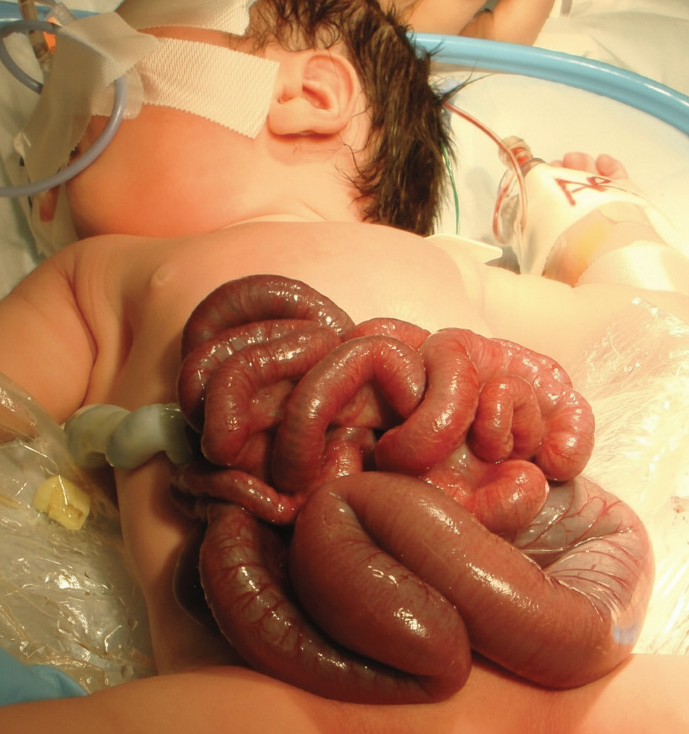
Which of the following statements regarding gastroschisis is TRUE?
In a case of undescended testis, after what age does further descent not occur?
A 3-year-old boy is referred after his initial pediatrician's assessment for an undescended testicle. On exam, his left testicle is normal and in place. He has no evidence of hernias. However, his right hemiscrotum is empty, and there is a testicle-sized mass palpable at the pubic tubercle. What is the most appropriate next step?
What is the approximate incidence of anastomotic leak following repair of esophageal atresia?
At what time is the optimal surgical timing for a sacrococcygeal teratoma?
Practice by Chapter
Neonatal Physiology
Practice Questions
Congenital Anomalies Overview
Practice Questions
Neonatal Intestinal Obstruction
Practice Questions
Necrotizing Enterocolitis
Practice Questions
Hirschsprung's Disease
Practice Questions
Anorectal Malformations
Practice Questions
Pediatric Hernias
Practice Questions
Pyloric Stenosis
Practice Questions
Biliary Atresia
Practice Questions
Pediatric Tumors
Practice Questions
Congenital Diaphragmatic Hernia
Practice Questions
Pediatric Trauma
Practice Questions
Want unlimited practice?
Get full access to all questions, explanations, and performance tracking.
Scan to download app