
Pediatric Surgery — MCQs

On this page
What is the end-stage treatment for Tetralogy of Fallot?
A newborn infant presents with bilious vomiting and epigastric distention immediately following birth. What is the most likely diagnosis?
Which of the following should contraindicate the performance of the Fontan procedure?
Cystic hygroma is known to occur in all of the following locations except?
What is the recommended treatment for annular pancreas?
A 15-year-old child presents with acute abdominal pain and a history of blood and mucous in the stool. A mass is palpable on physical examination. What is the MOST probable diagnosis?
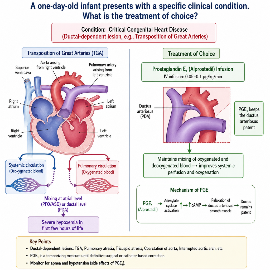
A one-day-old infant presents with a specific clinical condition. What is the treatment of choice?
What is the most common type of hypospadias?
Which of the following statements is NOT true about herniotomy?
Which of the following is true about Hirschsprung's disease?
Practice by Chapter
Neonatal Physiology
Practice Questions
Congenital Anomalies Overview
Practice Questions
Neonatal Intestinal Obstruction
Practice Questions
Necrotizing Enterocolitis
Practice Questions
Hirschsprung's Disease
Practice Questions
Anorectal Malformations
Practice Questions
Pediatric Hernias
Practice Questions
Pyloric Stenosis
Practice Questions
Biliary Atresia
Practice Questions
Pediatric Tumors
Practice Questions
Congenital Diaphragmatic Hernia
Practice Questions
Pediatric Trauma
Practice Questions
Want unlimited practice?
Get full access to all questions, explanations, and performance tracking.
Scan to download app