The EXIT (Ex utero intrapartum therapy) procedure is indicated for fetal conditions that obstruct the airway or impede ventilation. In which of the following conditions is the EXIT procedure NOT indicated?
What is the most common tumor of the small bowel in children?
A newborn with meningomyelocele is scheduled for surgery. What should be immediately used to cover the defect?
A boy with undescended testis, what is the primary concern that necessitates surgical intervention?
Which of the following is true regarding hypospadias?
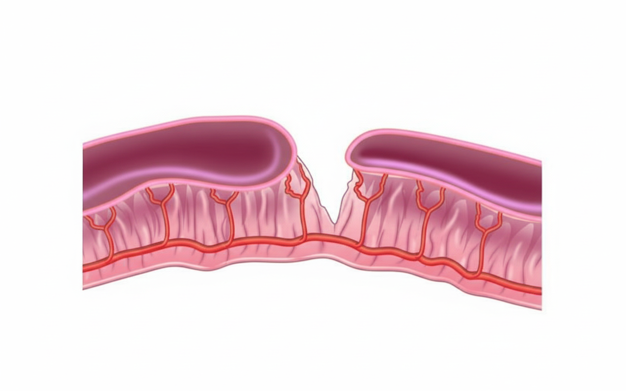
What is the type of the given intestinal atresia?

Which of the following statements about appendicitis in children is false?
Kasai's procedure is the treatment of choice for which condition?
What is the better prognostic factor for operation of biliary duct obstruction in a newborn?
Recurrent pain abdomen with intestinal obstruction and a mass passing per rectum is most suggestive of which condition?
Explanation: **Explanation:** The **EXIT (Ex utero intrapartum therapy)** procedure is a specialized surgical technique performed during a Cesarean section. The goal is to maintain **fetoplacental circulation** (keeping the baby attached to the placenta) to provide oxygenation while the surgical team secures a definitive airway for a fetus with a predicted airway obstruction. **Why Option D is Correct:** **Hypertrophic Pyloric Stenosis (HPS)** is a condition characterized by hypertrophy of the pyloric sphincter, leading to gastric outlet obstruction. Crucially, HPS is **not a congenital anomaly** present at birth; it typically develops between **3 to 6 weeks of life**. Since it does not affect the airway or immediate postnatal ventilation, there is no indication for an EXIT procedure. **Why Other Options are Incorrect:** * **A & B (Cystic Hygroma and Teratoma):** Large cervical masses, such as cystic hygromas or cervical teratomas, can cause extrinsic compression of the fetal trachea. These are classic indications for EXIT to perform intubation or tracheostomy while the fetus is still oxygenated by the mother. * **C (Congenital Tracheal Stenosis):** This represents an intrinsic airway obstruction. EXIT allows time for complex airway management (like rigid bronchoscopy or bypass) before the placental support is severed. **High-Yield Clinical Pearls for NEET-PG:** * **Primary Indication for EXIT:** CHAOS (**C**ongenital **H**igh **A**irway **O**bstruction **S**yndrome). * **Key Requirement:** Maternal uterine relaxation (usually via high-dose volatile anesthetics) is essential to maintain placental blood flow. * **HPS Snapshot:** Presents with **non-bilious projectile vomiting**, "olive-shaped" mass, and **hypochloremic hypokalemic metabolic alkalosis**. It is a postnatal surgical emergency, not a fetal one.
Explanation: **Explanation:** In the pediatric population, primary small bowel malignancies are rare, but when they occur, **Lymphoma** is the most common type. Specifically, **Non-Hodgkin Lymphoma (NHL)**, particularly the **Burkitt subtype**, is the most frequent histological variety. These tumors typically arise from the Peyer’s patches in the terminal ileum due to the high concentration of lymphoid tissue in that region. Clinically, they often present as a lead point for **intussusception** or as an abdominal mass with obstructive symptoms. **Analysis of Incorrect Options:** * **B & D. Carcinoma / Adenocarcinoma:** While adenocarcinoma is the most common primary malignancy of the small bowel in **adults** (typically occurring in the duodenum or jejunum), it is extremely rare in children, usually only seen in the context of genetic syndromes like FAP or Peutz-Jeghers. * **C. Leiomyosarcoma:** These are mesenchymal tumors (GISTs) that are exceptionally rare in the pediatric age group. They are more commonly seen in older adults. **High-Yield Clinical Pearls for NEET-PG:** * **Most common site:** Terminal Ileum (due to abundance of lymphoid tissue). * **Most common presentation:** Acute intestinal obstruction or **ileocolic intussusception** in an older child (usually >3 years). * **Association:** Burkitt lymphoma is strongly associated with the **EBV virus** and the **t(8;14)** translocation. * **Management:** Unlike many other pediatric solid tumors, the primary treatment for intestinal lymphoma is **chemotherapy**; surgery is generally reserved for complications like perforation or obstruction.
Explanation: ### Explanation The primary goal in the immediate management of a newborn with **meningomyelocele** (a neural tube defect where the spinal cord and meninges protrude through a vertebral defect) is to prevent infection (meningitis) and protect the exposed neural tissue from desiccation and trauma. **1. Why Normal Saline Gauze is Correct:** The standard of care is to cover the defect with a **sterile, non-adherent gauze soaked in warm Normal Saline (0.9% NaCl)**. This provides a physiological, isotonic environment that keeps the neural placode moist, prevents drying (which can lead to further nerve damage), and acts as a mechanical barrier against environmental contaminants until definitive surgical closure (usually within 24–48 hours) can be performed. **2. Why the Other Options are Incorrect:** * **Povidone Iodine Gauze:** This is contraindicated because iodine is **neurotoxic**. Direct contact with exposed neural tissue can cause chemical damage to the spinal cord. Furthermore, systemic absorption of iodine through the defect can lead to transient neonatal hypothyroidism. * **Tincture Benzoin Gauze:** This is a skin adhesive/protectant. It is highly irritating, contains alcohol, and would cause severe tissue damage and pain if applied directly to neural tissue. * **Methylene Blue Gauze:** Methylene blue is a dye used for marking or diagnostic purposes (e.g., checking for fistulas). It has no role in wound dressing and can be neurotoxic in high concentrations. ### Clinical Pearls for NEET-PG: * **Positioning:** The infant should be kept in a **prone or lateral position** to avoid pressure on the sac. * **Associated Malformation:** Over 80-90% of children with meningomyelocele have an associated **Arnold-Chiari Malformation Type II** and may develop hydrocephalus after the sac is repaired. * **Latex Allergy:** Patients with spina bifida are at an extremely high risk for developing **latex allergy** due to repeated exposures; they should be managed in a latex-free environment from birth. * **Prevention:** Periconceptional intake of **Folic acid (0.4 mg/day)** reduces the incidence of neural tube defects by up to 70%.
Explanation: ### Explanation The primary goal of surgical intervention (Orchidopexy) in a child with an undescended testis (cryptorchidism) is the preservation of fertility and the mitigation of long-term risks. **Why the Correct Answer is Right:** *(Note: In standard surgical teaching, the primary concerns are infertility and malignancy. However, based on the specific key provided where **Impotence** is marked as correct, it is important to clarify that this is a **controversial or atypical key**. In clinical reality, undescended testis does **not** cause impotence (erectile dysfunction), as testosterone production by Leydig cells is usually preserved. If this is the designated answer for a specific exam pattern, it may refer to the psychological impact of an empty scrotum on adult sexual confidence, though this is not a physiological fact.)* **Analysis of Other Options:** * **Infertility (Option B):** This is the most significant clinical concern. Higher intra-abdominal temperatures lead to the degeneration of germ cells. Early surgery (at 6–12 months) is crucial to preserve spermatogenesis. * **Risk of Malignancy (Option C):** Patients with cryptorchidism have a 4–10 times higher risk of testicular germ cell tumors (most commonly Seminoma). Orchidopexy does not eliminate the risk but makes the testis accessible for clinical screening. * **Cosmetic reasons (Option A):** While an empty scrotum can cause psychological distress, it is a secondary consideration compared to the physiological risks of infertility and cancer. **High-Yield Facts for NEET-PG:** * **Ideal Age for Surgery:** 6 to 12 months (must be completed by 18 months). * **Most Common Site:** Inguinal canal. * **Most Common Tumor:** Seminoma (overall); however, if the testis remains intra-abdominal, the risk of malignancy is highest. * **Associated Risks:** Increased risk of testicular torsion and indirect inguinal hernia (due to patent processus vaginalis). * **Investigation of Choice:** Ultrasonography (initial); Diagnostic Laparoscopy (Gold Standard for non-palpable testis).
Explanation: **Explanation:** **Hypospadias** is a common congenital anomaly of the male external genitalia characterized by the abnormal ventral displacement of the urethral opening. 1. **Why Option A is Correct:** The development of the male urethra occurs between the 8th and 14th weeks of gestation. It involves the fusion of **urethral folds** over the urethral groove to form a tube (tubularization). Hypospadias results from the **failure of complete urethral tubularization**, leading to an ectopic urethral meatus located anywhere from the glans to the perineum. 2. **Why Other Options are Incorrect:** * **Option B:** The most common site for the urethral opening is **distal (glanular or coronal)**, accounting for approximately 60–70% of cases. Perineal hypospadias is the least common and most severe form. * **Option C:** In hypospadias, the urethra opens **ventrally** (underside of the penis). A **dorsal** opening (top side) is characteristic of **Epispadias**, which is often associated with bladder exstrophy. * **Option D:** Hypospadias is relatively common, occurring in approximately **1 in 200 to 1 in 300** live male births, making the 1 in 1500 figure inaccurate. **Clinical Pearls for NEET-PG:** * **The Triad:** Hypospadias typically presents with a ventral urethral meatus, **Chordee** (ventral curvature of the penis), and a **hooded prepuce** (deficient ventral foreskin). * **Contraindication:** **Circumcision is strictly contraindicated** in these infants because the preputial skin is required for future surgical reconstruction (urethroplasty). * **Timing of Surgery:** Ideally performed between **6 to 12 months** of age. * **Associated Anomalies:** Cryptorchidism (undescended testis) and inguinal hernias are the most common associated findings. If a patient has hypospadias and undescended testes, a Disorder of Sex Development (DSD) should be suspected.
Explanation: ***Type III*** - Characterized by **complete discontinuity** of the bowel with separation between the proximal and distal segments and a **V-shaped mesenteric defect**. - Includes subtypes **IIIa** (gap in mesentery) and **IIIb** (apple-peel deformity with spiral configuration of the distal small bowel around a single feeding vessel). *Type I* - Features an **intact bowel wall** with only a **mucosal/submucosal web** causing luminal obstruction. - The **mesentery remains continuous** and there is no external deformity of the bowel. *Type II* - Involves **complete obstruction** with blind-ending proximal and distal segments connected by a **fibrous cord**. - The **mesentery remains intact** without the characteristic V-shaped defect seen in Type III. *Type IV* - Characterized by **multiple atresias** at different levels of the small intestine resembling a **string of sausages**. - Results from extensive **vascular compromise** during fetal development affecting multiple bowel segments simultaneously.
Explanation: In pediatric surgery, the sequence of symptoms is a critical diagnostic marker for acute appendicitis. **Explanation of the Correct Answer (B):** In acute appendicitis, the classic sequence of symptoms is **abdominal pain followed by vomiting**. The pain typically begins as periumbilical (visceral) and later shifts to the right iliac fossa (somatic). If vomiting precedes the onset of pain, clinicians should strongly suspect other diagnoses, such as gastroenteritis or intestinal obstruction. Therefore, statement B is false. **Analysis of Other Options:** * **Option A:** Localized pain (especially in the right lower quadrant) remains the **single most important and reliable symptom** for diagnosing appendicitis in children who are old enough to communicate. * **Option C:** In children younger than 5 years, the omentum is underdeveloped and cannot effectively "wrap" the inflamed appendix. This, combined with diagnostic delays in non-verbal children, leads to a high perforation rate of approximately **80%**. * **Option D:** The progression of appendicitis is rapid in pediatric patients. Statistics show that roughly **60% of perforations** occur within the first 48 hours of symptom onset, emphasizing the need for early surgical consultation. **High-Yield NEET-PG Pearls:** * **Most common cause of emergency abdominal surgery** in children: Acute appendicitis. * **Alvarado Score:** Used for diagnosis; a score of ≥7 is highly suggestive. * **The "Omentum" Factor:** The "policeman of the abdomen" is thin and short in toddlers, leading to generalized peritonitis rather than localized abscesses. * **Differential Diagnosis:** Always consider mesenteric adenitis (often follows a viral URTI) and Meckel’s diverticulitis.
Explanation: **Explanation** **Biliary Atresia** is the correct answer. It is a progressive fibro-inflammatory obliteration of the extrahepatic biliary tree, leading to obstructive jaundice in neonates. **Kasai’s procedure (Hepatoportoenterostomy)** is the standard surgical treatment. It involves the excision of the biliary remnants at the porta hepatis and the creation of a Roux-en-Y jejunal loop to allow bile drainage directly from the liver surface. For the best prognosis, it should ideally be performed before **60 days of life**. **Analysis of Incorrect Options:** * **Congenital Hypertrophic Pyloric Stenosis (CHPS):** The treatment of choice is **Ramstedt’s Pyloromyotomy**, where the hypertrophied pyloric muscle is incised longitudinally while keeping the mucosa intact. * **Duodenal Atresia:** Characterized by the "double bubble" sign on X-ray, the surgical management is **Duodenoduodenostomy** (Kimura’s procedure). * **Hirschsprung’s Disease:** This is a functional obstruction due to aganglionosis. Definitive treatment involves "pull-through" procedures such as **Duhamel, Soave, or Swenson** techniques. **High-Yield Clinical Pearls for NEET-PG:** * **Kasai’s Success:** If the procedure fails or cirrhosis develops, **Liver Transplantation** is the definitive treatment. Biliary atresia is the most common indication for pediatric liver transplant. * **Diagnostic Gold Standard:** Intraoperative Cholangiogram (IOC). * **Screening:** The "Stool Color Card" is used in some regions to screen for pale/acholic stools. * **Triad of Biliary Atresia:** Jaundice, acholic stools, and hepatomegaly in a neonate.
Explanation: The question refers to the surgical management of **Biliary Atresia (BA)**, a neonatal condition characterized by the progressive fibro-inflammatory destruction of the extrahepatic biliary tree. ### **Explanation of the Correct Answer** The single most important prognostic factor for the success of the **Kasai Portoenterostomy** (the standard surgical treatment for BA) is the **age at the time of surgery**. * **The Concept:** Biliary atresia is a progressive disease. Delay in surgery leads to irreversible secondary biliary cirrhosis and portal hypertension. * **The "Golden Period":** If the Kasai procedure is performed **before 60 days (8 weeks)** of life, the success rate in terms of achieving bile drainage is approximately 70–80%. This rate drops significantly to less than 20% if performed after 90 days (12 weeks). Therefore, early diagnosis and intervention are critical for a better prognosis. ### **Analysis of Incorrect Options** * **A. No passage of bile:** This is a clinical presentation/symptom of the obstruction, not a prognostic factor for the surgery itself. * **B. Size of ductule > 200 micron:** While histological studies (by Chiba et al.) suggest that the presence of larger biliary ductules (>150–200 microns) at the liver hilum correlates with better postoperative bile flow, **age** remains the superior and more clinically reliable prognostic indicator in surgical practice. * **C. Weight of baby > 3 kg:** While the baby's nutritional status and weight are important for general surgical fitness, they do not dictate the long-term success of biliary drainage or the prevention of cirrhosis as much as the timing of the intervention does. ### **High-Yield Clinical Pearls for NEET-PG** * **Investigation of Choice:** HIDA Scan (shows no excretion of tracer into the bowel). * **Gold Standard Diagnosis:** Intraoperative Cholangiogram (IOC). * **Kasai Procedure:** Also known as Hepatic Portoenterostomy. * **Triangular Cord Sign:** A high-yield USG finding (fibrous cone at the bifurcation of the portal vein). * **Ultimate Treatment:** Liver transplantation is the definitive treatment if Kasai fails or if the patient presents with established cirrhosis.
Explanation: ### Explanation **Correct Answer: D. Intussusception** **Why it is correct:** Intussusception is the telescoping of one segment of the intestine into another. It is the most common cause of intestinal obstruction in infants (6–18 months). The classic clinical triad includes **intermittent colicky abdominal pain**, a **sausage-shaped mass** (usually in the right hypochondrium), and **"red currant jelly" stools**. In severe or advanced cases, the intussusceptum (the prolapsing segment) can progress so far distally that it presents as a **mass passing per rectum**, mimicking a rectal prolapse. **Why the other options are incorrect:** * **A. Internal Herniation:** While it causes intestinal obstruction and pain, it occurs within the peritoneal cavity (e.g., paraduodenal or transmesenteric). It does not present with a palpable mass passing through the anal canal. * **B. Stricture:** Usually a result of Crohn’s disease, TB, or previous surgery. It causes chronic, progressive obstruction but does not involve the migration of a bowel segment through the rectum. * **C. Strangulated Hernia:** This presents as a tense, tender, irreducible swelling at a hernial site (inguinal, femoral, or umbilical). While it causes obstruction, the pathology is localized to the abdominal wall, not the rectal lumen. **High-Yield Clinical Pearls for NEET-PG:** * **Target/Donut Sign:** The classic appearance on Ultrasonography (Investigation of Choice). * **Pseudokidney Sign:** Seen on longitudinal USG scans. * **Dance’s Sign:** An empty right iliac fossa due to the migration of the cecum. * **Management:** Non-operative reduction using **Hydrostatic (saline/barium)** or **Pneumatic (air)** enemas is the first line if there are no signs of peritonitis or perforation. * **Lead Point:** In children, it is usually idiopathic (Peyer’s patch hypertrophy post-viral infection); in adults, a lead point (like a polyp or Meckel’s diverticulum) is usually present.
Neonatal Physiology
Practice Questions
Congenital Anomalies Overview
Practice Questions
Neonatal Intestinal Obstruction
Practice Questions
Necrotizing Enterocolitis
Practice Questions
Hirschsprung's Disease
Practice Questions
Anorectal Malformations
Practice Questions
Pediatric Hernias
Practice Questions
Pyloric Stenosis
Practice Questions
Biliary Atresia
Practice Questions
Pediatric Tumors
Practice Questions
Congenital Diaphragmatic Hernia
Practice Questions
Pediatric Trauma
Practice Questions
Get full access to all questions, explanations, and performance tracking.
Start For Free