What is the cause of hydrocele in infants?
What is the surgical procedure for hypertrophic pyloric stenosis of infancy?
Hypochloremia, hypokalemia, and alkalosis are typically seen in which of the following conditions?
Which of the following causes distension of the abdomen?
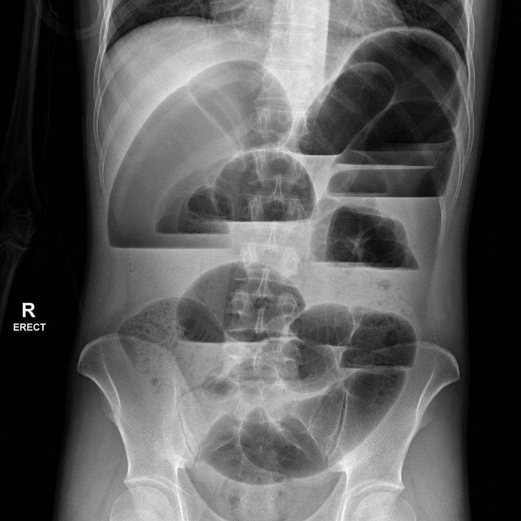
True statements about congenital megacolon include all of the following except:
In cryptorchidism, what is the hallmark age for histological changes to appear in the testis?
What is the commonest salivary gland tumor in children?
What is the standard surgical treatment for a thyroglossal cyst?
What is the best age for surgical correction of a combined unilateral cleft lip and hard palate?
A newborn presents with a mid-anterior abdominal wall defect. A characteristic feature of this swelling is spontaneous resolution by age 5 years. What is the most likely diagnosis?
Explanation: **Explanation:** The development of a hydrocele in infants is primarily a developmental anatomical issue rather than an acquired pathology. **1. Why Patent Processus Vaginalis (PPV) is correct:** During fetal development, the **processus vaginalis** is a peritoneal diverticulum that precedes the descent of the testis into the scrotum. Normally, the stalk of this process obliterates shortly before or after birth. If the processus remains **patent**, peritoneal fluid can track down and accumulate around the testis within the tunica vaginalis, resulting in a **communicating hydrocele**. This is the most common cause in the pediatric age group and is embryologically identical to the mechanism of an indirect inguinal hernia (the difference being the size of the opening). **2. Why other options are incorrect:** * **Patent gubernaculum:** The gubernaculum is a mesenchymal structure that guides testicular descent; it does not form a lumen or sac that could hold fluid. * **Impaired lymphatic drainage:** This is a common cause of **secondary (acquired) hydroceles** in adults (e.g., due to filariasis or post-surgical scarring), but it is not the primary mechanism in infants. * **Epididymal cyst:** This is a discrete, fluid-filled collection arising from the efferent ductules of the epididymis, usually presenting as a separate paratesticular mass rather than generalized scrotal swelling. **Clinical Pearls for NEET-PG:** * **Management:** Most congenital hydroceles resolve spontaneously by **1–2 years of age** as the PPV closes. Surgery (High Ligation) is indicated if it persists beyond age 2 or if a clinical hernia is present. * **Physical Exam:** Hydroceles are characterized by **transillumination** and the inability to "get above the swelling" (if communicating). * **Key Difference:** In a hydrocele, the fluid is in the *tunica vaginalis*; in a hernia, *bowel/omentum* is in the *processus vaginalis*.
Explanation: **Explanation:** **Hypertrophic Pyloric Stenosis (HPS)** is characterized by the hypertrophy of the circular muscle fibers of the pylorus, leading to gastric outlet obstruction. 1. **Why Ramstedt’s Operation is Correct:** The definitive surgical treatment for HPS is **Ramstedt’s pyloromyotomy**. In this procedure, a longitudinal incision is made through the hypertrophied serosa and muscularis layers down to the submucosa. The muscle is then spread until the bulging intact mucosa is seen. This relieves the obstruction without entering the lumen of the stomach. 2. **Why Other Options are Incorrect:** * **Truncal Vagotomy:** Used in the surgical management of peptic ulcer disease to reduce acid secretion; it has no role in pediatric pyloric obstruction. * **Heller’s Operation:** Specifically, Heller’s Cardiomyotomy is the surgical treatment for **Achalasia Cardia**, involving the division of muscle fibers at the lower esophageal sphincter. * **Delorme’s Operation:** A mucosal proctectomy used for the treatment of **rectal prolapse**. **High-Yield Clinical Pearls for NEET-PG:** * **Classic Presentation:** A 3–6 week-old male infant with **non-bilious, projectile vomiting** and a palpable "olive-shaped" mass in the epigastrium. * **Metabolic Abnormality:** Hypochloremic, hypokalemic, metabolic alkalosis with **paradoxical aciduria** (crucial for exams). * **Diagnosis:** Ultrasound is the investigation of choice (Pyloric thickness >4mm, length >14mm). * **Management Priority:** HPS is a **medical emergency, not a surgical one.** The priority is correcting dehydration and electrolyte imbalances before proceeding to Ramstedt’s operation.
Explanation: ### Explanation The metabolic hallmark of **Congenital Hypertrophic Pyloric Stenosis (CHPS)** is **Hypochloremic, Hypokalemic, Metabolic Alkalosis with Paradoxical Aciduria.** **1. Why Option A is Correct:** The pathophysiology is driven by persistent, non-bilious projectile vomiting. * **Hypochloremia & Alkalosis:** Vomitus contains high amounts of Gastric HCl. Loss of $H^+$ ions leads to metabolic alkalosis, while loss of $Cl^-$ ions leads to hypochloremia. * **Hypokalemia:** As the body tries to compensate for alkalosis, the kidneys exchange $K^+$ for $H^+$ to preserve serum pH. Furthermore, dehydration triggers the Renin-Angiotensin-Aldosterone System (RAAS); aldosterone causes $Na^+$ reabsorption at the expense of $K^+$ and $H^+$ excretion. * **Paradoxical Aciduria:** In severe dehydration, the kidney prioritizes volume expansion ($Na^+$ saving) over pH balance. It excretes $H^+$ ions instead of $Na^+$, leading to acidic urine despite systemic alkalosis. **2. Why Other Options are Incorrect:** * **Hirschsprung’s Disease:** Presents with distal bowel obstruction and failure to pass meconium. If vomiting occurs, it is usually bilious and late-onset, typically causing dehydration rather than this specific electrolyte pattern. * **Esophageal Atresia:** Presents with drooling and choking on first feed. Since gastric acid production is not "lost" via vomiting (as the obstruction is proximal to the stomach), this specific alkalosis does not develop. * **Jejunal Atresia:** Presents with **bilious vomiting**. Because the obstruction is distal to the ampulla of Vater, the vomitus contains alkaline bile and pancreatic juices, often leading to a neutral pH or metabolic acidosis. **3. High-Yield Clinical Pearls for NEET-PG:** * **Age of Presentation:** Typically 3–6 weeks of life. * **Physical Exam:** Palpable "olive-shaped" mass in the epigastrium and visible gastric peristalsis. * **Diagnosis:** Ultrasound is the gold standard (Pyloric muscle thickness >4mm, length >14mm). * **Management:** CHPS is a **medical emergency, not a surgical one.** Correct electrolytes and dehydration first. The definitive surgery is **Ramstedt’s Pyloromyotomy.**
Explanation: **Explanation:** Abdominal distension in pediatric surgery and general medicine often results from either a mechanical obstruction or functional ileus (paralysis of bowel motility). **1. Why Option D is Correct:** * **Hirschsprung’s Disease:** This is a mechanical cause of distension. The absence of ganglion cells in the distal colon (aganglionosis) prevents relaxation of the internal anal sphincter, leading to a functional obstruction, proximal colonic dilation, and marked abdominal distension. * **Hyperkalemia:** While hypokalemia is a classic cause of paralytic ileus, **severe hyperkalemia** can also lead to intestinal dysmotility and distension. It disrupts the normal electrical gradient of smooth muscle cells, potentially leading to muscle weakness or paralysis of the gut wall. * **Hypomagnesemia:** Magnesium is a critical cofactor for the Na+/K+-ATPase pump. Low levels impair smooth muscle contraction and are frequently associated with refractory paralytic ileus, leading to gas accumulation and distension. **2. Why Other Options are Incorrect:** * **Hypokalemia:** While hypokalemia *does* cause abdominal distension (paralytic ileus), the options provided (A, B, and C) are incorrect because they either repeat the same electrolyte twice or lack the specific combination found in the validated correct answer (D), which includes the surgical hallmark (Hirschsprung's) alongside metabolic triggers. **3. NEET-PG High-Yield Pearls:** * **Hirschsprung’s Disease:** The gold standard for diagnosis is a **Rectal Suction Biopsy** showing absence of ganglion cells and hypertrophy of nerve bundles (increased Acetylcholinesterase staining). * **Paralytic Ileus:** Always check electrolytes in a distended postoperative patient. The most common metabolic triggers are **Hypokalemia, Hypomagnesemia, and Hyponatremia.** * **X-ray Finding:** In Hirschsprung’s, look for the "inverted cone" appearance on a contrast enema, representing the transition zone between the narrow aganglionic segment and the dilated proximal normal colon.
Explanation: **Explanation:** Congenital Megacolon, or **Hirschsprung’s Disease**, is characterized by the absence of ganglion cells (Auerbach’s and Meissner’s plexuses) in the distal bowel, leading to a functional obstruction. **Why "Large Stool" is the Correct Answer (The Exception):** In Hirschsprung’s disease, the stool is typically **ribbon-like, pellet-like, or thin**, rather than large. This is because the stool must pass through a narrow, tonically contracted aganglionic segment. "Large, bulky stools" are instead a hallmark of **acquired megacolon** (functional constipation), where the rectum is dilated and filled with a massive fecal bolus. **Analysis of Other Options:** * **A. Dilatation and hypertrophy of pelvic colon:** This occurs in the **proximal** ganglionic segment. As it tries to overcome the distal obstruction, the normal colon undergoes compensatory hypertrophy and massive dilatation (the "megacolon"). * **B. Loud borborygmi:** These are hyperactive bowel sounds produced as the proximal dilated bowel struggles with vigorous peristalsis to push contents through the narrow distal segment. * **C. Symptoms appear within 3 days:** 90% of affected neonates fail to pass meconium within the first 24–48 hours of life. Early symptoms include abdominal distension and bilious vomiting. **NEET-PG High-Yield Pearls:** * **Gold Standard Diagnosis:** Rectal Suction Biopsy (shows absence of ganglion cells and increased acetylcholinesterase staining). * **Pathophysiology:** Failure of neural crest cell migration (proximal to distal). * **Associated Syndrome:** Down Syndrome (Trisomy 21) is the most common chromosomal association. * **Radiology:** Contrast enema shows a "Transition Zone" (narrow distal segment and dilated proximal segment). * **Classic Sign:** "Blast sign" or "Squirt sign" (explosive release of gas and stool upon digital rectal exam).
Explanation: **Explanation:** In cryptorchidism (undescended testis), the hallmark age for the onset of irreversible histological changes is **1 year**. **1. Why 1 Year is Correct:** The core pathophysiology involves the exposure of the testis to a higher core body temperature compared to the cooler scrotal environment. By age **1 year**, significant histological alterations begin to manifest, including: * **Loss of germ cells:** A decrease in the number of spermatogonia. * **Reduced tubular diameter:** Atrophy of the seminiferous tubules. * **Peritubular fibrosis:** Thickening of the basement membrane. Because these changes can lead to future infertility and increase the risk of malignancy, current guidelines recommend performing **Orchidopexy between 6 to 12 months of age** to preserve fertility potential. **2. Why Other Options are Incorrect:** * **4, 6, and 8 months:** While the testis is unlikely to descend spontaneously after 3–4 months of age, the microscopic damage to the germinal epithelium is generally not significant or irreversible at these early stages. These ages represent the window where surgical intervention is planned to *prevent* the damage that becomes prominent at the 1-year mark. **Clinical Pearls for NEET-PG:** * **Spontaneous Descent:** Most testes descend by 3 months; it is rare after 4 months. * **Most Common Site:** The inguinal canal is the most common location for an undescended testis. * **Malignancy Risk:** Cryptorchidism increases the risk of testicular cancer (most commonly **Seminoma**). Orchidopexy does not eliminate this risk but makes the testis accessible for screening. * **Infertility:** Bilateral cryptorchidism carries a much higher risk of infertility than unilateral cases.
Explanation: **Explanation:** In the pediatric population, salivary gland tumors are relatively rare, accounting for less than 10% of all head and neck tumors. However, when they occur, the distribution differs significantly from adults. **1. Why Mucoepidermoid Carcinoma is Correct:** While the majority of salivary gland masses in children are inflammatory (like mumps) or vascular (like hemangiomas), **Mucoepidermoid Carcinoma (MEC)** is the **most common malignant salivary gland tumor** and the **most common overall salivary gland neoplasm** in children and adolescents. It most frequently involves the parotid gland. Unlike adults, where benign tumors (Pleomorphic Adenoma) are more common than malignancies, the "Rule of 50" often applies in pediatrics: approximately 50% of solid salivary masses in children are malignant. **2. Analysis of Incorrect Options:** * **Pleomorphic Adenoma (Option C):** This is the most common benign salivary tumor in children. While frequent, current epidemiological data and surgical series indicate that MEC slightly edges it out as the most common epithelial neoplasm in the pediatric age group. * **Adenoid Cystic Carcinoma (Option B):** This is the second most common malignant salivary tumor in children. It is known for perineural invasion but is less frequent than MEC. * **Lymphoma (Option A):** While lymphomas can involve the intra-parotid lymph nodes, they are considered systemic or nodal malignancies rather than primary salivary gland epithelial tumors. **3. NEET-PG High-Yield Pearls:** * **Most common salivary gland tumor (Overall/Adults):** Pleomorphic Adenoma. * **Most common salivary gland tumor (Children):** Mucoepidermoid Carcinoma. * **Most common site:** Parotid gland (for both benign and malignant). * **Most common vascular lesion of the parotid in infants:** Hemangioma (often regresses spontaneously). * **Malignancy Risk:** The smaller the salivary gland, the higher the risk of malignancy (e.g., a sublingual gland mass is more likely to be malignant than a parotid mass).
Explanation: **Explanation:** The **Sistrunk operation** is the gold standard surgical treatment for a thyroglossal cyst. The thyroglossal duct is formed during the descent of the thyroid gland from the foramen cecum to its final position in the neck. Because the duct is intimately associated with the development of the **hyoid bone**, the cyst's tract often passes through or behind it. The Sistrunk procedure involves a wide excision of the cyst, the entire tract up to the foramen cecum, and the **central portion of the hyoid bone**. This radical approach reduces the recurrence rate to less than 3%, compared to over 50% with simple excision. **Analysis of Options:** * **B. Cyst excision:** Simple excision (enucleation) is inadequate because it leaves behind the proximal duct and the hyoid bone, leading to a very high rate of recurrence. * **C. Antibiotic therapy:** This is only indicated if the cyst is acutely infected (thyroglossal cyst abscess). It is a supportive measure to resolve inflammation before definitive surgery but does not treat the underlying anatomical defect. * **D. All of the above:** Incorrect, as Sistrunk is the specific definitive surgical standard. **High-Yield Clinical Pearls for NEET-PG:** * **Location:** Most common midline neck swelling in children; moves upward on **protrusion of the tongue** (due to attachment to the hyoid) and on deglutition. * **Ectopic Thyroid:** Always perform an ultrasound before surgery to ensure the cyst is not the patient's only functioning thyroid tissue. * **Malignancy:** Though rare (<1%), the most common cancer arising in a thyroglossal cyst is **Papillary Thyroid Carcinoma**.
Explanation: **Explanation:** The surgical management of cleft lip and palate follows a strict chronological sequence to balance anatomical restoration with facial growth. 1. **Why 15-18 months is correct:** In a **combined** unilateral cleft lip and hard palate, the timing is dictated by the more complex component—the **hard palate**. While the soft palate is often repaired earlier (6–12 months) to facilitate speech development, the hard palate repair is ideally deferred until **15–18 months**. This delay allows for significant transverse maxillary growth; operating too early on the hard palate can lead to severe midface hypoplasia and dental malocclusion due to surgical scarring. 2. **Analysis of incorrect options:** * **3-6 months (Option B):** This is the standard timing for **Cleft Lip** repair (often following the "Rule of 10s"). It is too early for palate repair as it carries a high risk of growth retardation of the maxilla. * **6-9 months (Option C):** This is the window typically reserved for **Soft Palate** repair (Staphylorrhaphy) to ensure the child develops proper velopharyngeal competence before they begin speaking. * **9-15 months (Option D):** While some centers repair the palate at 12 months, for a **combined** defect involving the hard palate, 15-18 months is the preferred consensus to optimize the balance between speech outcomes and maxillofacial growth. **High-Yield NEET-PG Pearls:** * **Rule of 10s (for Cleft Lip):** 10 weeks of age, 10 lbs weight, 10 g/dL Hemoglobin. * **Sequence of Repair:** Lip (3-6 months) → Soft Palate (6-12 months) → Hard Palate (15-18 months) → Alveolar Bone Grafting (9-11 years, during mixed dentition). * **Most common complication of palate surgery:** Velopharyngeal insufficiency and fistula formation.
Explanation: ### Explanation **Correct Answer: D. Umbilical hernia** **Why it is correct:** An umbilical hernia is a common pediatric condition caused by the failure of the umbilical ring to close after birth. It presents as a protrusion at the umbilicus that is covered by **skin and subcutaneous tissue**. The hallmark of a pediatric umbilical hernia is its high rate of **spontaneous resolution**. Approximately 80–90% of these hernias close by age 3 to 5 years as the abdominal wall muscles strengthen. Surgery is generally deferred until after age 5 unless the defect is very large (>2 cm) or complications occur. **Why the other options are incorrect:** * **A. Patent Urachus:** This is a persistent communication between the bladder and the umbilicus. It presents with continuous urine leakage from the umbilicus, not a swelling that spontaneously resolves. * **B. Omphalocele:** This is a major midline defect where abdominal viscera protrude through the umbilical ring, **covered only by a translucent sac** (amnion and peritoneum). It is a surgical emergency associated with chromosomal anomalies and does not resolve spontaneously. * **C. Ectopia Vesicae (Bladder Exstrophy):** This involves a defect in the lower abdominal wall and anterior bladder wall, leaving the bladder mucosa exposed. It requires complex reconstructive surgery and is often associated with epispadias. **High-Yield Clinical Pearls for NEET-PG:** * **Covering:** Unlike Gastroschisis (no sac) or Omphalocele (peritoneal sac), an Umbilical Hernia is always covered by **skin**. * **Management:** Reassurance is the first line. Surgery (Umbilical Herniorrhaphy) is indicated if the hernia persists beyond **age 4–5**, is symptomatic, or becomes incarcerated (rare). * **Associations:** Increased incidence in infants with **Down syndrome, Trisomy 13, 18, and Congenital Hypothyroidism.** * **Strangulation:** The risk of strangulation in pediatric umbilical hernias is extremely low compared to adults.
Neonatal Physiology
Practice Questions
Congenital Anomalies Overview
Practice Questions
Neonatal Intestinal Obstruction
Practice Questions
Necrotizing Enterocolitis
Practice Questions
Hirschsprung's Disease
Practice Questions
Anorectal Malformations
Practice Questions
Pediatric Hernias
Practice Questions
Pyloric Stenosis
Practice Questions
Biliary Atresia
Practice Questions
Pediatric Tumors
Practice Questions
Congenital Diaphragmatic Hernia
Practice Questions
Pediatric Trauma
Practice Questions
Get full access to all questions, explanations, and performance tracking.
Start For Free