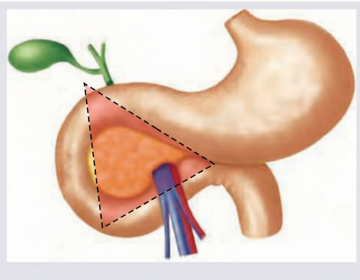
Which tumor is commonly seen in the area marked below?

Gastrinoma is classically associated with which of the following?
A patient with obstructive jaundice has the following imaging findings suggestive of pancreatic cancer. Which is the most appropriate method for obtaining tissue diagnosis?

Which of the following is not an indication for surgical intervention in acute pancreatitis?
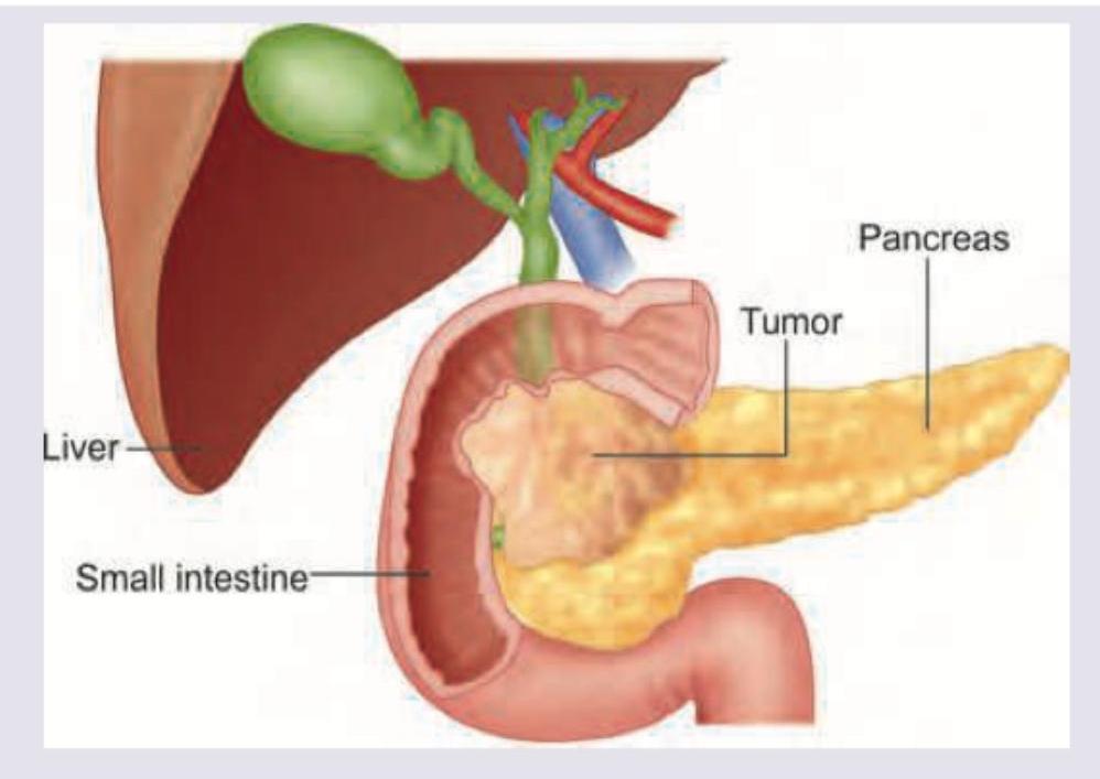
A 65-year-old patient presents with obstructive jaundice and 15 kg weight loss. An ultrasound shows a 4 cm mass in the head of the pancreas with dilated bile ducts. Further work up includes a helical CT scan. The study shows several lesions consistent with metastasis in the right and left lobes of the liver and encasement of gastroduodenal artery. The most appropriate treatment would be:
In acute pancreatitis, surgery is indicated in which one of the following conditions?
A 40-year old alcoholic male complains of acute pain in the epigastrium associated with vomiting for the last 10 days. On clinical examination, he is found to have a mass in the epigastrium. The most likely diagnosis is
During Pylorus preserving pancreatico-duodenectomy (PPPD) the following organs are removed except:
A 45 year old female presented with a cystic lesion in the lesser sac on CT scan. Endoscopic ultrasound guided aspiration showed amylase to be 500 IU and carcinoembryonic antigen as 500ng/ml. What was she suffering from?
Pancreatic pseudocysts developing complications are best managed by?
Explanation: ***Gastrinoma*** - **Gastrinomas** are predominantly found in the **Gastrinoma Triangle (Passaro's Triangle)**, bounded by the junction of the cystic and common hepatic ducts, the second and third portions of the duodenum, and the neck and body of the pancreas. - This anatomical region accounts for **90%** of gastrinomas, with the **duodenum** being the most common location within this triangle. *Insulinoma* - **Insulinomas** are distributed more evenly throughout the pancreas, with **90%** located in the pancreatic parenchyma rather than the duodenum. - They show **no predilection** for the gastrinoma triangle and can occur in the head, body, or tail of the pancreas with equal frequency. *Non-functional tumors* - **Non-functional pancreatic neuroendocrine tumors** typically occur in the **pancreatic body and tail** rather than the gastrinoma triangle. - They tend to be **larger at presentation** since they lack hormonal symptoms for early detection, unlike gastrinomas which cause **Zollinger-Ellison syndrome**. *VIPoma* - **VIPomas** predominantly occur in the **distal pancreas (body and tail)**, not in the gastrinoma triangle region. - They cause **WDHA syndrome** (watery diarrhea, hypokalemia, achlorhydria) and are rarely found in the duodenal or periampullary region.
Explanation: **Explanation:** **Gastrinoma** is a neuroendocrine tumor (most commonly found in the Gastrinoma Triangle) that secretes excessive amounts of gastrin, leading to **Zollinger-Ellison Syndrome (ZES)**. **Why Diarrhea is the Correct Answer:** Diarrhea occurs in approximately 30–50% of patients with gastrinoma and is often the presenting symptom. The underlying mechanisms include: 1. **High Acid Volume:** Massive gastric acid secretion exceeds the resorptive capacity of the small intestine. 2. **Mucosal Injury:** Excess acid causes direct inflammatory damage to the intestinal villi. 3. **Steatorrhea:** The low pH in the duodenum inactivates pancreatic lipase and causes the precipitation of bile acids, leading to fat malabsorption. **Analysis of Incorrect Options:** * **B. Constipation:** This is not a feature of ZES. The hypersecretory state leads to increased intestinal motility and fluid load. * **C. Achlorhydria:** Gastrinoma is characterized by **Hyperchlorhydria** (extreme acid production). Achlorhydria (lack of acid) is actually a stimulus for physiological hypergastrinemia, but in the context of a gastrinoma, the acid levels are pathologically high. * **D. Multiple superficial gastric ulcers:** While ZES causes ulcers, they are typically **deep, refractory, and often located in atypical sites** (e.g., distal duodenum or jejunum). Furthermore, they are more commonly duodenal than gastric. **High-Yield Clinical Pearls for NEET-PG:** * **Passaro’s Triangle (Gastrinoma Triangle):** Junction of cystic/common bile duct, junction of 2nd/3rd part of duodenum, and neck/body of pancreas. * **Diagnosis:** Best initial test is **Fasting Serum Gastrin** (>1000 pg/mL is diagnostic). The most specific provocative test is the **Secretin Stimulation Test**. * **Association:** 25% of cases are associated with **MEN-1 syndrome** (3Ps: Parathyroid, Pancreas, Pituitary). * **Localization:** **Somatostatin Receptor Scintigraphy (SRS)** is the imaging modality of choice.
Explanation: ***EUS guided trans-gastric biopsy*** - **Endoscopic Ultrasound (EUS)** provides high-resolution imaging of the pancreas and surrounding structures, allowing for precise targeting of lesions for biopsy. - EUS-guided fine-needle aspiration (FNA) can accurately diagnose pancreatic tumors causing **obstructive jaundice** with minimal risk of tumor seeding. *CECT guided biopsy* - While computed tomography (CECT) can identify pancreatic masses, **percutaneous CECT-guided biopsy** carries a higher risk of complications such as bleeding, pancreatitis, and tumor seeding, especially for lesions near vital structures. - This method is less accurate for small lesions and may not always provide sufficient tissue for complete diagnosis compared to EUS-FNA. *MRI guided biopsy* - **MRI-guided biopsy** is not a standard or preferred method for biopsying pancreatic lesions due to technical challenges and the availability of more efficient and less invasive alternatives like EUS-FNA. - While MRI is excellent for imaging tissue characteristics, its real-time guidance capabilities for biopsy are limited compared to ultrasound. *Laparoscopic biopsy* - **Laparoscopic biopsy** is a more invasive surgical procedure compared to EUS-FNA and is typically reserved for cases where less invasive methods have failed or when **surgical staging** is required. - It involves general anesthesia, longer recovery, and higher risks of complications, making it less appropriate for obtaining a primary tissue diagnosis in obstructive jaundice due to a pancreatic mass.
Explanation: ***Acute fluid collection*** - **Acute fluid collections** are common in acute pancreatitis and are often **sterile** and resolve spontaneously without intervention. - Early surgical intervention for uncomplicated acute fluid collections is generally **contraindicated** due to high morbidity and mortality. *Diagnostic dilemma* - When the diagnosis of acute pancreatitis is uncertain and other surgical emergencies, such as **perforated viscus** or **ischemic bowel**, cannot be ruled out, surgery may be necessary. - An **exploratory laparotomy** can help confirm the diagnosis and address any concurrent surgical pathology. *Pancreatic abscess* - A **pancreatic abscess** is a localized collection of pus in or near the pancreas, indicating **infected necrotic tissue**. - Surgical drainage and debridement are typically required to control the infection and prevent systemic sepsis. *Infected pancreatic necrosis* - **Infected pancreatic necrosis** is a severe complication of acute pancreatitis with high mortality, often requiring surgical debridement (necrosectomy). - While sterile necrosis may be managed conservatively, **infected necrosis** necessitates intervention to remove the source of infection.
Explanation: **_Endoscopic stenting of bile duct_** - The presence of **distant liver metastases** and **vascular encasement** makes the disease inoperable and renders curative surgery impossible. - **Endoscopic stenting** offers effective palliation for **obstructive jaundice**, improving quality of life by relieving symptoms such as itching and nausea, and preventing cholangitis. *Total pancreatectomy* - This is an **extensive surgical procedure** suitable for resectable pancreatic head tumors without metastatic disease. - It is **highly morbid** and not indicated in the presence of **liver metastases** and **vascular encasement**, as it would not be curative and carries significant risks. *Biliary and gastric bypass* - This procedure aims to relieve both **biliary obstruction** and potential gastric outlet obstruction, which can occur from pancreatic head tumors. - While it addresses symptoms, it is still a **surgical intervention** with associated risks and is generally reserved for patients with a longer life expectancy or when endoscopic stenting is unsuccessful or unfeasible. It is not the most appropriate initial palliative step given the metastatic disease. *Pancreaticoduodenectomy (Whipple procedure)* - The **Whipple procedure** is the standard curative surgical treatment for **resectable pancreatic head cancers**. - However, the patient's presentation with **liver metastases** and **gastroduodenal artery encasement** indicates unresectable disease, making this procedure inappropriate and potentially harmful.
Explanation: ***Infected pancreatic necrosis*** - **Infected pancreatic necrosis** is a severe complication of acute pancreatitis requiring surgical or percutaneous debridement (necrosectomy) to remove infected tissue and prevent sepsis. - The presence of infection in necrotic tissue significantly increases morbidity and mortality, making intervention crucial. *Acute pseudocyst* - An acute pseudocyst is usually managed conservatively and only requires intervention if it is **symptomatic**, rapidly expanding, or becomes infected. - Surgical drainage is typically reserved for large, symptomatic, or complicated pseudocysts that persist beyond 6 weeks. *Acute fluid collection* - **Acute fluid collections** are generally self-limiting and resolve without intervention. - They are typically asymptomatic and represent an early stage of fluid accumulation, often preceding pseudocyst formation. *Sterile pancreatic necrosis* - **Sterile pancreatic necrosis** is usually managed with supportive care, as surgical intervention in the absence of infection does not improve outcomes and may increase complications. - The key distinction is the absence of infection—surgery is indicated only when necrosis becomes infected.
Explanation: **Pancreatic pseudocyst** - The presence of an **epigastric mass** in an **alcoholic male** with acute epigastric pain and vomiting for 10 days is highly suggestive of a pancreatic pseudocyst, which commonly develops after **acute pancreatitis**. - Pseudocysts are collections of **pancreatic fluid** encased by a non-epithelial inflammatory wall, often presenting as a palpable mass within days to weeks after an acute episode. - The **alcoholic history** and **acute presentation** strongly support this diagnosis. *Carcinoma of head of the pancreas* - While pancreatic carcinoma can cause epigastric pain, **vomiting** and an **acute course** over 10 days are less typical. - It is usually associated with **weight loss**, **jaundice** (if the head is involved), and a more insidious onset. *Perforated peptic ulcer with sub-hepatic abscess* - A perforated peptic ulcer presents with **sudden, severe epigastric pain** that rapidly worsens, often with signs of **peritonitis** and acute abdomen. - A sub-hepatic abscess would typically be accompanied by **fever**, **leukocytosis**, and signs of sepsis. *Hepatoma in left lobe of liver* - A hepatoma would usually present with a **liver mass**, potentially **jaundice**, weight loss, and abnormal liver function tests, rather than an acute onset of severe epigastric pain and vomiting. - The mass would typically be felt in the **right upper quadrant** or epigastrium, but the clinical picture does not align with an acute pancreatic process.
Explanation: ***Stomach*** - In a **pylorus-preserving pancreaticoduodenectomy (PPPD)**, the **pylorus** and a portion of the **stomach** are deliberately preserved. - This distinguishes it from the classic Whipple procedure, where the **antrum** and pylorus of the stomach are removed. *Distal Bile Duct* - The **distal bile duct** is routinely resected in both standard and pylorus-preserving Whipple procedures to ensure adequate margins and remove potential **lymph node** metastases. - This is necessary because pancreatic head tumors often involve or compress the **distal common bile duct**. *Gall bladder* - The **gallbladder** is invariably removed during a PPPD to provide access to the **common bile duct** and facilitate the creation of a **choledochojejunostomy**. - Its removal prevents future complications like **cholecystitis** or **choledocholithiasis** secondary to altered bile flow. *Head of pancreas* - The **head of the pancreas** is the primary target for resection in a PPPD as this is typically the location of the **tumor**. - This involves removing the mass along with surrounding pancreatic tissue to achieve clear **surgical margins**.
Explanation: ***Mucinous neoplasm of pancreas*** - **Markedly elevated CEA** (500 ng/ml, well above the threshold of 192 ng/ml) is highly specific for **mucinous cystic neoplasms** (MCN or IPMN). - The presence of **elevated amylase** (500 IU) indicates communication with the pancreatic ductal system, which can occur with **intraductal papillary mucinous neoplasms (IPMN)** or MCN with ductal involvement. - **CEA >192 ng/ml has >90% specificity** for distinguishing mucinous from non-mucinous lesions. - This patient likely has either an **MCN** (mucinous cystadenoma/cystadenocarcinoma) or **IPMN** with malignant potential requiring surgical evaluation. *Pseudocyst pancreas with ductal communication* - Pseudocysts typically have **high amylase** but **low CEA (<5 ng/ml)**. - A CEA of 500 ng/ml essentially **rules out a simple pseudocyst**. - Pseudocysts lack epithelial lining and do not produce CEA. *Chronic pseudocyst* - Similar to acute pseudocyst, chronic pseudocysts have **high amylase but low CEA**. - The markedly elevated CEA (500 ng/ml) makes this diagnosis incorrect. - Would expect CEA <5 ng/ml in pseudocyst fluid. *Pancreatic adenocarcinoma* - Solid pancreatic adenocarcinoma can have elevated CEA, but typically presents as a **solid mass**, not a cystic lesion. - Cyst fluid analysis would show **malignant cells on cytology** and typically **low amylase**. - Does not present as a pure cystic lesion in the lesser sac.
Explanation: ***Surgery*** - When pancreatic pseudocysts develop **complications** (infection, hemorrhage, rupture, gastric outlet/biliary obstruction), definitive management is required. - Surgical internal drainage procedures (**cyst-gastrostomy**, **cyst-jejunostomy**, or **cyst-duodenostomy**) provide durable treatment by creating a permanent communication between the mature pseudocyst and the GI tract. - Surgery is particularly indicated when the pseudocyst has a **mature wall (>6 weeks)**, is **large (>6 cm)**, or when endoscopic approaches are not feasible or have failed. - While endoscopic drainage (EUS-guided) is increasingly used as first-line therapy, surgery remains the gold standard for complicated pseudocysts requiring definitive management, especially with complex anatomy or failed minimally invasive approaches. *Conservative treatment* - Conservative management with observation, pain control, and nutritional support is appropriate only for **asymptomatic, small (<6 cm)**, and **uncomplicated pseudocysts** with high likelihood of spontaneous resolution. - Once complications develop, conservative treatment is **inadequate** and poses risks of further deterioration. *Radiologically guided interventions* - Percutaneous drainage may be used for **infected pseudocysts** or as a temporizing measure, but carries high risk of **external fistula formation** (25-50%) and **recurrence**. - Does not provide internal drainage and is generally less effective than surgical or endoscopic internal drainage for complicated pseudocysts. - Not considered definitive management when complications are present. *External drainage* - External percutaneous catheter drainage is primarily a **temporizing measure** for critically ill patients or infected pseudocysts not amenable to other approaches. - High risk of **pancreaticocutaneous fistula** formation and does not address the underlying pancreatic duct communication. - Requires subsequent definitive management in most cases; not appropriate as primary treatment for complicated pseudocysts.
Pancreatic Anatomy and Physiology
Practice Questions
Acute Pancreatitis
Practice Questions
Chronic Pancreatitis
Practice Questions
Pancreatic Pseudocysts
Practice Questions
Pancreatic Adenocarcinoma
Practice Questions
Cystic Neoplasms of Pancreas
Practice Questions
Neuroendocrine Tumors of Pancreas
Practice Questions
Pancreatic Trauma
Practice Questions
Pancreatectomy Techniques
Practice Questions
Whipple Procedure
Practice Questions
Pancreatic Anastomosis
Practice Questions
Complications of Pancreatic Surgery
Practice Questions
Get full access to all questions, explanations, and performance tracking.
Start For Free