
Pancreatic Surgery — MCQs

On this page
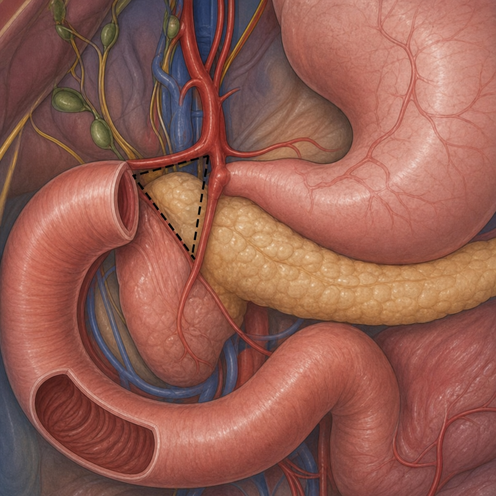
What is the name of this triangle?
True about pancreatic pseudocysts is:
A patient with jaundice is found to have a pancreatic head mass. What is the best diagnostic test?
All are true about Whipple procedure except?
A 50-year-old male with a history of alcohol abuse presents with epigastric pain radiating to the back. A CT scan of the abdomen shows a pseudocyst in the pancreas. What is the most appropriate management?
A 60-year-old man presents with jaundice and abdominal pain. An ultrasound reveals a pancreatic mass compressing the common bile duct. Further imaging confirms a resectable tumor. Which treatment plan would be the most appropriate for this patient?
A 58-year-old alcoholic male presents with jaundice and imaging reveals a pancreatic mass. Which of the following factors are MOST important in determining whether this tumor is resectable?
What is a key feature of the Whipple procedure?
In a patient with locally advanced pancreatic cancer, what factors should be considered to decide between immediate surgery and neoadjuvant chemotherapy?
A 55-year-old male with chronic pancreatitis presents with a 4 cm pseudocyst causing gastric outlet obstruction. What considerations are essential in deciding between endoscopic drainage and surgical cystogastrostomy?
Practice by Chapter
Pancreatic Anatomy and Physiology
Practice Questions
Acute Pancreatitis
Practice Questions
Chronic Pancreatitis
Practice Questions
Pancreatic Pseudocysts
Practice Questions
Pancreatic Adenocarcinoma
Practice Questions
Cystic Neoplasms of Pancreas
Practice Questions
Neuroendocrine Tumors of Pancreas
Practice Questions
Pancreatic Trauma
Practice Questions
Pancreatectomy Techniques
Practice Questions
Whipple Procedure
Practice Questions
Pancreatic Anastomosis
Practice Questions
Complications of Pancreatic Surgery
Practice Questions
Want unlimited practice?
Get full access to all questions, explanations, and performance tracking.
Scan to download app