
Pancreatic Surgery — MCQs

On this page
A chronic alcoholic presents with repeated episodes of severe intractable abdominal pain. On evaluation, the pancreatic duct was found to be dilated and stones were noted in the tail of the pancreas. What is the most appropriate management?
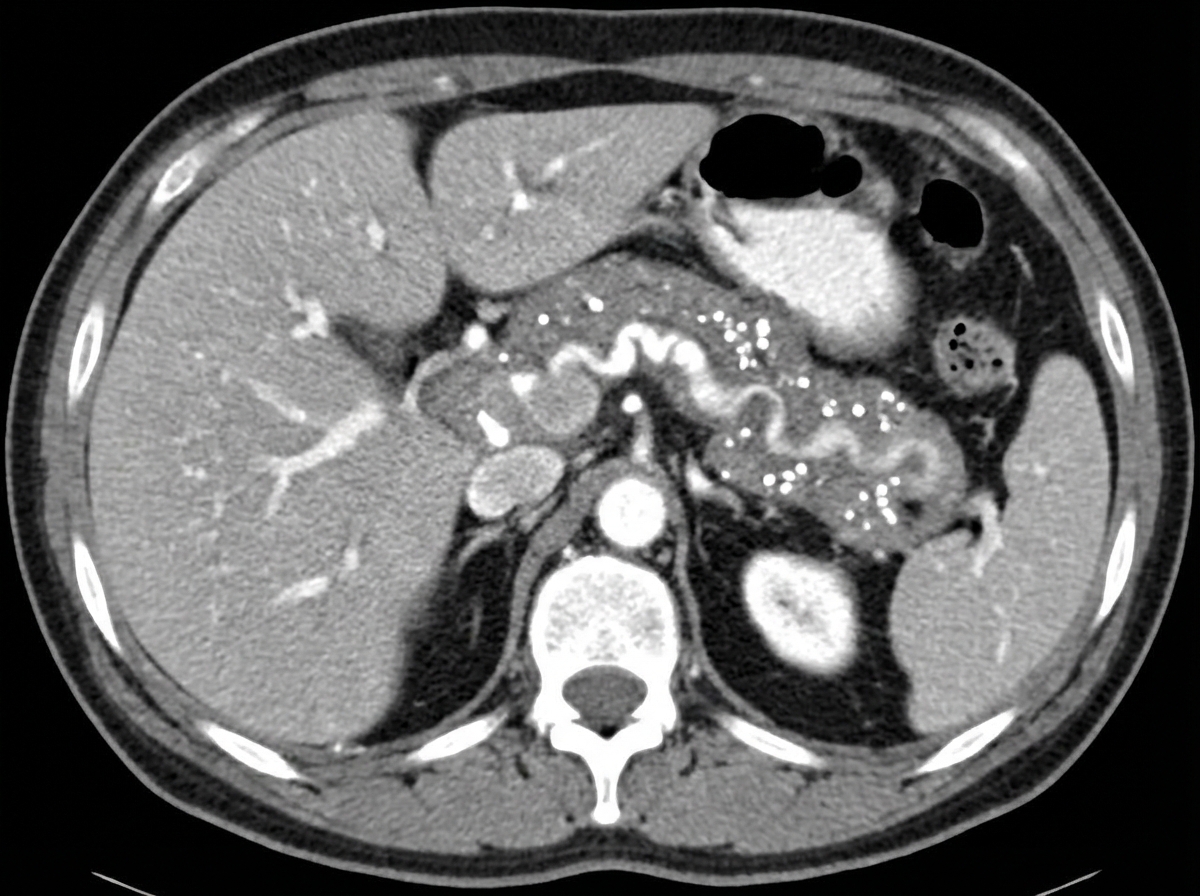
A 30-year-old known alcoholic patient presented with severe abdominal pain. On examination, a rigid abdomen is noted. The patient has a history of multiple episodes of abdominal pain over the last 4-5 years and has lost 5 kg of weight in the past 6 months. Serum lipase and amylase levels are normal. CECT abdomen was performed. What is the most likely diagnosis?
What is the most common functional neuroendocrine tumor of the pancreas?
A 40-year-old male presents with acute upper abdominal pain, shock, feeble pulse, and tachycardia. Examination reveals epigastric tenderness. Gastric aspiration shows no blood and provides relief. Abdominal X-ray is negative for free air under the diaphragm. Investigations show TLC 13,500, serum bilirubin 2.0 mg/dL, and serum amylase 800 IU/L. What is the most likely diagnosis?
A patient presents with a three-day history of epigastric pain radiating to the back. Serum amylase levels are normal, but ultrasound of the abdomen reveals gall bladder stones and an enlarged pancreas. A CT scan confirms the diagnosis. Which of the following is the most likely diagnosis?
What is the most common complication of chronic pancreatitis?
A 60-year-old man with a history of heavy alcohol consumption presented with symptoms suggestive of acute pancreatitis that began 4 days ago. He continued to consume alcohol and was admitted for further evaluation. He is currently experiencing severe vomiting and complains of dizziness upon standing. Examination reveals epigastric and right hypochondrium tenderness, with a reddish discoloration noted in the flanks. Which of the following statements regarding the patient is most accurate?
Which of the following is not a complication of a pseudopancreatic cyst?
Serum amylase level is increased in all of the following conditions except?
A 64-year-old man complains of abdominal pain, pruritus, 4-lb weight loss, and anorexia. There are multiple scratch marks on the skin of the extremities and flank. The bilirubin is 1.0 mg/dL. To determine if the condition is due to cholestasis, blood should be tested for which of the following?
Practice by Chapter
Pancreatic Anatomy and Physiology
Practice Questions
Acute Pancreatitis
Practice Questions
Chronic Pancreatitis
Practice Questions
Pancreatic Pseudocysts
Practice Questions
Pancreatic Adenocarcinoma
Practice Questions
Cystic Neoplasms of Pancreas
Practice Questions
Neuroendocrine Tumors of Pancreas
Practice Questions
Pancreatic Trauma
Practice Questions
Pancreatectomy Techniques
Practice Questions
Whipple Procedure
Practice Questions
Pancreatic Anastomosis
Practice Questions
Complications of Pancreatic Surgery
Practice Questions
Want unlimited practice?
Get full access to all questions, explanations, and performance tracking.
Scan to download app