
Pancreatic Surgery — MCQs

On this page
A 47-year-old executive consults his physician with complaints of feeling tired and several months of abdominal pain and "dark-colored" urine. Physical examination reveals slight jaundice and a palpable, non-tender gallbladder. Which of the following disorders is most likely, given this presentation?
Which of the following CT scan findings indicate unresectable criteria for cancer of the pancreas?
What is the complication least likely to occur in a pseudocyst of the pancreas?
Which of the following statements regarding a pseudocyst of the pancreas is FALSE?
The boundaries of the gastrinoma triangle are all of the following EXCEPT:
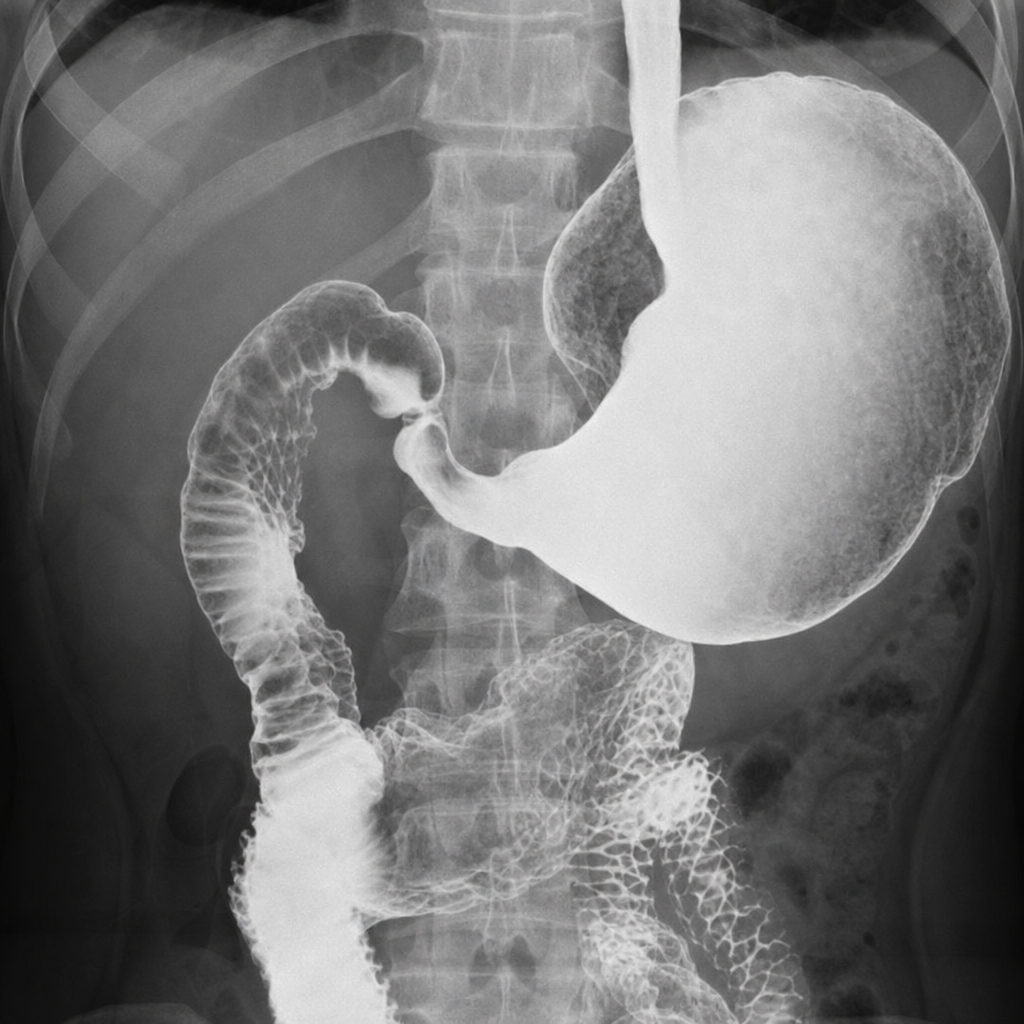
What is the operation of choice for the presented condition?
Which of the following is NOT a recognized cause of acute pancreatitis?
What is the prime cause of death from a pancreatic pseudocyst?
Complications of chronic pancreatitis include the following, except:
What are the common causes of acute pancreatitis?
Practice by Chapter
Pancreatic Anatomy and Physiology
Practice Questions
Acute Pancreatitis
Practice Questions
Chronic Pancreatitis
Practice Questions
Pancreatic Pseudocysts
Practice Questions
Pancreatic Adenocarcinoma
Practice Questions
Cystic Neoplasms of Pancreas
Practice Questions
Neuroendocrine Tumors of Pancreas
Practice Questions
Pancreatic Trauma
Practice Questions
Pancreatectomy Techniques
Practice Questions
Whipple Procedure
Practice Questions
Pancreatic Anastomosis
Practice Questions
Complications of Pancreatic Surgery
Practice Questions
Want unlimited practice?
Get full access to all questions, explanations, and performance tracking.
Scan to download app